Methodologies for bladder cancer detection with Raman based urine cytology†
Abstract
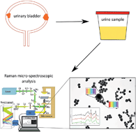
Bladder cancer has the highest recurrence rate of any cancer. The American Urological Association recommends cystoscopic surveillance every 3–6 months for 3 years, and at least once a year thereafter, particularly for high-risk patients; however, cystoscopy is invasive, expensive, and is not without insignificant morbidity for the patient. Urine cytology is often used as an adjunct to cystoscopy; however, it has a low sensitivity in detecting low grade bladder cancers. Recent studies have investigated the application of Raman micro-spectroscopy for the detection of bladder cancer via urine cytology, and it has been demonstrated to significantly improve the diagnostic sensitivity of urine cytology for low grade bladder cancer under ideal experimental conditions. In this paper we attempt to move Raman micro-spectroscopy a step closer to the clinic by systematically examining the potential of this technology to classify low and high grade bladder cancer cell lines under the stringent clinical conditions that can be expected in the standard pathology laboratory, in terms of consumables, protocols, and instrumentation. We show that the use of glass slides, traditional fixing agents, lengthy exposure to urine, red blood cell lysing agents, as well as common cell deposition methods, do not significantly impact on the diagnostic potential of Raman based urine cytology. This study suggests that urine samples prepared with the ThinPrep® UroCyte™ method and analysed with Raman micro-spectroscopy could provide a useful alternative to cystoscopy for long term bladder cancer surveillance.


 Please wait while we load your content...