Controllable synthesis of polydopamine nanoparticles in microemulsions with pH-activatable properties for cancer detection and treatment†
Abstract
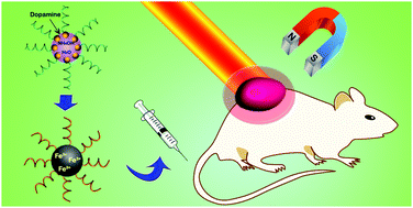
Polydopamine nanoparticles (PDA NPs) which combine diagnostic and therapeutic functions are potentially useful in biomedicine. However, it is difficult to synthesize PDA NPs of a relatively small size (≤50 nm in diameter) using the traditional polymerization of dopamine monomers in an alkaline water–ethanol solution at room temperature. Herein, PDA NPs with average diameters ranging from 25 nm to 43 nm are prepared in a way which is similar to the silica-like reverse microemulsion process. The size of the PDA NPs can be modulated by changing the amount of dopamine monomers in the microemulsion. After conjugation with ferric ions (Fe3+), the poly(ethylene glycol) modified Fe–PDA NPs (termed as PEG–Fe–PDA NPs) exhibited pH-activatable magnetic resonance imaging (MRI) contrast and high photothermal performance. The combination of a small dimension and the pH-activatable MRI contrast can greatly facilitate tumor accumulation and increase the tumor imaging sensitivity against animal models in vivo. Completely inhibited tumor growth was achieved by the PEG–Fe–PDA NPs mediated by photothermal therapy with MRI guidance.


 Please wait while we load your content...