Synthesis, molecular structure and electrochemical properties of nickel(ii) benzhydrazone complexes: influence of ligand substitution on DNA/protein interaction, antioxidant activity and cytotoxicity†
Abstract
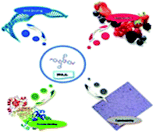
A series of new nickel(II) benzhydrazone complexes having the general formula [Ni(L)2] (where L = thiophene aldehyde benzhydrazone) have been synthesized via the reaction of Ni(OAc)2·4H2O with 2 equivalents of benzhydrazone ligands in a DMF/ethanol medium. The complexes have been characterized by analytical, spectral (FT-IR, UV-vis, NMR and ESI-mass) and single-crystal X-ray crystallography methods. All the complexes exhibit quasi-reversible one-electron reduction responses (NiII–NiI) within the E1/2 range from −0.71 to −0.77 V versus SCE. The structure of one of the complexes has been determined by a single-crystal X-ray diffraction study, which shows that coordination of the benzhydrazone ligands to the nickel occurs via azomethine nitrogen and imidolate oxygen atoms as monobasic bidentate donors with two units of the ligand in a square-planar geometry. The DNA-binding interactions of the complexes with calf thymus DNA have been investigated by absorption, emission, electrochemical, circular dichroism and viscosity measurements, which revealed that the complexes could interact with DNA via intercalation. The protein-binding interactions of the complexes with BSA were investigated by UV-vis, fluorescence and synchronous fluorescence methods, which indicated that stronger binding of the complexes with BSA and a static quenching mechanism was observed. Moreover, the potential for free-radical scavenging of all the complexes was also determined using DPPH, hydroxyl and nitric oxide radicals under in vitro conditions. Furthermore, the cytotoxicity of all the complexes was examined in vitro on the human cervical cancer cell line HeLa, MCF-7 and the normal mouse embryonic fibroblast cell line NIH-3T3 under identical conditions and they exhibited good IC50 values. These values were further supported by a neutral red uptake assay using HeLa cell lines. AO-EB/DAPI staining assays and flow cytometry analysis revealed that the complexes induce cell death only by apoptosis.


 Please wait while we load your content...