Investigation of photoinduced electron transfer on TiO2 nanowire arrays/porphyrin composite via scanning electrochemical microscopy†
Abstract
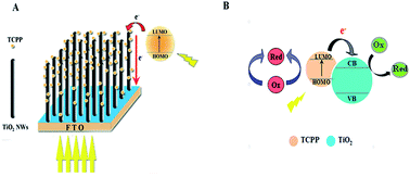
In the present study, vertically aligned single-crystalline TiO2 nanowire arrays with various lengths grown on transparent substrate have been successfully prepared using a facile hydrothermal method. They were then sensitized with a photoactive compound, 5,10,15,20-tetrakis(4-carboxylphenyl)porphyrin. The material was fully characterized by scanning electron microscopy (SEM), X-ray diffraction (XRD) and ultraviolet-visible absorption spectroscopy (UV-vis). A UV-vis/scanning electrochemical microscope (SECM) platform was developed by utilizing TiO2 nanowire arrays, and the influence of wire length as well as wavelength and intensity of light on the electron transfer rate was discussed in detail through the generated probe approach curves. The results showed that the electron transfer rate at the surface of the TiO2/porphyrin composite was accelerated by increasing the length of the nanowire, probably due to more porphyrin loading and the match of the conduction band edge of TiO2 with the LUMO energy level of the porphyrin molecules, as evidenced by the DFT theoretical calculations. The maximum rate of electron transfer was reached at a wavelength of 546 nm and an illumination intensity of 90%. The efficient PET system constructed in this study will, we believe, provide useful information to help people investigate the mechanism of the charge transfer process of artificial photosynthesis.


 Please wait while we load your content...