Ordering in bio-inorganic hybrid nanomaterials probed by in situ scanning transmission X-ray microscopy†

Abstract
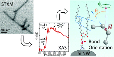
Phospholipid bilayer coated Si nanowires are one-dimensional (1D) composites that provide versatile bio-nanoelectronic functionality via incorporation of a wide variety of biomolecules into the phospholipid matrix. The physiochemical behaviour of the phospholipid bilayer is strongly dependent on its structure and, as a consequence, substantial modelling and experimental efforts have been directed at the structural characterization of supported bilayers and unsupported phospholipid vesicles; nonetheless, the experimental studies conducted to date have exclusively involved volume-averaged techniques, which do not allow for the assignment of spatially resolved structural variations that could critically impact the performance of the 1D phospholipid-Si NW composites. In this manuscript, we use scanning transmission X-ray microscopy (STXM) to probe bond orientation and bilayer thickness as a function of position with a spatial resolution of ∼30 nm for Δ9-cis 1,2-dioleoyl-sn-glycero-3-phosphocholine layers prepared Si NWs. When coupled with small angle X-ray scattering measurements, the STXM data reveal structural motifs of the Si NWs that give rise to multi-bilayer formation and enable assignment of the orientation of specific bonds known to affect the order and rigidity of phospholipid bilayers.
 Please wait while we load your content...

