Assisted deposition of nano-hydroxyapatite onto exfoliated carbon nanotube oxide scaffolds
Abstract
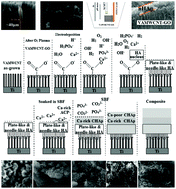
Electrodeposited nano-hydroxyapatite (nHAp) is more similar to biological apatite in terms of microstructure and dimension than apatites prepared by other processes. Reinforcement with carbon nanotubes (CNTs) enhances its mechanical properties and increases adhesion of osteoblasts. Here, we carefully studied nHAp deposited onto vertically aligned multi-walled CNT (VAMWCNT) scaffolds by electrodeposition and soaking in a simulated body fluid (SBF). VAMWCNTs are porous biocompatible scaffolds with nanometric porosity and exceptional mechanical and chemical properties. The VAMWCNT films were prepared on a Ti substrate by a microwave plasma chemical vapour deposition method, and then oxidized and exfoliated by oxygen plasma etching (OPE) to produce graphene oxide (GO) at the VAMWCNT tips. The attachment of oxygen functional groups was found to be crucial for nHAp nucleation during electrodeposition. A thin layer of plate-like and needle-like nHAp with high crystallinity was formed without any need for thermal treatment. This composite (henceforth referred to as nHAp–VAMWCNT–GO) served as the scaffold for in vitro biomineralization when soaked in the SBF, resulting in the formation of both carbonate-rich and carbonate-poor globular-like nHAp. Different steps in the deposition of biological apatite onto VAMWCNT–GO and during the short-term biomineralization process were analysed. Due to their unique structure and properties, such nano-bio-composites may become useful in accelerating in vivo bone regeneration processes.


 Please wait while we load your content...