Identification of dopaminergic neurons of the substantia nigra pars compacta as a target of manganese accumulation†‡
Abstract
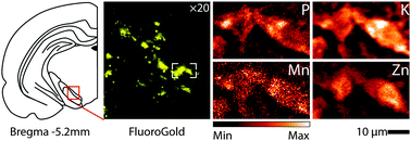
Manganese serves as a cofactor to a variety of proteins necessary for proper bodily development and function. However, an overabundance of Mn in the brain can result in manganism, a neurological condition resembling Parkinson's disease (PD). Bulk sample measurement techniques have identified the globus pallidus and thalamus as targets of Mn accumulation in the brain, however smaller structures/cells cannot be measured. Here, X-ray fluorescence microscopy determined the metal content and distribution in the substantia nigra (SN) of the rodent brain. In vivo retrograde labeling of dopaminergic cells (via FluoroGold™) of the SN pars compacta (SNc) subsequently allowed for XRF imaging of dopaminergic cells in situ at subcellular resolution. Chronic Mn exposure resulted in a significant Mn increase in both the SN pars reticulata (>163%) and the SNc (>170%) as compared to control; no other metal concentrations were significantly changed. Subcellular imaging of dopaminergic cells demonstrated that Mn is located adjacent to the nucleus. Measured intracellular manganese concentrations range between 40–200 μM; concentrations as low as 100 μM have been observed to cause cell death in cell cultures. Direct observation of Mn accumulation in the SNc could establish a biological basis for movement disorders associated with manganism, specifically Mn caused insult to the SNc. Accumulation of Mn in dopaminergic cells of the SNc may help clarify the relationship between Mn and the loss of motor skills associated with manganism.


 Please wait while we load your content...