Use of solid-state nanopores for sensing co-translocational deformation of nano-liposomes†
Abstract
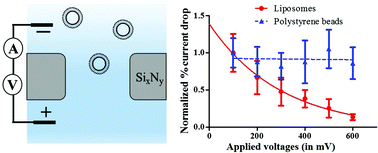
Membrane deformation of nano-vesicles is crucial in many cellular processes such as virus entry into the host cell, membrane fusion, and endo- and exocytosis; however, studying the deformation of sub-100 nm soft vesicles is very challenging using the conventional techniques. In this paper, we report detecting co-translocational deformation of individual 1,2-dioleoyl-sn-glycero-3-phosphocholine (DOPC) nano-liposomes using solid-state nanopores. Electrokinetic translocation through the nanopore caused the soft DOPC liposomes (85 nm diameter) to change their shape, which we attribute to the strong electric field strength and physical confinement inside the pore. The experiments were performed at varying transmembrane voltages and the deformation was observed to mount up with increasing applied voltage and followed an exponential trend. Numerical simulations were performed to simulate the concentrated electric field strength inside the nanopore and a field strength of 14 kV cm−1 (at 600 mV applied voltage) was achieved at the pore center. The electric field strength inside the nanopore is much higher than the field strength known to cause deformation of 15–30 μm giant membrane vesicles. As a control, we also performed experiments with rigid polystyrene beads that did not show any deformation during translocation events, which further established our hypothesis of co-translocational deformation of liposomes. Our technique presents an innovative and high throughput means for investigating deformation behavior of soft nano-vesicles.

- This article is part of the themed collection: From nanopores to nanochannels

 Please wait while we load your content...