Iron oxide nanorods as high-performance magnetic resonance imaging contrast agents†
Abstract
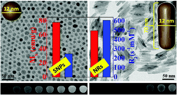
An efficient magnetic resonance imaging (MRI) contrast agent with a high R2 relaxivity value is achieved by controlling the shape of iron oxide to rod like morphology with a length of 30–70 nm and diameter of 4–12 nm. Fe3O4 nanorods of 70 nm length, encapsulated with polyethyleneimine show a very high R2 relaxivity value of 608 mM−1 s−1. The enhanced MRI contrast of nanorods is attributed to their higher surface area and anisotropic morphology. The higher surface area induces a stronger magnetic field perturbation over a larger volume more effectively for the outer sphere protons. The shape anisotropy contribution is understood by calculating the local magnetic field of nanorods and spherical nanoparticles under an applied magnetic field (3 Tesla). As compared to spherical geometry, the induced magnetic field of a rod is stronger and hence the stronger magnetic field over a large volume leads to a higher R2 relaxivity of nanorods.


 Please wait while we load your content...