
Manganese leads to an increase in markers of oxidative stress as well as to a shift in the ratio of Fe(ii)/(iii) in rat brain tissue
Abstract
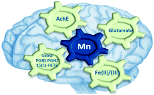
Occupationally or environmentally caused chronic exposure to Manganese (Mn) can lead to a degeneration of dopaminergic neurons inducing a Parkinson-like complaint called manganism. Deciphering the ongoing neurodegenerative mechanisms in the affected brain is still a major task for understanding the complex modes of action. Therefore, we applied a non-toxic, oral feeding in rats simulating a chronic exposure to Mn. Analysis of brain extracts by electrospray ionization Fourier transform resonance mass spectrometry (ESI-FT-ICR-MS) revealed an increase in markers of oxidative stress like glutathione disulfide (GSSG), prostaglandins, and 15(S)-HETE, a marker of lipid peroxidation. Furthermore, acetylcholinesterase (AchE) activity and glutamate concentrations were elevated in brain samples of Mn-supplemented rats, suggesting oxidative stress in the brain tissue. Application of ion chromatography coupled to inductively coupled plasma-optical emission spectrometry (IC-ICP-OES) further showed a shift of Fe(III) towards Fe(II) in the brain samples enabling for example the action of the Fenton reaction. This is the first time that changes in the Fe-species distribution could be related to Mn-induced neuroinflammation and is therefore enlarging the knowledge of this complex neurodegenerative condition. The combination of our findings provides substantial evidence that Mn-induced neuroinflammation leads to oxidative stress triggered by multifactorial pathophysiological processes.
 Please wait while we load your content...