Fine structural features of nanoscale zero-valent iron characterized by spherical aberration corrected scanning transmission electron microscopy (Cs-STEM)†
Abstract
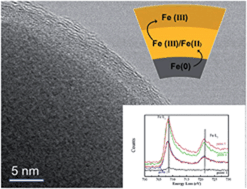
An angstrom-resolution physical model of nanoscale zero-valent iron (nZVI) is generated with a combination of spherical aberration corrected scanning transmission electron microscopy (Cs-STEM), selected area electron diffraction (SAED), energy-dispersive X-ray spectroscopy (EDS) and electron energy-loss spectroscopy (EELS) on the Fe L-edge. Bright-field (BF), high-angle annular dark-field (HAADF) and secondary electron (SE) imaging of nZVI acquired by a Hitachi HD-2700 STEM show near atomic resolution images and detailed morphological and structural information of nZVI. The STEM-EDS technique confirms that the fresh nZVI comprises of a metallic iron core encapsulated with a thin layer of iron oxides or oxyhydroxides. SAED patterns of the Fe core suggest the polycrystalline structure in the metallic core and amorphous nature of the oxide layer. Furthermore, Fe L-edge of EELS shows varied structural features from the innermost Fe core to the outer oxide shell. A qualitative analysis of the Fe L2,3 edge fine structures reveals that the shell of nZVI consists of a mixed Fe(II)/Fe(III) phase close to the Fe (0) interface and a predominantly Fe(III) at the outer surface of nZVI.


 Please wait while we load your content...