Live-cell vibrational imaging of choline metabolites by stimulated Raman scattering coupled with isotope-based metabolic labeling†
Abstract
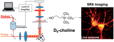
Choline is a small molecule that occupies a key position in the biochemistry of all living organisms. Recent studies have strongly implicated choline metabolites in cancer, atherosclerosis and nervous system development. To detect choline and its metabolites, existing physical methods such as magnetic resonance spectroscopy and positron emission tomography are often limited by the poor spatial resolution and substantial radiation dose. Fluorescence imaging, although with submicrometer resolution, requires introduction of bulky fluorophores and thus is difficult in labeling the small choline molecule. By combining the emerging bond-selective stimulated Raman scattering microscopy with metabolic incorporation of deuterated choline, herein we have achieved high resolution imaging of choline-containing metabolites in living mammalian cell lines, primary hippocampal neurons and the multicellular organism C. elegans. Different subcellular distributions of choline metabolites are observed between cancer cells and non-cancer cells, which may reveal a functional difference in the choline metabolism and lipid-mediated signaling events. In neurons, choline incorporation is visualized within both soma and neurites, where choline metabolites are more evenly distributed compared to proteins. Furthermore, choline localization is also observed in the pharynx region of C. elegans larvae, consistent with its organogenesis mechanism. These applications demonstrate the potential of isotope-based stimulated Raman scattering microscopy for future choline-related disease detection and development monitoring in vivo.


 Please wait while we load your content...