
Quantitative bioimaging by LA-ICP-MS: a methodological study on the distribution of Pt and Ru in viscera originating from cisplatin- and KP1339-treated mice†
Abstract
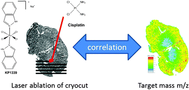
Laser ablation-inductively coupled plasma-mass spectrometry (LA-ICP-MS) was used to study the spatially-resolved distribution of ruthenium and platinum in viscera (liver, kidney, spleen, and muscle) originating from mice treated with the investigational ruthenium-based antitumor compound KP1339 or cisplatin, a potent, but nephrotoxic clinically-approved platinum-based anticancer drug. Method development was based on homogenized Ru- and Pt-containing samples (22.0 and 0.257 μg g−1, respectively). Averaging yielded satisfactory precision and accuracy for both concentrations (3–15% and 93–120%, respectively), however when considering only single data points, the highly concentrated Ru sample maintained satisfactory precision and accuracy, while the low concentrated Pt sample yielded low recoveries and precision, which could not be improved by use of internal standards (115In, 185Re or 13C). Matrix-matched standards were used for quantification in LA-ICP-MS which yielded comparable metal distributions, i.e., enrichment in the cortex of the kidney in comparison with the medulla, a homogenous distribution in the liver and the muscle and areas of enrichment in the spleen. Elemental distributions were assigned to histological structures exceeding 100 μm in size. The accuracy of a quantitative LA-ICP-MS imaging experiment was validated by an independent method using microwave-assisted digestion (MW) followed by direct infusion ICP-MS analysis.
 Please wait while we load your content...