
Visualizing the localization of transfection complexes during graphene nanoparticle-based transfection†
Abstract
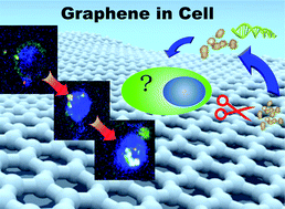
Recent research has shown that ultra-small graphene oxide (USGO) particles functionalized with polyethylenimine (PEI) can be used to transfect human cell lines with very high efficiency and low toxicity. In this study, confocal fluorescence microscopy is used to provide more insight into the nature of the complexes formed by DNA and USGO_PEI and to track their localization during the transfection process. The results indicate that the DNA enters the nucleus in complex with the USGO_PEI, which remains inside the nucleus. Also, it is observed that USGO_PEI and plasmid DNA form large aggregates in the transfection medium. Both observations raise concerns that this type of nanomaterial might need further improvements for therapeutic use.
 Please wait while we load your content...

