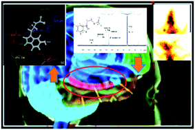
Functional imaging, for understanding the pathophysiology of neuropsychiatric disorders and neurocognition pathways, can be achieved by targeting receptors in the central neural system (CNS). The multitude of physiological functions elicited by the interaction of serotonin with its receptors and the relation between the level and ‘state’ of the serotonin receptor (5-HT1A) and neuropsychiatric diseases makes this receptor an attractive target for imaging and therapeutics. Thus, for imaging of “active” 5-HT1A a ligand with an agonistic mode of binding has been designed and evaluated as a SPECT radiotracer. In this study, serotonin, the natural ligand, has been modified as the dithiocarbamate and radiolabelled with 99mTc. The efficacy of the complex as an imaging agent has been evaluated in terms of radiochemical purity, stability, lipophilicity, biodistribution pattern and scintigraphy. For understanding the binding mode, the ligand has been docked on the 5-HT1A receptor homology models built using the crystal structure of the human β2-adrenergic receptor in the presence (PDB 3D4S) and absence (PDB 2RH1) of cholesterol. The docking results are in line with the agonist mode of binding for the ligand and strongly supported by the experimental data. Preliminary results indicate excellent radiolabelling above 95%, a biphasic blood clearance pattern with t1/2 (fast) of 45 minutes and t1/2 (slow) of 5.9 hours and a predominantly hepatobiliary excretion route. The regional brain uptake studies indicate localization of the complex in the hippocampus region. Thus, a novel dithiocarbamate based on the scaffold of serotonin has been synthesized and evaluated as a potential neuroimaging agent.

You have access to this article
 Please wait while we load your content...
Something went wrong. Try again?


 Please wait while we load your content...