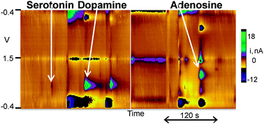
Methods to determine neurochemical concentrations in small samples of tissue are needed to map interactions among neurotransmitters. In particular, correlating physiological measurements of neurotransmitter release and the tissue content in a small region would be valuable. HPLC is the standard method for tissue content analysis but it requires microliter samples and the detector often varies by the class of compound being quantified; thus detecting molecules from different classes can be difficult. In this paper, we develop capillary electrophoresis with fast-scan cyclic voltammetry detection (CE-FSCV) for analysis of dopamine, serotonin, and adenosine content in tissue punches from rat brain slices. Using field-amplified sample stacking, the limit of detection was 5 nM for dopamine, 10 nM for serotonin, and 50 nM for adenosine. Neurotransmitters could be measured from a tissue punch as small as 7 μg (7 nL) of tissue, three orders of magnitude smaller than a typical HPLC sample. Tissue content analysis of punches in successive slices through the striatum revealed higher dopamine but lower adenosine content in the anterior striatum. Stimulated dopamine release was measured in a brain slice, then a tissue punch collected from the recording region. Dopamine content and release had a correlation coefficient of 0.71, which indicates much of the variance in stimulated release is due to variance in tissue content. CE-FSCV should facilitate measurements of tissue content in nanoliter samples, leading to a better understanding of how diseases or drugs affect dopamine, serotonin, and adenosine content.

You have access to this article
 Please wait while we load your content...
Something went wrong. Try again?


 Please wait while we load your content...