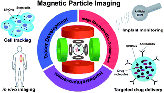
Magnetic particle imaging (MPI) is an emerging biomedical imaging technology that allows the direct quantitative mapping of the spatial distribution of superparamagnetic iron oxide nanoparticles. MPI's increased sensitivity and short image acquisition times foster the creation of tomographic images with high temporal and spatial resolution. The contrast and sensitivity of MPI is envisioned to transcend those of other medical imaging modalities presently used, such as magnetic resonance imaging (MRI), X-ray scans, ultrasound, computed tomography (CT), positron emission tomography (PET) and single photon emission computed tomography (SPECT). In this review, we present an overview of the recent advances in the rapidly developing field of MPI. We begin with a basic introduction of the fundamentals of MPI, followed by some highlights over the past decade of the evolution of strategies and approaches used to improve this new imaging technique. We also examine the optimization of iron oxide nanoparticle tracers used for imaging, underscoring the importance of size homogeneity and surface engineering. Finally, we present some future research directions for MPI, emphasizing the novel and exciting opportunities that it offers as an important tool for real-time in vivo monitoring. All these opportunities and capabilities that MPI presents are now seen as potential breakthrough innovations in timely disease diagnosis, implant monitoring, and image-guided therapeutics.
You have access to this article
 Please wait while we load your content...
Something went wrong. Try again?
Please wait while we load your content...
Something went wrong. Try again?