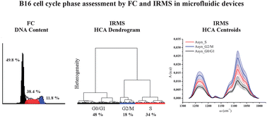
The knowledge of cell cycle phase distribution is of paramount importance for understanding cellular behaviour under normal and stressed growth conditions. This task is usually assessed using Flow Cytometry (FC) or immunohistochemistry. Here we report on the use of FTIR microspectroscopy in Microfluidic Devices (MD-IRMS) as an alternative technique for studying cell cycle distribution in live cells. Asynchronous, S- and G0-synchronized B16 mouse melanoma cells were studied by running parallel experiments based on MD-IRMS and FC using Propidium Iodide (PI) staining. MD-IRMS experiments have been done using silicon-modified BaF2 devices, where the thin silicon layer prevents BaF2 dissolution without affecting the transparency of the material and therefore enabling a better assessment of the Phosphate I (PhI) and II (PhII) bands. Hierarchical Cluster Analysis (HCA) of cellular microspectra in the 1300–1000 cm−1 region pointed out a distribution of cells among clusters, which is in good agreement with FC results among G0/G1, S and G2/M phases. The differentiation is mostly driven by the intensity of PhI and PhII bands. In particular, PhI almost doubles from the G0/G1 to G2/M phase, in agreement with the trend followed by nucleic acids during cellular progression. MD-IRMS is then proposed as a powerful method for the in situ determination of the cell cycle stage of an individual cell, without any labelling or staining, which gives the advantage of possibly monitoring specific cellular responses to several types of stimuli by clearly separating the spectral signatures related to the cellular response from those of cells that are normally progressing.
You have access to this article
 Please wait while we load your content...
Something went wrong. Try again?
Please wait while we load your content...
Something went wrong. Try again?