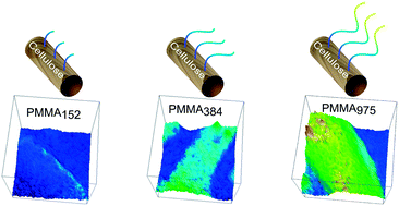
Visualization of poly(methyl methacrylate) (PMMA) grafts on cellulose via high-resolution FT-IR microscopy imaging†
Abstract
Cellulose surfaces grafted with PMMA of different graft lengths were characterized via high-resolution

* Corresponding authors
a
KTH Royal Institute of Technology, School of Chemical Science and Engineering, Department of Fibre and Polymer Technology, Teknikringen 56-58, Stockholm, Sweden
E-mail:
mavem@kth.se
Fax: +468 790 8283
Tel: +468 790 7225
b
Preparative Macromolecular Chemistry, Institut für Technische Chemie und Polymerchemie, Karlsruhe Institute of Technology (KIT), Engesserstr. 18, Karlsruhe, Germany
E-mail:
christopher.barner-kowollik@kit.edu
Fax: +49 721 608 45640
Tel: +49 721 608 45641
Cellulose surfaces grafted with PMMA of different graft lengths were characterized via high-resolution

 Please wait while we load your content...
Something went wrong. Try again?
S. Hansson, T. Tischer, A. S. Goldmann, A. Carlmark, C. Barner-Kowollik and E. Malmström, Polym. Chem., 2012, 3, 307 DOI: 10.1039/C1PY00338K
To request permission to reproduce material from this article, please go to the Copyright Clearance Center request page.
If you are an author contributing to an RSC publication, you do not need to request permission provided correct acknowledgement is given.
If you are the author of this article, you do not need to request permission to reproduce figures and diagrams provided correct acknowledgement is given. If you want to reproduce the whole article in a third-party publication (excluding your thesis/dissertation for which permission is not required) please go to the Copyright Clearance Center request page.
Read more about how to correctly acknowledge RSC content.
 Fetching data from CrossRef.
This may take some time to load.
Loading related content
