Issue 11, 2012

Chitin nanofibers: preparations, modifications, and applications

Abstract
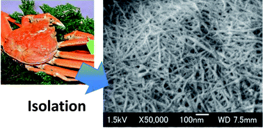
Chitin nanofibers are prepared from the exoskeletons of crabs and prawns by a simple mechanical treatment after the removal of proteins and minerals. The obtained nanofibers have fine nanofiber networks with a uniform width of approximately 10–20 nm and a high aspect ratio. The method used for chitin-nanofiber isolation is also successfully applied to the cell walls of mushrooms. They form a complex with glucans on the fiber surface. A grinder, a Star Burst atomization system, and a high speed blender are all used in the mechanical treatment to convert chitin to nanofibers. Mechanical treatment under acidic conditions is the key to facilitate fibrillation. At pH 3–4, the cationization of amino groups on the fiber surface assists nano-fibrillation by electrostatic repulsive force. By applying this finding, we also prepared chitin nanofibers from dry chitin powder. Chitin nanofibers are acetylated to modify their surfaces. The acetyl DS can be controlled from 1 to 3 by changing the reaction time. An acetyl group is introduced heterogeneously from the surface to the core. Nanofiber morphology is maintained even in the case of high acetyl DS. Optically transparent chitin nanofiber composites are prepared with 11 different types of acrylic resins. Due to the nano-sized structure, all of the composites are highly transparent. Chitin nanofibers significantly increase the Young's moduli and the tensile strengths and decrease the thermal expansion of all acrylic resins due to the reinforcement effect of chitin nanofibers. Chitin nanofibers show chiral separation ability. The chitin nanofiber membrane transports the D-isomer of glutamic acid, phenylalanine, and lysine from the corresponding racemic amino acid mixtures faster than the corresponding L-isomer. The chitin nanofibers improve clinical symptoms and suppress ulcerative colitis in a DSS-induced mouse model of acute ulcerative colitis. Moreover, chitin nanofibers suppress myeloperoxidase activation in the colon and decrease serum interleukin-6 concentrations.

Graphical abstract: Chitin nanofibers: preparations, modifications, and applications

Article information

Article type
Feature Article
Submitted
17 Feb 2012
Accepted
22 Mar 2012
First published
26 Mar 2012
This article is Open Access
Creative Commons BY license

Nanoscale, 2012,4, 3308-3318

Chitin nanofibers: preparations, modifications, and applications

S. Ifuku and H. Saimoto, Nanoscale, 2012, 4, 3308 DOI: 10.1039/C2NR30383C

This article is licensed under a Creative Commons Attribution 3.0 Unported Licence. You can use material from this article in other publications without requesting further permissions from the RSC, provided that the correct acknowledgement is given.

Read more about how to correctly acknowledge RSC content.

Social activity

Spotlight

Advertisements