Issue 11, 2012

Thioflavin-S staining coupled to flow cytometry. A screening tool to detect in vivoprotein aggregation

Abstract

Amyloid deposits are associated with an increasing number of human disorders, including Alzheimer's and Parkinson's diseases. Recent studies provide compelling evidence for the existence of amyloid-like conformations in the insoluble bacterial inclusion bodies (IBs) produced during the recombinant expression of amyloidogenic proteins. This makes prokaryotic cells a physiologically relevant system to study the mechanisms of in vivo amyloid deposition. We show here that the application of flow cytometry to detect Thioflavin-S (Th-S) fluorescence provides a fast, robust, quantitative, non-invasive method to screen for the presence of in vivo intracellular amyloid-like aggregates in bacteria, with potential application in the analysis of the impact of genetic mutations or chemical compounds on the aggregation of disease-associated polypeptides.
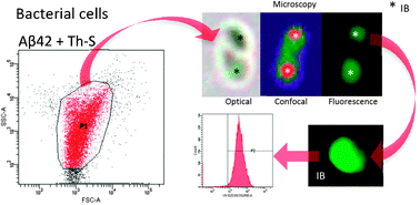
Graphical abstract: Thioflavin-S staining coupled to flow cytometry. A screening tool to detect in vivo protein aggregation

Article information

Article type
Method
Submitted
30 May 2012
Accepted
23 Jul 2012
First published
24 Jul 2012

Mol. BioSyst., 2012,8, 2839-2844

Thioflavin-S staining coupled to flow cytometry. A screening tool to detect in vivo protein aggregation

A. Espargaró, R. Sabate and S. Ventura, Mol. BioSyst., 2012, 8, 2839 DOI: 10.1039/C2MB25214G

To request permission to reproduce material from this article, please go to the Copyright Clearance Center request page.

If you are an author contributing to an RSC publication, you do not need to request permission provided correct acknowledgement is given.

If you are the author of this article, you do not need to request permission to reproduce figures and diagrams provided correct acknowledgement is given. If you want to reproduce the whole article in a third-party publication (excluding your thesis/dissertation for which permission is not required) please go to the Copyright Clearance Center request page.

Read more about how to correctly acknowledge RSC content.

Spotlight

Advertisements