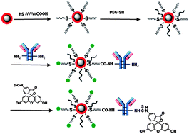
Multimodal imaging based on complementary detection principles has great potential for improving the accuracy of tumor diagnosis. Fe3O4 core/Au shell nanoparticles (Fe3O4@Au NPs) can be used as an effective multimodal contrast agent due to combining the advantageous and highly complementary features of the Fe3O4 core and gold shell. In the present work, we have developed a novel Fe3O4@Au NP based probe for targeting and multimodal imaging of cancer cells. The prepared Fe3O4@Au NPs have been characterized by transmission electron microscopy (TEM), visible spectra and SERS. The potential use of the prepared Fe3O4@Au NPs as a multimodal contrast probe for magnetic resonance imaging (MRI), microwave-induced thermoacoustic imaging and photoacoustic imaging was then evaluated. Importantly, when conjugated with a cancer cell targeted molecular and fluorescent dye, the Fe3O4@Au NPs could be internalized by the corresponding cancer cells selectively and sensitively, and fluorescence imaging was realized at the same time. The bio-modified Fe3O4@Au NPs incorporating multiple functionalities into one single nano-structured system can be effectively used for targeting and multimodal imaging of cancer cells simultaneously.

You have access to this article
 Please wait while we load your content...
Something went wrong. Try again?


 Please wait while we load your content...