Lite Version|Standard version

To gain access to this content please
Log in via your home Institution.
Log in with your member or subscriber username and password.
Download

In this paper we show how the 3-dimensional structural arrangement of particles in a colloidosome is derived from a single scanning electron microscopy (SEM) image. We exploit the fact that particles are located on the surface of a sphere to directly quantify the 3-dimensional positions of all particles from a single 2-dimensional SEM image. The 3-dimensional particle positions are used to quantify the structural arrangement of the particles by performing both a Delaunay triangulation (DT) as well as a Voronoi tessellation (VT) of the colloidosome surface, which we use to validate the procedure for determining the 3-dimensional particle positions. In addition to the DT and the VT, we calculate the radial pair correlation function g(r). We explain how the pair-distribution is modified to suit a spherical surface and how the structural order relates to similar data on colloidal films. Our results illustrate that even for a colloidosome in its ground state, the resulting ordering is short-ranged (in our case 4 particle diameters), which is in contrast to densely packed colloidal films.
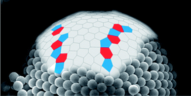
Graphical abstract: Reconstruction of the 3D structure of colloidosomes from a single SEM image

Page: ^ Top