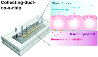
Fluid-shear-stress-induced translocation of aquaporin-2 and reorganization of actin cytoskeleton in renal tubular epithelial cells
Abstract
In vivo, renal tubular

* Corresponding authors
a
Interdisciplinary Program in Nano-Science and Technology, Seoul National University, Seoul 151-742, Korea
E-mail:
sky4u@snu.ac.kr
b School of Mechanical and Aerospace Engineering, Seoul National University, Seoul 151-742, Korea
c Interdisciplinary Program of Bioengineering, Seoul National University, Seoul 151-742, Korea
d
Department of Biochemistry and Cell Biology, School of Medicine, Kyungpook National University, Taegu 700-422, Korea
E-mail:
thkwon@knu.ac.kr
In vivo, renal tubular

 Please wait while we load your content...
Something went wrong. Try again?
 Fetching data from CrossRef.
This may take some time to load.
Loading related content
