Issue 13, 2009

Hierarchical pattern of microfibrils in a 3D fluorapatite–gelatine nanocomposite: simulation of a bio-related structure building process

Abstract

The shape development of a biomimetic fluorapatite–gelatine nanocomposite on the μm scale is characterised by a fractal mechanism with the origin being intrinsically coded in a (central) elongated hexagonal-prismatic seed. The 3D superstructure of the seed is distinctively overlaid by a pattern consisting of gelatine microfibrils. The orientation of the microfibrils is assumed to be controlled by an intrinsic electrical field generated by the nanocomposite during development and growth of the seed. In order to confirm this assumption and to get more detailed information on orientational relations of the complex nanocomposite we simulated the pattern formation process up to the μm scale. The results from experimental studies and simulation results on an atomistic level support a model scenario wherein the elementary building blocks for the aggregation are represented by elongated hexagonal-prismatic objects (A-units), with the embedded collagen triple-helices in their centers. The interactions of the A-units are consequently modelled by three contributions: the crystal energy part (originating from the pair-wise interactions of the “apatite shells” of the prismatic units), the electrostatic interaction (originating from the unit charges located at the ends of the collagen triple helices), and the interaction energy of the A-units mediated by the solvent. The next level of complexity is related to the fact that micro fibrils were found in the fluorapatite–gelatine nanocomposites. They consist of bundles of triple helical protein molecules, which are embedded within the 3D-hexagonal prismatic arrangement of the A-units. In our approach we consider the microfibrils as chains of flexible dipoles with effective dipole moments. The crystal growth processes is modelled as an energetically controlled stepwise association of elementary building blocks of different kind on a 3D-grid. The remarkable and excellent qualitative agreement between the simulated fibril patterns and the observations made by SEM and TEM support the concept of an intrinsic electric field driven morphogenesis of the fluorapatite–gelatine nanocomposite. The simulated fibril pattern also bears the chance to make fresh attempts in order to find explanations for experimental observations which are not understood up to now.

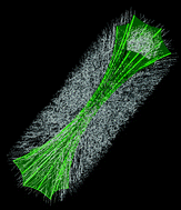
Graphical abstract: Hierarchical pattern of microfibrils in a 3D fluorapatite–gelatine nanocomposite: simulation of a bio-related structure building process

Article information

Article type
Paper
Submitted
08 Oct 2008
Accepted
08 Jan 2009
First published
06 Feb 2009

Phys. Chem. Chem. Phys., 2009,11, 2186-2194

Hierarchical pattern of microfibrils in a 3D fluorapatite–gelatine nanocomposite: simulation of a bio-related structure building process

R. Paparcone, R. Kniep and J. Brickmann, Phys. Chem. Chem. Phys., 2009, 11, 2186 DOI: 10.1039/B817723F

To request permission to reproduce material from this article, please go to the Copyright Clearance Center request page.

If you are an author contributing to an RSC publication, you do not need to request permission provided correct acknowledgement is given.

If you are the author of this article, you do not need to request permission to reproduce figures and diagrams provided correct acknowledgement is given. If you want to reproduce the whole article in a third-party publication (excluding your thesis/dissertation for which permission is not required) please go to the Copyright Clearance Center request page.

Read more about how to correctly acknowledge RSC content.

Social activity

Spotlight

Advertisements