Issue 3, 2009

Elemental bio-imaging of melanoma in lymph node biopsies

Abstract

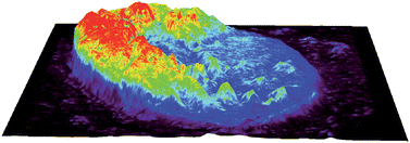
The spatial distribution of trace elements in human lymph nodes partially infiltrated by melanoma cells was determined by elemental bio-imaging. Imaging of 31P within the nodal capsule and normal lymph node tissue showed a clear demarcation of the tumour boundary, with a significant decrease in relative 31P concentration within the tumour. The location of the tumour boundary was confirmed by haematoxylin and eosin staining of serial sections and observation by light microscopy. Further enhancement of the tumour boundary was achieved by imaging the 31P/34S ratio. 31P/66Zn ratio images showed a decreasing ratio beyond the tumour boundary that extended into peritumour normal lymph node tissue.

Graphical abstract: Elemental bio-imaging of melanoma in lymph node biopsies

Article information

Article type
Paper
Submitted
24 Jul 2008
Accepted
07 Nov 2008
First published
27 Nov 2008

Analyst, 2009,134, 450-453

Elemental bio-imaging of melanoma in lymph node biopsies

D. Hare, F. Burger, C. Austin, F. Fryer, R. Grimm, B. Reedy, R. A. Scolyer, J. F. Thompson and P. Doble, Analyst, 2009, 134, 450 DOI: 10.1039/B812745J

To request permission to reproduce material from this article, please go to the Copyright Clearance Center request page.

If you are an author contributing to an RSC publication, you do not need to request permission provided correct acknowledgement is given.

If you are the author of this article, you do not need to request permission to reproduce figures and diagrams provided correct acknowledgement is given. If you want to reproduce the whole article in a third-party publication (excluding your thesis/dissertation for which permission is not required) please go to the Copyright Clearance Center request page.

Read more about how to correctly acknowledge RSC content.

Social activity

Spotlight

Advertisements