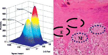
Distinction of malignant melanoma and epidermis using IR micro-spectroscopy and statistical methods
Abstract

* Corresponding authors
a
Department of Physics and the Cancer Research Center, Ben-Gurion University (BGU), Beer-Sheva, Israel
E-mail:
shaulm@bgu.ac.il
Fax: +972-8-647 2903
Tel: +972-8-646 1749
b Department of Pathology, Soroka University Medical Center (SUMC), Beer-Sheva, Israel

 Please wait while we load your content...
Something went wrong. Try again?
Z. Hammody, S. Argov, R. K. Sahu, E. Cagnano, R. Moreh and S. Mordechai, Analyst, 2008, 133, 372 DOI: 10.1039/B712040K
To request permission to reproduce material from this article, please go to the Copyright Clearance Center request page.
If you are an author contributing to an RSC publication, you do not need to request permission provided correct acknowledgement is given.
If you are the author of this article, you do not need to request permission to reproduce figures and diagrams provided correct acknowledgement is given. If you want to reproduce the whole article in a third-party publication (excluding your thesis/dissertation for which permission is not required) please go to the Copyright Clearance Center request page.
Read more about how to correctly acknowledge RSC content.
 Fetching data from CrossRef.
This may take some time to load.
Loading related content
