Abstract
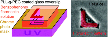
The original micropatterning technique on gold, although very efficient, is not accessible to most biology labs and is not compatible with their techniques for image acquisition. Other solutions have been developed on silanized glass coverslips. These methods are still hardly accessible to biology labs and do not provide sufficient reproducibility to become incorporated in routine biological protocols. Here, we analyzed cell behavior on micro-patterns produced by various alternative techniques. Distinct cell types displayed different behavior on micropatterns, while some were easily constrained by the patterns others escaped or ripped off the patterned adhesion molecules. We report methods to overcome some of these limitations on glass coverslips and on plastic dishes which are compatible with our experimental biological applications. Finally, we present a new method based on UV crosslinking of adhesion


 Please wait while we load your content...