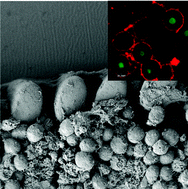
A novel 3D mammalian cell perfusion-culture system in microfluidic channels†
Abstract
Mammalian cells cultured on 2D surfaces in microfluidic channels are increasingly used in

Maintenance work is planned from 09:00 BST to 12:00 BST on Saturday 28th September 2024.
During this time the performance of our website may be affected - searches may run slowly, some pages may be temporarily unavailable, and you may be unable to access content. If this happens, please try refreshing your web browser or try waiting two to three minutes before trying again.
We apologise for any inconvenience this might cause and thank you for your patience.
* Corresponding authors
a
Institute of Bioengineering and Nanotechnology, 31 Biopolis Way, The Nanos, Singapore
E-mail:
hyu@ibn.a-star.edu.sg, nmiyuh@nus.edu.sg
Fax: +65-6516 8261
Tel: +65-6516 3466
b NUS Graduate Programme in Bioengineering, NUS Graduate School for Integrative Sciences and Engineering, National University of Singapore, Singapore
c Department of Physiology, Yong Loo Lin School of Medicine, National University of Singapore, Singapore
d Singapore-MIT Alliance, E4-04-10, 4 Engineering Drive 3, Singapore
e NUS Tissue-Engineering Programme, DSO Labs, National University of Singapore, Singapore
f Department of Haematology-Oncology, National University Hospital, Singapore
g Division of Bioengineering, Faculty of Engineering, National University of Singapore, Singapore
h Department of Orthopaedic Surgery, Yong Loo Lin School of Medicine, National University of Singapore, Singapore
Mammalian cells cultured on 2D surfaces in microfluidic channels are increasingly used in

 Please wait while we load your content...
Something went wrong. Try again?
Y. Toh, C. Zhang, J. Zhang, Y. M. Khong, S. Chang, V. D. Samper, D. van Noort, D. W. Hutmacher and H. Yu, Lab Chip, 2007, 7, 302 DOI: 10.1039/B614872G
To request permission to reproduce material from this article, please go to the Copyright Clearance Center request page.
If you are an author contributing to an RSC publication, you do not need to request permission provided correct acknowledgement is given.
If you are the author of this article, you do not need to request permission to reproduce figures and diagrams provided correct acknowledgement is given. If you want to reproduce the whole article in a third-party publication (excluding your thesis/dissertation for which permission is not required) please go to the Copyright Clearance Center request page.
Read more about how to correctly acknowledge RSC content.
 Fetching data from CrossRef.
This may take some time to load.
Loading related content
