

 Open Access Article
 This Open Access Article is licensed under a
 Creative Commons Attribution 3.0 Unported Licence
Clinical translation and landscape of stimuli-responsive nanomedicines and microscale therapeutics
Dmytro Kobzev
 *a,
Olesia Kulyk
b,
Roman A. Barmin
a,
Anatoliy Tataretsb,
Roger M. Pallaresa,
Fabian Kiesslinga,
Twan Lammers
*a and
Quim Peña
*a
*a,
Olesia Kulyk
b,
Roman A. Barmin
a,
Anatoliy Tataretsb,
Roger M. Pallaresa,
Fabian Kiesslinga,
Twan Lammers
*a and
Quim Peña
*a
aInstitute for Experimental Molecular Imaging, Center for Biohybrid Medical Systems, RWTH Aachen University Clinic, Aachen 52074, Germany. E-mail: dkobzev@ukaachen.de; tlammers@ukaachen.de; jpena@ukaachen.de
bInstitute of Functional Materials Chemistry of State Scientific Institution “Institute for Single Crystals” of National Academy of Sciences of Ukraine, Kharkiv, 61072, Ukraine
First published on 29th May 2026
Abstract
Stimuli-responsive materials enable temporal and spatial control over drug delivery and action. Traditional triggerable therapeutics are largely based on small molecules, like prodrugs and photodynamic therapy agents. Advances in nanotechnology and micromaterials have greatly expanded the field, as evidenced by clinically translated hyperthermia-generating iron oxide nanoparticles, radiotherapy-enhancing hafnium oxide nanoparticles, and ultrasound-responsive microbubbles. We here analyze the (pre-)clinical landscape of trigger-responsive therapeutics between 2014 and 2024, encompassing over 90![[thin space (1/6-em)]](https://www.rsc.org/images/entities/char_2009.gif) 000 publications and 1000 clinical trials. External stimuli include light, ultrasound, radiation, magnetic field and temperature. Key internal stimuli are pH, redox and enzymes. Our analysis shows that light is by far the most popular external stimulus (44% of papers; 361 trials). Among internal stimuli, which account for 46% of papers (558 trials), redox and enzyme activation are the most explored ones. In recent years, interest in radiation (114 trials), ultrasound (33 trials), temperature (14 trials), and magnetic actuation (3 trials) is increasing, typically involving nano- and microscale platforms. In the second part of our paper, we examine translational trajectories and identify key barriers that are limiting the clinical progress of stimuli-responsive therapeutics. Important issues to address to help promote clinical translation include: (1) inaccurate medical need identification; (2) overly complex material design; (3) limited tissue penetration; (4) limited device accessibility; (5) economic constraints; and (6) challenging clinical adoption. We conclude by providing practical and practicable solutions to address these key limitations, going from nano- and micro-formulation design to development, translation and implementation, together aiming to increase the clinical impact of stimuli-responsive therapeutics.
000 publications and 1000 clinical trials. External stimuli include light, ultrasound, radiation, magnetic field and temperature. Key internal stimuli are pH, redox and enzymes. Our analysis shows that light is by far the most popular external stimulus (44% of papers; 361 trials). Among internal stimuli, which account for 46% of papers (558 trials), redox and enzyme activation are the most explored ones. In recent years, interest in radiation (114 trials), ultrasound (33 trials), temperature (14 trials), and magnetic actuation (3 trials) is increasing, typically involving nano- and microscale platforms. In the second part of our paper, we examine translational trajectories and identify key barriers that are limiting the clinical progress of stimuli-responsive therapeutics. Important issues to address to help promote clinical translation include: (1) inaccurate medical need identification; (2) overly complex material design; (3) limited tissue penetration; (4) limited device accessibility; (5) economic constraints; and (6) challenging clinical adoption. We conclude by providing practical and practicable solutions to address these key limitations, going from nano- and micro-formulation design to development, translation and implementation, together aiming to increase the clinical impact of stimuli-responsive therapeutics.
 Dmytro Kobzev | Dmytro Kobzev obtained his Master's degree in biotechnology and bioengineering from the National Technical University “Kharkiv Polytechnic Institute” (Ukraine) in 2018. He received a PhD in chemistry from SSI “Institute for Single Crystals” (Ukraine) in 2024 and a PhD in bioengineering from Ariel University (Israel) in 2024 focusing on polymethine dyes and targeted drug delivery. His research interests include organic synthesis, fluorescent dyes, photophysics, and antibody–drug conjugates for diagnostics and cancer therapy. He is currently a postdoctoral researcher at RWTH Aachen University (Germany), focuses on bioconjugation and ultrasound-responsive materials for biomedical applications. |
 Olesia Kulyk | Olesia Kulyk obtained her PhD in organic chemistry in 2014 from Karazin Kharkiv National University (Ukraine). In 2015, she joined the group of Prof. D. Bonifazi (first University of Namur, then Cardiff University), where she worked on multichromophoric supramolecular systems and artificial light-harvesting assemblies. She later held a research position at Ariel University (Israel), focusing on polymethine dyes for antimicrobial and anticancer photodynamic therapy. She is currently a deputy director and senior researcher at SSI “Institute for Single Crystals” of the NAS of Ukraine. Her research focuses on organic dyes, supramolecular chemistry, and fluorescent materials for biomedical applications. |
 Roman A. Barmin | Roman Barmin is a PhD student at RWTH Aachen University Hospital, supervised by Dr Roger M. Pallares and Prof. Twan Lammers. He holds a BSc in Physical Chemistry and Engineering from Moscow State University and an MSc in Biophotonics from the Skolkovo Institute of Science and Technology. He has experience in both academia and industry through his involvement in, and management of, (inter)national science and higher-education initiatives. His research focuses on ultrasound-responsive materials, with broader interests in designing simple, translatable nano- and microscale colloidal systems for biomedical use. |
 Twan Lammers | Twan Lammers obtained a DSc in Radiation Oncology from Heidelberg University in 2008 and a PhD in Pharmaceutical Technology from Utrecht University in 2009. In the same year, he started the Nanomedicine and Theranostics group at RWTH Aachen University Clinic. In 2014, he was promoted to full professor. His group aims to individualize and improve disease treatment by combining drug targeting with imaging. He is associate editor for the Journal of Controlled Release and Drug Delivery and Translational Research, is a past president of the Controlled Release Society, and currently serves as secretary of the European Society for Molecular Imaging. |
 Quim Peña | Quim Peña obtained his PhD in Chemistry from the UAB (Spain) and AMU (France) in 2019, focusing on metal-based anticancer compounds. He is now a Junior Group-Leader in the Department of Nanomedicine and Theranostics at the Institute for Experimental Molecular Imaging (RWTH Aachen University Hospital). His research integrates pharmaceutical and prodrug chemistry strategies with nanomedicine engineering for controlled (metallo-)drug delivery and improved (immuno-)therapy efficacy, tolerability, and translation. Quim is part of the Management Committee of the EU4MOFs European Network on tech-transfer of metal–organic porous materials and actively involved in scientific societies like the CRS, currently serving on the YSC Board. |
1. Introduction
Stimuli-responsive materials have garnered considerable interest for diagnostic and therapeutic purposes. With several products already in clinical use, such as the light-responsive drug Photofrin1 and the radiation-enhancing nanodrug Hensify,2,3 this type of materials are engineered to respond to external (light,4–6 temperature,7,8 radiation,3,9 magnetic field10–12 and ultrasound13,14) or internal stimuli (pH,15,16 redox17 and enzymes16,18), resulting in the induction of direct material-mediated therapeutic effects16,17,19 or triggering the release of active pharmaceutical ingredients (API). Many clinically relevant stimuli-responsive therapeutics are based on small molecules, such as fluorescent dyes and prodrugs.5,9,14 Yet, the increasing clinical relevance of nano- and micro-technologies in the past decades20,21 has also promoted the development and use of novel stimuli-responsive platforms for therapeutic purposes, like superparamagnetic iron oxide nanoparticles (SPION).10–12 Stimuli-responsive platforms allow to tailor pharmacokinetics and biodistribution profiles, enhance target-site accumulation, and increase stimuli-material responsiveness and performance control, thereby resulting in improved therapeutic outcomes.22The conceptual roots of stimuli-responsive materials can be traced back more than a century to the early foundations of light-based medical therapies. Back in the 1880s, the physician Niels Finsen pioneered the use of phototherapy to treat lupus vulgaris, a skin condition caused by tuberculosis.23 His work, which earned the Nobel Prize in Medicine in 1903, laid the foundation for modern light-based therapies and led to the development of photodynamic therapy (PDT). In the 1960s, PDT was clinically established and has been used since then in the treatment of various cancers, including esophageal (e.g., Photofrin) and prostate (e.g., Foscan and TOOKAD),1 as well as other diseases such as actinic keratosis (e.g., Metvix)24 and macular degeneration (e.g., Visudyne).25 In fact, PDT global market share was valued at USD 4.6 billion in 2024, and it is predicted to double in the next 10 years,26,27 highlighting its continuously increasing interest. Other recent light-based treatments, such as photothermal therapy (PTT)28 and photoimmunotherapy (PIT),29 are currently under clinical evaluation (e.g., gold nanoparticle-based product AuroShell30 and phthalocyanine-antibody conjugate ASP-1929).29
Beyond light, materials sensitive to other external stimuli have also reached clinical evaluation. These include the temperature-sensitive liposomal doxorubicin product ThermoDox,31 with several clinical trials ongoing in combination with microwave irradiation, focused ultrasound, and radiation therapy,31 and materials responsive to ultrasound, magnetic fields, or radiation. The latter three stimuli have been proportionally less explored than light for therapeutic purposes, but have become increasingly relevant in recent years, especially in combination with nano- and microtechnology (Fig. 1). Prominent examples include ultrasound-responsive microbubbles,13 used as contrast agents for clinical ultrasound imaging since the 1990s and investigated for therapeutic purposes from the 2000s onwards;13,32 SPION formulations like NanoTherm, clinically approved for the treatment of brain tumors since 2010;33–35 and radiation-enhancing hafnium (Hf) oxide nanoparticles like Hensify, approved in 2019 for the treatment of locally-advanced soft tissue sarcoma.3
 | ||
| Fig. 1 Representative examples of clinically relevant stimuli-responsive materials and products. Over the years, many different stimuli-responsive materials have been designed and evaluated for the treatment of several diseases, including cancer, inflammation, and infectious diseases. These mostly include small-molecule fluorescent dyes and prodrugs, which can also be delivered in nanoscale carriers based on lipids, polymers or antibodies, but also nano- and microscale platforms intrinsically responsive to external stimuli (e.g., magnetic- or radiation-responsive metal-based nanoparticles and ultrasound-responsive microbubbles). Abbreviations: NP – nanoparticles, SPION – superparamagnetic iron oxide nanoparticles, Hf – hafnium, ADC – antibody–drug conjugates, HPMA – 2-hydroxypropyl methacrylate. | ||
Besides external stimuli, materials responding to internal triggers such as pH,15,16 redox,17 and enzymatic processes16,18 are also extensively developed. Many of these nano- or microparticle systems are designed to deliver small-molecule API in response to different and, ideally, specific (patho)physiological conditions, including high levels of cathepsins (enzyme)16,18,36 or glutathione (redox),17 or low pH.15 Prominent clinical successes include antibody–drug conjugates (ADC), with at least 11 out of 14 products on the market leveraging internal stimuli, such as the pH-sensitive ADC Mylotarg and Trodelvy, and the enzyme-cleavable Enhertu and Tivdak.16 The global ADC market size accounted for USD 11.43 billion in 2024 and is predicted to triple by 2034.37 Instead, other widely explored systems sensitive to internal stimuli like anticancer polymer–drug conjugates have so far failed to reach FDA/EMA approval.38
Despite the extensive research efforts dedicated to the development of stimuli-responsive (nano)materials, many of which have shown high preclinical promise, their clinical translation has been hampered by several challenges. In addition to the added design complexity associated to the stimuli-responsiveness nature, which can complicate scale-up manufacturing and regulatory standardization, other translational aspects such as the lack of specific device availability in many hospitals, the (logistical) complexity of integrating such treatments into standard regimens, and the high associated costs of some devices to generate stimuli like magnetic fields, have also contributed to limiting their clinical impact.
Here, we aim to examine the current landscape of stimuli-responsive materials for medicinal applications, with a particular focus on externally activated nano- and microscale therapeutics and on their translational potential via scientometric analysis of existing preclinical and clinical data (Fig. 1). By identifying the most frequently studied and clinically tested stimuli-responsive material platforms for therapeutic purposes, and by analyzing their design principles, mechanisms of action, and specific pharmaceutical and clinical challenges, we provide insights into the developmental trajectories and translational bottlenecks associated with each stimulus type, and we propose strategies to promote their translation and maximize clinical impact.
2. Design and preclinical landscape of stimuli-responsive materials
Stimuli-responsive therapeutics rely on activatable small molecules or functional units,5,9 which can also be integrated into nano- or microscale platforms.7,19,39 They are structurally designed to contain a built-in trigger—such as a π-electron system,5,14 transition metal center,5,40 labile chemical bond,41 or self-assembling/disassembling motif7,39—that undergoes a predictable and controlled transition upon exposure to a source of energy (stimulus). Depending on the molecular design, this change can be reversible5 or irreversible,41 and result in the activation of a range of actions, including structural reorganization,7,39 molecular excitations (e.g., through interactions with singlet–triplet excited state),5 (metal) catalytic activity,42 or payload (API) release.16,41 Altogether, these features can be exploited to spatiotemporally control biological and therapeutic effects, minimizing off-target toxicity and enhancing treatment efficacy.22 These processes are ultimately governed by the interaction between the applied energy source and specific molecular or material features, which define the mechanism of action for each stimulus modality.The most relevant classes of stimuli-responsive small molecules consist of fluorescent dyes5,14 and, to a lesser extent, also (chemotherapeutic) prodrugs.9 These structures can be used alone or formulated into nano-/microscale carriers such as liposomes,7 lipid nanoparticles,43 micelles,44,45 hydrogels,39 synthetic polymers,46,47 polycarbohydrates (e.g., chitosan and dextran),48,49 antibodies and ADC,16,19 and (inorganic) solid nanoparticles.50,51 There are also source-specific nano- and micro-materials that display intrinsic responses upon the application of a stimulus due to specific compositions and sizes, or the incorporation of reactive functional units in their structure. Examples of these include quantum dots (light),52 microbubbles53,54 and nanobubbles55 (ultrasound), and metal(loid)-based nanoparticles based on gold56,57 (light), iron (SPION;10,11,58 magnetic field), and hafnium,2,3 gadolinium59 or boron-1060 (radiation).
Five major external stimuli can be categorized, each representing a distinct energy source that acts as a driving force to activate the stimuli-responsive material and trigger biological effects. These stimuli include light, temperature, ultrasound, radiation and magnetic field. In parallel, endogenous stimuli such as changes in pH, redox potential and enzymatic levels are also relevant in the design of therapeutic materials. Some external stimuli have already led to the development of distinct clinical strategies. For instance, light-activated therapies encompass PDT,4 PTT28 and PIT;29 and ultrasound-based therapies include sonodynamic therapy (SDT),14 but also sonopermeation approaches, which combine microbubbles and ultrasound to enhance chemo-, nano-, and immunotherapies.53,61 Radiotherapy efficiency can be enhanced by radiation-responsive materials,2,59 and magnetic-induced hyperthermia (MIH) harnesses magnetic nanoparticles to generate localized heat upon exposure to an alternating magnetic field, leading to targeted tissue ablation.10,11
In general, the stimuli-responsiveness capability of the materials can be therapeutically exploited in two different ways: either (a) directly inducing biological effects to cells and tissues after external stimulus application (Fig. 2A), or (b) acting as a passive carrier system and releasing active payloads (e.g., chemotherapeutics) upon internal or external stimuli (Fig. 2B). Regarding the former, the mechanism of action can vary slightly depending on the source of energy (Fig. 2A), encompassing (i) the production of cell-damaging radicals like reactive oxygen species (ROS) (for PDT,4 PIT,29 SDT14 and RT62,63) and radiation-induced nucleic acid damage (for RT63); and (ii) the conversion of the source energy into heat by the material (for PTT56 and MIH64), subsequently leading to biomolecule and tissue ablation. These mechanisms not only promote cell death at the target site, but can also result in the induction of systemic immune responses, with potential for immunotherapy applications.65
 | ||
| Fig. 2 Modes of action of stimuli-responsive materials for therapeutic purposes. (A) Stimuli-activatable therapies. Stimuli-responsive materials can induce direct biological and therapeutic effects mediated by the interaction of the material with the external stimuli. These mostly include radical and reactive oxygen species (ROS) generation (for PDT, PIT, SDT and RT), which induce biomolecule/DNA damage and oxidative stress, radiation-induced DNA damage (for RT), and heat-mediated cell ablation (for PTT and MIH). (B) Stimuli-triggered drug release. Stimuli-responsive materials can act as passive carrier systems, delivering active payloads (from small chemotherapeutics to macromolecules like proteins and nucleic acids) upon external or internal stimuli. This process can be promoted (1) by inducing changes in the (nano)carrier structure (e.g., swelling or disassembling of hydrogels, liposomes or micelles), and (2) cleavage of the linker between the drug and the carrier (e.g., in antibody- and polymer–drug conjugates). Abbreviations: PDT – photodynamic therapy, PIT – photoimmunotherapy, SDT – sonodynamic therapy, RT – radiation therapy, PTT – photothermal therapy, MIH – magnetic-induced hyperthermia, DPPG – dipalmitoylphosphatidylglycerol, DOPE – dioleoyl phosphatidylethanolamine. | ||
Regarding stimuli-triggered drug release (Fig. 2B), the stimulus does not directly activate the therapeutic agent itself, but it induces structural and chemical changes in the carrier material, promoting the delivery of the active payloads. This can involve assembling/disassembling of the delivery platform (e.g., as observed in hydrogels,39 liposomes7 and micelles44), or cleavage of a linker between the carrier material and the drug (e.g., in ADC41 and polymer conjugates46,66). In contrast to external stimuli-activated therapies, which can directly mediate biological effects through the material itself, stimuli-triggered drug release only facilitates payload delivery, enabling the therapeutic agent (API) to act independently at the target site.
To understand how the different stimuli have impacted materials research and development for therapeutic applications over the last years, we first analyzed the landscape of related publications between 1980 and 2024 (Fig. 3). Five main external stimuli were considered (light, temperature/heat, ultrasound, radiation and magnetic field), and three common internal stimuli (pH, enzyme levels and redox activity). This analysis covered small molecules and nano- and micro-scale structures, and involved 91691 publications and 1083 trials, by using the keyword search shown in Scheme S1.
 | ||
| Fig. 3 Landscape of stimuli-responsive therapeutics in preclinical research. (A) and (B) Number of research publications over the years, starting from 1980 until 2024 (a total of 131199 publications) (A), and zoom-in between 2014 and 2024 (91691 publications) (B). (C) Distribution of the scope and type of materials responsive to light, ultrasound, magnetic field, radiation, temperature/heat, and internal stimuli (pH, enzymatic levels and redox activity) in the last decade. The blue color corresponds to small molecules, and the red color to nano-/microscale materials. Data were obtained in October 2025, from Digital Science's Dimensions platform, available at https://app.dimensions.ai. Keywords used for the search are shown in Scheme S1. Abbreviations: LNP – lipid nanoparticles, ADC – antibody–drug conjugates, NP – nanoparticles, QD – quantum dots, 5-ALA – 5-aminolevulinic acid. | ||
Based on the data presented in Fig. 3A and B, light- and internal stimuli-responsive materials have thus far dominated the field (46% and 44%, respectively), together accounting for approximately 90% of all the publications on stimuli-responsive therapeutics in the last 40 years. Both categories have exhibited comparable growth trajectories, with an exponential increase over the last two decades, reaching about 6000 publications each only in 2024.
Comparatively, the other stimulus modalities have received significantly less attention for therapeutic purposes, although some emerging trends are noteworthy. Publications on ultrasound-responsive therapeutic materials have tripled over the past five years, reaching about 600 publications. The research interest in magnetic field-responsive therapeutics has increased about 5-fold in the last decade (∼300 publications, despite the slight recession observed in 2024). The research growth in the latter case is likely encouraged by the approvals of several SPION formulations during the early 1990s and 2000s for the treatment of anemia and imaging,58 and, quite relevantly, by the approval of the magnetic-responsive SPION product NanoTherm in 2010 as a medical device for the treatment of glioblastoma.35 On the other hand, radiation therapy-potentiation using nanoscale radioenhancers like Hensify3 or small-molecule chemotherapeutic agents like cisplatin, which have been reported to radiosensitize cells,9,67 has shown a steady growth reaching ∼250 publications annually in the 2020s. The attention to temperature-responsive materials peaked in 2021, but has since stagnate (∼200 publications annually), which might be partly influenced by the failed phase III clinical trials of the thermosensitive liposomal doxorubicin formulation ThermoDox in 2013 (HEAT trial),68 and later in 2020 (intermediate results of OPTIMA trial).69
We then analyzed the scope and type of materials explored for each stimulus in preclinical research (Fig. 3C). The searches included small molecules (e.g., fluorescent dyes and (chemo)therapeutic agents) and relevant nano- and micromaterial classes (liposomes, lipid nanoparticles, micelles, hydrogels, polymers, polysaccharides, ADC, and non-metal and metal solid nanoparticles). As a general observation, the impact of nanotechnology and microtechnology is evident from the data. In light-responsive and radiation-responsive materials, this accounts for more than half of the retrieved publications (59% and 64%, respectively), and it becomes predominant in ultrasound-responsive materials (83%), and even more in heat-, magnetic field-, and internal stimuli-responsive materials. Regarding the latter, it is important to note that in addition to the impact of nanotechnology in the development of internal stimuli-responsive therapeutics (e.g., antibody- and polymer–drug conjugates), small molecule prodrugs responsive to pH, enzyme levels or redox have also been widely explored, with several clinical products like ixazomib citrate (cancer), pretomanid (tuberculosis), or omidenepag isopropyl (glaucoma and hypertension).70,71 However, many of these prodrugs are insoluble or present suboptimal pharmacokinetic properties, thus often being evaluated preclinically together with nanoscale delivery systems. This feature strongly influenced the results obtained using our keyword search query (Scheme S1), making it difficult to accurately estimate the preclinical landscape and research relevance of only internally activatable small-molecule prodrugs (with many of these reported as part of nanoparticle formulations).
2.1. Light-responsive materials
Light-responsive systems primarily rely on photon absorption by chromophores, leading (1) to excited-state reactions that generate ROS (PDT/PIT)4 or (2) to non-radiative relaxation processes that convert light into heat (PTT).56 Small molecules are highly relevant in the design of light-responsive materials, representing close to 40% of the total number of publications in the field, as compared to the 59% covered by nanomaterials. Classical photosensitizers remain prominent for PDT, with π electron-conjugated small molecules like porphyrins, phthalocyanines, 5-aminolevulinic acid (5-ALA, a naturally occurring porphyrin precursor in the human body), Methylene Blue, and xanthene derivatives (e.g., Rose Bengal) collectively accounting for about 33% of the publications on light-responsive materials (Fig. 3C). Regarding nanomaterial classes, metal nanoparticles represent 22% of the total number of publications on light-activated materials (Fig. 3C), with gold being among the most studied metals. Gold nanoparticles are known to play a central role in PTT, relying on heat generation through plasmonic resonance excitation for targeted cell ablation.56,57 Liposomes, lipid nanoparticles, natural (polysaccharides) and synthetic polymers, and hydrogels, present in about 25% of the total number of publications on light-sensitive materials, are widely used as carrier materials of light-responsive molecules like porphyrins, Methylene Blue or phthalocyanines for both PTT and PDT. Photosensitizer-carrying ADC have given rise to the PIT field,72 which combines the advantages of immunotherapy with PDT for synergistic effects.73 Although PIT is only present in 4% of the total publications on light-responsive therapeutic systems, its interest has been growing over the last years, partly driven by the preclinical promise observed with several ADC bearing IR700 and 2ICy7 dyes.72,74 In particular, the outcomes of the clinical trial involving the IR700-bearing antibody (discussed in Section 3) can lead to further developments in the field of light-sensitive ADC.Despite the promising preclinical data, light penetration in human tissue is limited to a depth of approximately 1.5 cm, which significantly restricts the clinical use of light-triggered therapies to superficial conditions (e.g., actinic keratosis).75 Thus, there has been a growing interest towards the development of new materials sensitive to near-infrared (NIR) light, which exhibit deeper tissue penetration capabilities, particularly to NIR-I (700–900 nm wavelength) and NIR-II (900–1700 nm wavelength) optical windows.6 These materials could act in more biologically transparent regions, effectively reaching deeper-seated tissues (up to 4 cm).76 Such materials include small molecules like polymethines (6%; such as iodinated cyanines for bacterial eradication77 and cancer treatment74), BODIPY,78 or metal coordination complexes (e.g., ruthenium compounds), as well as donor–acceptor–donor polymers,79 carbon nanoparticles,80 quantum dots,81,82 and metal nanoparticles.83 Despite the extensive preclinical development in NIR-I- and NIR-II-activatable therapeutics, only the ruthenium complex TLD1433 and the IR700-bearing antibody have reached clinical trials to date.84
2.2. Ultrasound-responsive materials
While phototherapies rely on light sources (i.e., electromagnetic waves), ultrasound renders waves that are mechanical by their nature. Hence, they can enable deeper tissue penetration, up to 10 cm, making it well-suited for targeting and treating deep-seated lesions.85 However, both sound scattering and cavitation control must be considered to fully realize its potential.86,87 Depending on the parameters (intensity, frequency and acoustic pressure), ultrasound can induce thermal or mechanical effects, the latter via cavitation.86,87 To promote mechanical effects, microbubbles, which are gas-filled vesicles of typically 1–5 µm in diameter, are particularly effective and represent the most extensively studied material in the field,88 with ∼35% of related publications. Originally developed as contrast agents for the diagnosis of cardiac and hepatic lesions, microbubbles oscillate in response to ultrasound.87 These oscillations not only enhance imaging signals but can also “massage” vascular endothelium, hence, temporarily opening biological barriers like the blood–brain barrier and tumor vasculature through sonoporation or sonopermeation.89,90 Since the 2000s, microbubble-mediated therapy has sparked intense preclinical research, reaching clinical evaluation for the treatment of glioblastoma,91,92 inoperable pancreatic cancer93 and neurodegenerative diseases.94Lipid-coated microbubbles such as SonoVue (Bracco) and Definity (Lantheus) are already employed for intravenous co-administration with various therapeutics in clinical trials, depending on the cancer type and study protocol. However, microbubbles with thicker shell, coated with albumin (Albunex, Optison) or synthetic polymers like poly(butyl cyanoacrylate), offer higher payload capacity.47,95 In this setup, microbubbles serve as cavitation nuclei, while the loaded agents provide additional targeting or therapeutic functions. Since microbubbles are confined to the vascular compartment and are rapidly cleared within a few minutes, they can offer a transient and localized delivery platform once activated with ultrasound.96,97 Therefore, loaded microbubbles may improve the safety of drugs that cause off-target toxicity upon systemic administration. Nanobubbles98 and phase-change nanodroplets99 (jointly contributing to 5% of the publications) are currently being investigated as alternatives to microbubbles, aiming to achieve longer circulation times while preserving cavitation responsiveness and enabling deeper target-tissue penetration.
Although far less investigated than microbubbles, other nanoparticle platforms like liposomes can also be engineered to selectively respond to ultrasound by introducing gas in their cavities or excipients in the formulation that alter the acoustic properties of the liposome core medium.100 In particular, the latter has recently been demonstrated to be effectively activated acoustically, enabling controlled and site-targeted drug delivery and neuromodulation.101
Finally, small-molecule fluorescent dyes, accounting for 17% of publications in the field, are commonly employed in SDT.14 These dyes are mainly based on porphyrins and their precursor 5-ALA, Methylene Blue, and xanthenes like Rose Bengal. Recently, heptamethine cyanines have also shown promising preclinical outcomes in SDT.102 While the exact mechanisms of SDT remain not fully understood, it is hypothesized that these molecules follow a reaction cascade similar to PDT, locally generating ROS in deep-seated tissues, beyond the reach of conventional light-based approaches.103
2.3. Radiation-responsive materials
Radiation-responsive systems primarily function by absorbing ionizing radiation and generating secondary electrons and reactive species, which enhance DNA damage and oxidative stress in tissues. Metal nanoparticles, which account for about 43% of all publications in this category (Fig. 3C), are the most extensively studied materials. Elements with high atomic (Z) number, such as gold, hafnium, and gadolinium, exhibit strong photoelectric absorption and Auger electrons, producing secondary electrons, which can interact with surrounding water and biomolecules to generate ROS and induce oxidative stress, amplifying radiation-induced DNA damage, and consequently promoting cell death.2,3,56,59 However, under clinically relevant irradiation conditions in the megavoltage energy range, where Compton scattering dominates and differences in photon attenuation become weakly dependent on atomic number, the therapeutic efficacy of these materials cannot be attributed solely to enhanced photon absorption. Instead, as the radiation beam propagates through the tissues, it undergoes spectral degradation (beam softening), leading to the generation of low-energy photons and secondary electrons that interact more efficiently with high-Z elements.104,105 Their radiosensitizing effects therefore likely involve a combination of physical, chemical, and biological mechanisms, including interactions with these secondary radiation components that increase local secondary electron production and promote electron cascades yielding a higher proportion of short-range, low-energy electrons. These processes result in highly localized energy deposition and enhanced radiolysis-mediated ROS generation, which can amplify downstream biological responses, such as DNA damage complexity, increasing radiation effectiveness.106,107 Clinically relevant examples include AGuIX and Hensify radio-enhancing nanoparticles for cancer therapy, which will be further discussed in Section 3. Among non-metal-based nanoparticles, boron-10 has shown particularly promising radiosensitizing properties. Although mechanistically distinct from the previously mentioned radioenhancers, boron-10 enables targeted radiation dose delivery via neutron capture, which has also been reported to activate the immune system and improve treatment outcomes in combination with immunotherapy.108,109Interestingly, some chemotherapeutic drugs, such as cisplatin, carboplatin, fluorouracil, gemcitabine and capecitabine, have also been demonstrated to potentiate radiation therapy performance,9,110 despite not being directly responsive to radiation or designed to primarily act as radiosensitizers. Although in most cases the interplay between DNA, nuclear proteins, and drugs under radiation remains unclear, studies suggest that cisplatin binding to DNA increases its reactivity towards near-zero-eV (∼0.5 eV) electrons, which are abundant during radiotherapy, preventing effective DNA-damage repair after ionizing radiation.111 Similarly, there are other small molecules, such as Methylene Blue,112 vitamin D,113 and DNA repair inhibitors like Latexin114 and mTOR,115 also reported to potentiate radiotherapy. However, data in these cases are scarce, mechanisms are still unclear113 or the clinical significance is compromised.115 Fluorescent dyes are also explored as radiopotentiating molecules in radiodynamic therapy (RDT), with focus on porphyrin derivatives. While the underlying mechanisms remain poorly understood, several radiation-induced activation pathways have been proposed, including Cherenkov radiation, interactions with secondary electrons, and biochemical or biomolecular sensitization, ultimately contributing to increased tumor susceptibility to irradiation.116
Antibodies and ADC (∼6%, Fig. 3C) are being also explored in chemoradiotherapy settings, either similarly to conventional chemotherapeutic drugs117 or in combination therapy regimens, such as immune checkpoint inhibitors co-administered with hafnium oxide nanoparticles.118
It is well known that the hypoxic tumor microenvironment limits ROS-related therapeutic mechanisms, reducing (radio)therapy efficacy.119 A direct strategy to counter this involves oxygen switches,120 such as hydrogen peroxide (which is part of 4% of the total number of publications in radiation-sensitive materials, as indicated in Fig. 3C). Hydrogen peroxide can be, for example, (co-)formulated with sodium hyaluronate for intra-tumoral injections to oxygenate hypoxic regions and enhance ROS generation upon radiation.62
2.4. Magnetic field-responsive materials
Magnetic field-responsive materials operate through magnetic relaxation processes that convert alternating magnetic field energy into heat, enabling localized hyperthermia or triggered drug release. They primarily rely on iron oxide-based nanoparticles (55% of MRI-related therapeutic applications, Fig. 3C), mostly magnetite (Fe3O4).58 Superparamagnetic iron oxide nanoparticles (SPION), extensively explored for iron-deficiency anemia treatment and magnetic resonance imaging applications,58 can generate heat under the application of an alternating magnetic field, making them also ideal candidates for magnetic hyperthermia and magnetically guided drug delivery.11 Surface modification of SPION enables the modulation of colloidal stability and biocompatibility (e.g., by functionalizing them with biocompatible polymers like carbohydrates), while magneto-thermal conversion efficiency is primarily determined by the properties of the SPION core. This efficiency, often measured by the specific absorption rate, depends on magnetic parameters such as saturation magnetization, magnetic anisotropy, and relaxation mechanisms (e.g., Néel and Brownian).10,121 High specific absorption rate values are typically achieved by optimizing particle core size (below 30 nm), shape, and crystalline structure of SPION to enhance magnetic responses.122Mixed ferrites (MFe2O4, where M stands for cobalt, manganese, nickel or zinc) are also investigated (∼9% of publications in the field, Fig. 3C) to improve magnetic performance over conventional magnetite (Fe3O4), with properties varying based on the incorporated dopant.123–125 For instance, manganese doping enhances magnetic anisotropy and saturation magnetization, thereby improving thermal response under an alternating magnetic field.124 Cobalt doping, on the other hand, significantly enhances magnetic anisotropy and coercivity, enabling more efficient heat generation via Néel relaxation.125 These dopants help align the magnetic relaxation time with the field frequency used in clinical hyperthermia, maximizing heat production while maintaining biocompatibility when administered at clinically relevant concentrations.10,123–125
In addition to these, nanostructures like synthetic polymers and polysaccharides are also relevant in magnetically activated materials (17% of publications in the field, Fig. 3C). They often act as coating agents for SPION, preventing aggregation and improving their colloidal stability, biocompatibility, and resistance to oxidation.126,127 Finally, several works have shown how SPION can also be integrated in hydrogels, liposomes or micelles, resulting in hybrid (nano-)constructs and facilitating their thermal-mediated reorganization or destruction when a magnetic field is applied.128–130 Finally, hybrid materials integrating SPIONs into stents or vascular grafts are gaining attention for their theranostic potential. In such systems, SPIONs not only enable real-time tracking via MRI and magnetically guided localization but also serve as active therapeutic agents, either through magnetic hyperthermia or by facilitating externally triggered drug release. This multifunctional approach has shown promise in enhancing both patient-centered treatment precision and efficacy in vascular applications.131
2.5. Temperature-responsive materials
Conceptually, heat-responsive systems do not rely on direct energy–material interactions, but on temperature thresholds that trigger phase transitions or structural changes in the carrier or linker, controlling drug release. Unlike the previous external stimuli, temperature itself cannot be categorized as a direct source of energy, but rather as the mediator triggering the therapeutic outcome. Typically, electromagnetic or mechanical waves (i.e., external stimuli such as light, ultrasound, magnetic field and radiation) are exploited to locally generate heat and to either directly ablate target lesions or trigger drug release. Instead, we here focus specifically on thermal-triggered drug release.31,132 As shown in the research landscape analysis in Fig. 3C, hydrogels (41%), liposomes (27%), and polymers (13%) dominate the research on heat-responsive materials, highlighting their potential for precise, temperature-controlled drug release.31,133,134These materials exhibit a sharp thermal responsiveness due to phase transitions or conformational rearrangements near their lower critical solution temperature. For instance, poly(N-isopropylacrylamide) undergoes a coil-to-globule transition around 32 °C, resulting in abrupt changes in hydrophilicity and volume that can trigger drug release from nanocarriers.132 Polyethylene oxide-based non-ionic surfactants, Pluronics or Poloxamers, exhibit sharp sol–gel transition at 28–29 °C.135 Similarly, thermosensitive liposomes destabilize near 42 °C as lipid bilayers transition from a gel to liquid-crystalline phase (e.g., in the cancer nanomedicine ThermoDox), enhancing permeability and enabling burst release of encapsulated drugs such as doxorubicin.31
2.6. Internal stimuli-responsive materials
Internal stimuli-responsive therapeutics are engineered to exploit endogenous (patho-)physiological features (such as abnormal changes in pH, redox potential, or enzymatic activity) to achieve controlled drug delivery and therapeutic effects. Among these, antibody–drug conjugates (ADC) dominate the field, accounting for 42% of related publications (Fig. 3C). ADC achieve selective targeting through antibody recognition of disease biomarkers, coupled with linkers that release cytotoxic payloads intracellularly after exposure to a specific internal trigger, such as high enzymatic activity or low pH.16,19,66 ADC can also facilitate the so-called bystander effect, a phenomenon in which the drug payload can diffuse into and kill neighboring cells, even those that do not express the target antigen on their surface.136 Target antigens of the approved ADC drugs are typically specific proteins overexpressed in cancer cells, including HER2,137 Trop2,138 nectin-4 and EGFR in solid tumors, and CD19, CD22, CD33, CD30, BCMA and CD79b in hematological malignancies.16 Clinically approved examples of ADC include Enhertu (HER2+ target) and Tivdak (tissue factor target), which use cathepsin-sensitive linkers, and Trodelvy (Trop2 target), which relies on acid-cleavable carbamate chemistry.16,19To overcome resistance and safety limitations of conventional ADC, bispecific ADC targeting two epitopes or antigens to enhance internalization and specificity are gaining popularity.16 Probody-drug conjugates and bispecific ADC are emerging formats designed to improve tumor selectivity and reduce off-target toxicity. Probody-drug conjugates use a protease-cleavable peptide mask that blocks antigen binding in healthy tissues and is removed in the tumor microenvironment.139 Since tumors can develop resistance to cytotoxic drugs, alternative ADC strategies involve conjugating multiple payloads that act via distinct mechanisms (e.g., a TOP1 inhibitor targeting DNA replication, and a RNA polymerase II inhibitor blocking transcription), resulting in dual payload ADC, which are recently moving into first oncology clinical trials.140 Tumor heterogeneity and antigen loss can further limit ADC efficacy by promoting the outgrowth of antigen-negative subpopulations.139 In this context, stimuli-responsive ADC, including cleavable linkers enabling bystander effects and bispecific targeting, may help mitigate such evolutionary selection, although complete prevention of resistance remains challenging.
Beyond ADC, a diverse set of nanocarriers has been developed to respond to internal triggers. Examples of these encompass metal nanoparticles (11%), polymers (10%), hydrogels (9%), micelles (8%), liposomes (4%) and lipid nanoparticles (3%) (Fig. 3C). In these systems, the drug can be either directly conjugated to the carrier via a cleavable linker7,46,66 or non-covalently encapsulated.7,39,44,46 Three main stimuli are explored (changes in pH, redox and enzyme levels), and two main strategies are followed to develop internally triggerable therapeutics. These include the incorporation of stimuli-sensitive functional groups into the structure of the carrier, destabilizing it upon stimulus application and promoting drug release (particularly when the drug is non-covalently encapsulated), or conjugating the drug to the carrier via a sensitive, cleavable bond.
Redox-sensitive nanocarriers commonly exploit disulfide bonds that cleave in reductive, glutathione-rich environments, for instance, in tumor cells17,46,66,141 or inflammation sites. Instead, pH-responsive systems can be stratified by their activation site, namely, the slightly acidic tumor microenvironment (pH ∼ 6.5–7.2), endosomes (pH ∼ 5.5–6.8) or lysosomes (pH ∼ 4.5–5.5); each requiring distinct structural adaptations. pH-sensitive liposomes aiming for drug release in the tumor microenvironment can incorporate DOPE lipids (1,2-dioleoyl-sn-glycero-3-phosphoethanolamine). These liposomal formulations have already shown preclinical efficacy for doxorubicin delivery due to the destabilization of the liposomal membrane at pH ∼ 6.5 and the subsequent release of the drug.142 Lipid nanoparticles, for instance, contain ionizable lipids with tertiary amines (pKa ∼ 6.0–6.7) that facilitate endosomal escape after protonation at endosomal pH, together promoting effective intracellular delivery of RNA therapeutics.143,144 Acid-labile linkers such as hydrazones145,146 and acetals147 are also widely used to trigger drug release during lysosomal trafficking.
Enzyme-cleavable nanomaterials, especially those responsive to a cathepsin-cleavable valine–citrulline (Val–Cit) linker,148 matrix metalloproteinase-cleavable linkers149 or glycosidic bond cleavage,66,150 have also shown preclinical and clinical promise. However, the translation of such systems remains minimal compared to ADC. For instance, earlier-generation systems, such as drug-HPMA copolymers conjugates with matrix metalloproteinases-cleavable linkers, underwent clinical testing but failed to progress further,38 underscoring the challenge of moving enzyme-responsive designs beyond the proof-of-concept stage. Several reasons can be alluded to, primarily related to the drug-polymer conjugate design and linker choice, as well as the intrinsic heterogeneity of enzyme levels in biological systems.
Finally, and albeit not traditionally considered as “stimuli-responsive therapeutics”, it is worth noting that several nanomedicines that have been already approved for clinical use are in fact responsive to internal stimuli. Besides the abovementioned pH-dependency of lipid nanoparticles for efficient encapsulation and intracellular delivery of RNA (e.g., in Onpattro and Comirnaty),143 liposomes and polymeric micelles (e.g., Doxil, DaunoXome, Nanoxel) also exploit the acidic pH of lysosomes to trigger drug release via decomposition of the shell.151 Together, these examples underscore that internal stimuli-responsiveness can be embedded in clinically successful formulations when paired with a clear biological rationale and adequate material design.
3. Translational status of stimuli-responsive therapeutics
To assess the clinical relevance of these materials, we analyzed clinical trials involving stimuli-responsive therapeutics from the 1980s until 2024 (Fig. 4). For that, we used the same keyword strategy (Scheme S1) applied in the research landscape analysis (Fig. 3). Consistent with the trends in preclinical studies, materials responsive to internal stimuli and light dominate the clinical landscape (52% and 33%, respectively), together accounting for about 85% of all the clinical trials in this space. Several of the corresponding product candidates have progressed to late-stage trials, including phase III and IV (Table 1). Radiation-responsive materials, primarily involving radiosensitizers, account for about 10% of clinical trials, while ultrasound-responsive systems for therapeutic purposes represent ∼3% (although both include studies in advanced clinical phases, e.g., NCT05317858, NCT04667494, NCT04892173). In contrast, heat- and magnetic-field-responsive materials have remained clinically underrepresented over the last four decades. | ||
| Fig. 4 Evolution of clinical trials with stimuli-responsive therapeutics. (A) Number of clinical trial cases starting from 1980 until 2024 for the different classes of stimuli-responsive therapeutics. (B) Distribution of clinical trial phases of the studies between 2014 and 2024 (1083 trials). Data were obtained in October 2025, from the online database of clinical research studies, available at https://clinicaltrials.gov/. The data were analyzed and manually sorted out to exclude false positive cases, unrelated to stimuli-responsive therapeutics (e.g., devices or only diagnostic interventions). Abbreviations: US – ultrasound, MF – magnetic field. | ||
| Stimulus | Therapeutic/material (Brand name) | Active componenta | Therapy | Indication | Clinical trial phase | Clinical trial number | |
|---|---|---|---|---|---|---|---|
| a In the case of small molecules, the stimuli-active component stands for therapeutics.b Stimuli-activated therapeutics that induce direct biological effects after interaction of the material with the external stimuli.c Drug release triggered by the stimuli, promoted via either structural changes in the (nano)carrier or cleavage of drug-carrier linkers. The active component here refers to the drug/API molecule exerting the therapeutic action.d In this case, the drug molecule is not directly reacting to the stimuli but rather potentiates the therapeutic effect of the stimuli via sensitizing cells and tissues, sometimes even considered as a sensitizing agent. | |||||||
| Light | Small molecule | Ru(II) coordination complex (Ruvidar)b | — | Photodynamic therapy | Non-muscle invasive bladder cancer | II | NCT03945162 |
| 5-aminolevulinic acid;5-ALAb | — | Photodynamic therapy | Actinic keratosis | IV | NCT05359419, NCT03642535 | ||
| Hemoporfinb | — | Photodynamic therapy | Port-wine stain | IV | NCT04106258 | ||
| Methylene Blueb | — | Photodynamic therapy | Peri-implant disease | IV | NCT04187053 | ||
| Porphyrin derivativeb | — | Photodynamic therapy | Bile duct cancer | IV | NCT05551299 | ||
| Indocyanine Greenb | — | Photodynamic therapy | Periodontitis | II/III IV | NCT04857346 NCT04964167 | ||
| Nano/Micro | IR700-cetuximabb (ASP-1929) | IR700 (Phthalocyanine derivative) | Photoimmunotherapy | Head and neck cancer | III | NCT03769506 | |
| Visudyneb | Verteporfin | Photodynamic therapy | Pancreatic cancer | II | NCT03033225 | ||
| Indocyanine Green-chitosan nanoparticlesb | Indocyanine Green | Photodynamic therapy | Periodontitis | N/A | NCT06523244 | ||
| 5-ALA-loaded hydrogelb | 5-aminolevulinic acid (5-ALA) | Photodynamic therapy | Wound healing | N/A | NCT06445699 | ||
| Gold–silica nanoparticles (AuroShell)b | Gold–silica nanoparticles | Photothermal therapy | Prostate cancer | I/II | NCT04240639 | ||
| Gold-silver-cuprous oxide-containing nanogelb | Gold-silver-cuprous oxide composite | Photothermal therapy | Microbial keratitis | I | NCT05268718 | ||
| Methylene Blue-loaded chlorhexidine gelb | Methylene Blue | Photodynamic therapy | Periodontitis | N/A | NCT06469294 | ||
| Ultrasound | Small molecule | 5-Aminolevulinic acidb | — | Sonodynamic therapy | Glioma | II | NCT04845919 |
| Sinoporphyrinb | — | Sonodynamic therapy | Atherosclerosis | I/II | NCT03457662 | ||
| Nano/Micro | Microbubbles (Definity)b | Perflutren (C3F8) | Sonopermeation-mediated delivery of carboplatin | Glioblastoma | III | NCT05902169 | |
| Sonopermeation-mediated delivery of pembrolizumab | Brain metastases of non-small cell lung cancer | III | NCT05317858 | ||||
| Microbubbles (SonoVue)b | Sulphur hexafluoride (SF6) | Sonopermeation-mediated delivery of FOLFIRINOX | Pancreatic ductal adenocarcinoma | II | NCT04146441 | ||
| Sonopermeation-mediated delivery of bevacizumab | Glioblastoma | III | NCT06496971 | ||||
| Radiation | Small molecule | Hydrogen peroxidec (KORTUC) | — | Radiotherapy | Breast cancer | II | NCT03946202 |
| 5-aminolevulinic acidc | — | Radiodynamic therapy | Glioblastoma | I/II | NCT05590689 | ||
| Cisplatincd | — | Chemo-radiotherapy (plus pembrolizumab) | Head and neck cancer | III | NCT03040999 | ||
| Cisplatin and paclitaxelcd | — | Chemo-radiotherapy | Non-small cell lung cancer | III | NCT06545747 | ||
| Cisplatin and nimorazolecd | — | Chemo-radiotherapy | Head and neck cancer | III | NCT01880359 | ||
| Cisplatin and gemcitabinecd | — | Chemo-radiotherapy (plus sintilimab) | Nasopharyngeal cancer | III | NCT03700476 | ||
| Pirfenidonecd | — | Radiotherapy | Head and neck squamous cell carcinoma | II | NCT06142318 | ||
| Ropidoxuridinec | — | Radiotherapy | Glioblastoma | II | NCT06359379 | ||
| Tranilastcd | — | Radiotherapy | Nasopharyngeal carcinoma | II | NCT05626829 | ||
| Nano/Micro | Hafnium(IV) oxide nanoparticles (Hensify; NBTXR3)b | Hafnium(IV) oxide | Radiotherapy | Head and neck squamous cell carcinoma | III | NCT04892173 | |
| Soft tissue sarcoma | II/III | NCT02379845 | |||||
| Gadolinium nanoparticles (AGuIX)b | Gadolinium(III) -DOTAGA complex | Radiotherapy | Brain metastases | II | NCT03818386 | ||
| Metal–organic framework (RiMO-301)b | Hafnium complex | Radiotherapy | Advanced tumors | I | NCT03444714 | ||
| Fluorocarbon emulsion (NVX-108)d | Dodecafluoropentane (C5F12) | Radiotherapy | Glioblastoma | I | NCT02189109 | ||
| Magnetic field | Nano/Micro | SPION (NanoTherm)b | Fe3O4 nanoparticles | Magnetic hyperthermia | Focal prostate cancer | IIB | NCT05010759 |
| Glioblastoma | N/A | NCT06271421 | |||||
| SPIONb | Fe3O4 nanoparticles | Magnetic hyperthermia | Osteosarcoma | I | NCT04316091 | ||
| SPIONb (NTT agent) | γ-Fe2O3 nanoparticles | Magnetic hyperthermia | Pancreatic ductal adenocarcinoma | N/A | Not assigned | ||
| Heat | Nano/Micro | Liposomal doxorubicin (ThermoDox) | Doxorubicinc | Chemotherapy | Hepatocellular carcinoma | III | NCT02112656 |
| Mitomycin C-loaded hydrogel (UGN-103) | Mitomycin Cc | Chemotherapy | Urothelial cancer | III | NCT06331299 | ||
| 5-fluorouracil-loaded hydrogel | 5-Fluorouracilc | Chemotherapy | Colorectal cancer | II | NCT06385418 | ||
| Nitazoxanide-loaded Hydrogel | Nitazoxanidec | Antimicrobial therapy | Periodontitis | II | NCT04768530 | ||
| Octenidine-loaded hydrogel | Octenidinec | Antimicrobial therapy | Periodontitis | N/A | NCT06522438 | ||
| Internal stimuli | Nano/Micro | Mirvetuximab soravtansine (MIRV) | DM4c | Chemotherapy (redox-sensitive) | Ovarian, peritoneal, fallopian tube cancer | III | NCT04209855 |
| Bispecific antibody–drug conjugate (BL-B01D1) | Toxin Ed-04c | Chemotherapy (enzyme-sensitive) | Advanced solid tumors | I | NCT05194982 | ||
| Probody-drug conjugate (CX-2029) | Monomethyl auristatin Ec | Chemotherapy (enzyme-sensitive) | Advanced solid tumors | I/II | NCT03543813 | ||
| Nimesulide-loaded polymeric micelles (CA102N) | Nimesulidec | Chemotherapy (pH-sensitive) | Colorectal cancer | II | NCT06039202 | ||
| Epirubicin-loaded polymeric micelles (NC-6300) | Epirubicinc | Chemotherapy (lysosomal degradation) | Advanced solid tumors, sarcomas | I/II | NCT03168061 | ||
Relying on the retrieved data (Fig. 4), we then examined the most common conditions treated with stimuli-responsive materials in clinical trials over the past decade, and cancer emerges as the most clinically assessed indication (Fig. 5).
 | ||
| Fig. 5 Distribution of the indications treated with stimuli-responsive materials in clinical therapy trials. The distribution of the most common conditions treated in clinical trials with stimuli-responsive materials during 10 years (2014–2024), categorized by the different stimuli (light, ultrasound, radiation, temperature, magnetic field, and internal stimuli). Data were obtained in October 2025, from the online database of clinical research studies, available at https://clinicaltrials.gov/. The data (1083 trials) were analyzed and manually sorted to exclude false positive cases, unrelated to stimuli-responsive therapeutics (e.g., devices, or only diagnostic interventions). Abbreviations: CCA – cholangiocarcinoma (bile duct cancer), GC – gastric cancer, CRC – colorectal cancer, LC – liver cancer, PC – pancreatic cancer, OC – ovarian cancer, UC – urothelial cancer, GOJ – gastro-esophageal junction. Note: pink-colored points indicate cancerous diseases, cyan color represents non-cancerous diseases (specifically, vision disorders, dental and skin conditions, infections, cardiovascular diseases), and purple color represents other non-cancerous conditions. | ||
Regarding the specific stimuli, light-based therapies are predominantly applied to superficial or accessible (surgical) sites due to the limited tissue penetration of light, with dental conditions, skin cancers and vision disorders among the most targeted indications. Phototherapies are also under investigation for treating bacterial, viral, and fungal infections. Interestingly, some deep-seated cancers, such as urogenital, bile duct and lung cancers, are also being addressed with light-based therapies, but often in tandem with surgical procedures using fiber-optic devices (e.g., transperineal,152 bronchoscopic,153 or endoscopic154 techniques). Clinical trials with ultrasound-based therapies are focused mostly on brain cancers and cardiovascular diseases (Fig. 5). In this context, microbubbles are used to transiently open the blood–brain barrier for enhanced drug delivery, while sonoresponsive fluorescent dyes, including 5-ALA and porphyrins, are being evaluated for SDT. Radioenhancers are most explored for prostate and lung cancers, which are localized and amenable to intratumoral injections, and heat-responsive materials have shown promise in treating gastrointestinal cancers. Although the use of magnetic fields as a standalone therapeutic trigger is relatively recent, there is an upward trend in clinical trials in hard-to-treat malignancies, including brain, pancreas, and prostate cancers. Instead, materials responsive to internal stimuli are being deployed across a broad spectrum of solid and hematologic malignancies, with a special focus on breast, lung, urogenital, and gastrointestinal tumors.
To better evaluate the preclinical-to-clinical translation of stimuli-responsive therapeutics, we then specifically analyzed the distribution of the scope and type of materials explored for each stimulus in clinical trials (Fig. 6), as we did for the preclinical landscape (Fig. 3C). Overall, the relevance of nano- and micro-technology in clinically tested stimuli-responsive materials varies notably across stimulus type (Fig. 6 and Table 1). Their representation is relatively limited in light- (12%) and radiation-responsive (31%) clinical studies, where small-molecule therapeutics based on porphyrins, polymethines or 5-ALA still dominate the clinical trial landscape. In contrast, microbubbles dominate ultrasound-responsive therapies (67%), while clinical studies involving heat, magnetic fields, and internal stimuli essentially rely on nanotherapeutics.
 | ||
| Fig. 6 Landscape of stimuli-responsive material classes in clinical therapy trials. Distribution of the scope and type of materials responsive to light, ultrasound, radiation, heat, magnetic field, and internal stimuli (pH, enzymatic, redox) clinically evaluated for therapeutic purposes from 2014 until 2024. The blue color corresponds to small molecules, and the red color to nano- and microscale materials. Data were obtained in October 2025, from the online database of clinical research studies, available at https://clinicaltrials.gov/. The data (1083 trials) were analyzed and manually sorted out to exclude false positive cases, unrelated to stimuli-responsive therapeutics (e.g., devices or only diagnostical interventions). Abbreviations: ADC – antibody–drug conjugates, NP – nanoparticles, 5-ALA – 5-aminolevulinic acid, SPION – superparamagnetic iron oxide nanoparticles. Note: the term “Organic molecules” (in the Radiation chart) refers to organic compounds that are not chemotherapeutics, such as anti-inflammatory agents. | ||
3.1. Phototherapies
Phototherapies, including PDT, PTT, and PIT, have been evaluated in more than 360 clinical trials between 2014–2024, spanning applications in oncology, dermatology, and infectious diseases, assisting in the treatment of cancer and periodontal diseases, root canal disinfection, port-wine stain eradication and wound healing. As shown in Fig. 6 and Table 1, well-known photosensitizers, clinically approved over two decades ago, such as 5-ALA, porphyrins (Visudyne, a liposomal verteporfin formulation and the only clinically approved liposomal photosensitizer for age-related macular degeneration to date,25 Photofrin and Foscan), Indocyanine Green, and Methylene Blue, remain the focus of numerous trials (e.g., NCT04964167 and NCT05359419). In fact, about 90% of current clinical studies still rely on these agents or analogs, owing to their proven clinical safety, efficacy and established regulatory profile (Fig. 6). For instance, Visudyne is being repurposed for oncological applications, including pancreatic cancer (e.g., NCT03033225), prostate cancer (e.g., NCT03067051, NCT06807359), breast cancer (e.g., NCT02872064), and glioblastoma (e.g., NCT04590664). Most of these trials are currently on the safety and dose escalation stage. Verteporfin (photosensitizer, and the active compound of Visudyne) has also attracted interest for light-independent applications as a YAP/TAZ pathway inhibitor.155 In terms of small molecule innovation, a few novel photosensitizers have entered clinical trials, including IR700 (a phthalocyanine derivative for NIR PIT of head and neck cancer) and Ruvidar (also known as TLD-1433, a ruthenium(II) complex for PDT of non-muscle invasive bladder cancer, NCT03945162). Notably, intravesical 5-ALA (Metvix, CysView) is clinically approved for fluorescence-guided surgery and photodynamic diagnosis for bladder cancer.156 Despite the fact that there is a substantial number of preclinical studies incorporating photosensitizers in nanocarriers or using intrinsically activatable metal nanoparticles for light-based therapies (more than 18000 publications in the last decade, Fig. 3C), their clinical translation has been limited. Gold nanoparticles56 are among the most frequently studied light-activated nanomedicines, yet they represent only about 1% in clinical trials of phototherapeutic materials (Fig. 6). Other metal-based systems, such as silver157 and copper nanostructures, integrated in a gel matrix with gold nanoparticles, have also reached early-phase trials for the treatment of indications like severe drug-resistant bacterial keratitis (NCT05268718, phase I).
Ruvidar, a ruthenium(II)-based PDT agent activated by a 532 nm-laser, is currently in phase II trials for non-muscle invasive bladder cancer that has not responded to Bacillus Calmette-Guérin therapy (NCT03945162), the standard first-line immunotherapy for this condition.84 Administered intravesically as a lyophilized suspension, Ruvidar has shown complete response rates of 54%, 38%, and 37% at 6, 12, and 15 months, respectively.162 For context, the clinically approved anti-PD1 therapy pembrolizumab achieves a 46% complete response rate at 12 months.163 While cross-trial comparisons should be interpreted with caution, these data suggest differences in response profiles over time between treatment modalities. If approved, Ruvidar would be the first intravesical, light-activated therapy for non-muscle invasive bladder cancer.164
AuroShell, a gold nanoparticle consisting of a 120 nm silica core coated with a thin layer of gold (∼15 nm), has been developed for PTT and extensively investigated in clinical trials, particularly for localized prostate cancer56 (Table 1). A phase I/II clinical trial using intravenously administered AuroShell prior to magnetic resonance or ultrasound imaging-guided laser irradiation demonstrated effective tumor ablation with minimal side effects (NCT04240639). The patients with localized prostate cancer experienced a complete tumor reduction confirmed by negative targeted biopsies with no abnormalities observed on the magnetic resonance imaging at the treated area,30 with follow-up at 6 months and 1 year, confirming durable response. These findings suggest that, if tested vs. standard-of-care and approved, AuroShell-based PTT may offer a viable focal treatment alternative to radical prostatectomy in selected patients.30 Beyond prostate cancer, AuroShell has also shown promise in preclinical models of lymphoma, where PTT enhanced immune responses by reducing T-cell exhaustion and improving synergy with immunotherapies.165
3.2. Ultrasound-mediated therapies
These therapies are gaining clinical relevance owing to their non-invasive nature, substantial penetration depth, and spatial precision.85,87 Notably, 50% of the clinical trial entries between 2014–2024 have been initiated only in the past three years, reflecting the accelerated interest in this therapy. Of the 33 trials registered, microbubbles account for 67% of the cases, followed by 5-ALA (18%) and porphyrins (15%), as shown in Fig. 6. Microbubble-mediated therapies build on decades of clinical use in cardiac imaging. Since the early 2000s, albumin-coated Optison and lipid-shelled SonoVue and Definity have entered clinical use.166 While the thicker protein shell of Optison may limit its oscillation behavior and, hence, sonopermeation efficiency, lipid-based formulations are generally considered more suitable for therapeutic applications owing to their greater acoustic responsiveness.167 Both agents are filled with fluorinated gases to reduce gas dissolution and prolong circulation time. However, they differ in shell composition: SonoVue primarily utilizes 16-carbon lipids, whereas Definity uses 18-carbon lipids, which confer greater mechanical stability and may support better stable cavitation without collapsing.168,169 Definity is more commonly employed in preclinical studies of blood–brain barrier opening (46 references vs. 20 for SonoVue, based on our search), which might be attributed to the narrower size distribution (1–3 µm vs. 1–10 µm). The narrower distribution, combined with greater mechanical stability, may reduce the risk of tissue damage during sonication, which is important in sensitive tissues like the brain.170,171 Microbubbles are increasingly investigated to enhance drug delivery across the blood–brain barrier.172 Numerous phase I and II trials have demonstrated their safety and feasibility, paving the way for ongoing phase III trials (NCT06496971 and NCT05317858). Other phase I trials are also investigating ultrasound-mediated radiosensitization in patients with malignant melanoma (NCT05620290), breast cancer (NCT04431674),173 and head and neck cancers (NCT04431648).174Recent progress in brain-directed ultrasound platforms has sparked interest in SDT,175,176 and clinical trials are underway using systems such as ExAblate Neuro 4000 in glioblastoma and glioma patients (NCT04845919, NCT05362409, NCT04559685, NCT04469699), with one terminated due to funding challenges (NCT05370508) and one completed (NCT04845919).
SonoVue was investigated in the phase II trial NCT04146441 for the treatment of patients with inoperable pancreatic ductal adenocarcinoma receiving standard chemotherapy (FOLFIRINOX or nab-paclitaxel + gemcitabine). In the experimental arm, treatment was combined with a novel dual-frequency ultrasound transducer enabling simultaneous imaging (4.5 MHz) and therapy (0.35 MHz) at a mechanical index of 0.5.179 SonoVue agent was administered via nine intravenous boluses during a 35-minute ultrasound session. While safety was maintained, the median survival only increased modestly from 9.8 to 11.7 months in the sonopermeation-treated group. The authors attributed the limited therapeutic improvement to several factors, including variability in treatment timing, potentially overly destructive ultrasound parameters, and a low number of treatment cycles.93,180,181
Sinoporphyrin sodium has been recently evaluated in a phase II trial for SDT, targeting plaque inflammation in patients with symptomatic femoropopliteal peripheral artery disease (NCT03457662). After intravenous administration, a fraction of the sonosensitizer sinoporphyrin sodium accumulates in atherosclerotic plaques, where ultrasound activation triggers macrophage apoptosis and reduces inflammation.182 In a randomized, double-blind, sham-controlled study, localized ultrasound (1 MHz, up to 2.1 W cm−2) was applied to arterial plaques 4 hours after intravenous injection of sinoporphyrin sodium at 0.2 mg kg−1.183 Compared to sham treatment, SDT significantly reduced arterial inflammation (PET/CT: −0.72 vs. −0.19 target-to-background ratio; P < 0.001), improving walking functional performance (+118.8 seconds), increased ankle-brachial index, and lesion stenosis. These benefits persisted for up to 6 months, highlighting SDT as a promising non-invasive, macrophage-targeted therapy for cardiovascular lesions.
3.3. Radiation-mediated therapies
The analysis of 114 clinical trials conducted between 2014 and 2024 shows that chemotherapeutic drugs remain the most common adjuncts to radiation therapy (64%), followed by ADC (16%) involved in combinatorial regimens (Fig. 6). Metal nanoparticles, primarily based on hafnium and gadolinium, account for 15% of trials and have gained momentum mostly in the last decade. Since the discovery of X-rays in 1895, radiotherapy has been widely used as a monotherapy, leveraging its intrinsic ability to induce DNA damage and cytotoxicity. In recent years, however, chemoradiotherapy has emerged as a standard-of-care for several solid tumors, including head and neck, lung, esophageal, and rectal cancers.184–187 In glioblastoma, the combination of radiotherapy with temozolomide is commonly used post-surgery.188,189 These regimens capitalize on the radiosensitizing properties attributed to some chemotherapeutic agents such as cisplatin, carboplatin, fluorouracil, and gemcitabine, which, despite not being structurally responsive to radiation directly, have been reported to amplify DNA damage and enhance radiation therapy outcomes in combination treatment regimens.9 For instance, concurrent chemoradiotherapy (cisplatin and gemcitabine) combined with sintilimab improved event-free survival, with a higher rate observed in the sintilimab group compared to the standard therapy group (86% vs. 76%) in the treatment of locoregionally advanced nasopharyngeal carcinoma (CONTINUUM phase III trial; NCT03700476).190 In contrast, the KEYNOTE-412 phase III trial (NCT03040999)191 showed no event-free survival benefit from adding pembrolizumab to cisplatin-based chemoradiotherapy in head and neck squamous cell carcinoma, outlining the impact of the cancer type on the efficacy of such combined treatments. The clinical relevance of immune checkpoint inhibitors, particularly anti-PD1/L1 antibodies,192 also propelled combinational studies with chemoradiotherapy (NCT03700476, NCT03040999). Radiodynamic therapy is also being explored clinically, leveraging the favorable safety profile and prior clinical use of 5-ALA (Gliolan, orally administered porphyrin precursor), which acts as a radiosensitizer under ionizing radiation in glioblastoma (NCT05590689), though no results have been posted yet. Earlier trials (2000s) include motexafin gadolinium, a redox-active radiosensitizer, although its development was discontinued in 2007.193 Instead, radioenhancers like Hensify and AGuIX nanoparticles require an external radiation source to locally amplify cytotoxic effects at the tumor site, minimizing radiation exposure to healthy organs and reducing systemic and off-target toxicity.2,3,56,59AGuIX, a ∼5 nm-sized gadolinium-chelated polysiloxane-based nanoparticle, acts as both a radiosensitizer and contrast agent for magnetic resonance imaging.59 Administered intravenously, AGuIX accumulates in tumors and metastases and increases radiation-induced DNA damage. Its clinical evaluation has primarily focused on brain metastases, with a completed phase I trial (NCT02820454), where 13 of 14 evaluable patients showed tumor stabilization or reduction following treatment with radiotherapy in combination with AGuIX, with good tolerability and median overall survival of 5.5 months, although the absence of a control group precludes drawing any conclusions on the specific therapeutic benefit of AGuIX. Nonetheless, this combination was reported to be safe and feasible in patients with brain metastases.198 Ongoing phase II studies (NCT03818386, NCT04899908) aim to assess its efficacy in combination with whole-brain radiation therapy. Clinical results have demonstrated the accumulation of AGuIX nanoparticles in the brain and metastatic lesions.198 AGuIX is also being studied for proton therapy in a phase II trial for recurrent tumors (NCT04784221), however the study was terminated. Despite promising clinical developments, the company NH TherAguix (developer of AGuIX) entered judicial reorganization in 2025, raising uncertainties about the platform's future commercialization.199
RiMO-301, a hafnium-based nanoscale metal–organic framework also used as a radiosensitizer, entered clinical trials in 2023. It is intratumorally administered and, as Hensify, has been reported to be well-tolerated and to show preliminary activity in combination with radiotherapy. The study reported a 38.5% objective response rate (1 complete response and 4 partial responses of 13 patients) in a phase I trial for RiMO-301 in combination with palliative radiation for advanced tumors (NCT03444714).200 When considering additional combination with the PD1 inhibitor pembrolizumab, RiMO-301 objective response rate increased to 66.7% (4 of 6 patients), suggesting potential for combination strategies, although no final conclusions on efficacy can be drawn from a phase I study.
As a note, the two examples listed below cannot be technically considered as stimuli-responsive therapeutics, since they do not directly respond to or interact with the applied stimulus. Yet, they represent a widely established strategy, clinically evaluated in combinatorial settings, to potentiate radiotherapy in hypoxic cells and, thus, they are often considered as radiosensitizing agents.
Dodecafluoropentane emulsion, originally developed for oxygen therapy, was evaluated in a phase I trial (NCT02189109) for its potential to (re)oxygenate the typically hypoxic microenvironment of glioblastoma through systemic injection prior to radiotherapy. The treatment was well tolerated, and the median overall survival was 19.4 months with a 90% confidence limit, suggesting a potential survival advantage over the historical benchmark of 14.6 months with standard fractionated radiotherapy and temozolomide.201 However, due to the small cohort size (11 patients), further studies are necessary to determine the median progression-free survival.
Hydrogen peroxide renders a standalone approach to generate ROS and enhances the cytotoxic effects of radiation therapy for radioresistant tumors upon intratumoral injection in patients with locally advanced or recurrent breast cancer (NCT03946202), however this phase II study has not been completed yet. In a preliminary phase I trial, a subset of patients reported moderate pain after injection and skin toxicity comparable to RT alone when treated with KORTUC (0.5% of hydrogen peroxide in 0.83% sodium hyaluronate gel) + radiation, with 11 out of 12 demonstrating at least a partial or complete tumor response at a median follow-up of 12 months in this specific cohort.202 For disease stages 1 and 2, the administration of hydrogen peroxide (with the brand name KORTUC) achieved a 97.1% local control rate at 5 years, with minimal recurrence and metastasis.203 In contrast, outcomes in patients with disease stages 3 and 4 were less favorable (214 patients), with complete and partial response rates of 15% and 32%, respectively, and significantly lower median progression-free survival, underscoring reduced efficacy in advanced-stage disease.204
3.4. Magnetic field-mediated therapies
Clinical trials involving magnetic field-responsive therapeutics are mostly based on SPION. SPION were initially approved as magnetic resonance imaging contrast agents in 1996 for liver imaging (e.g., Resovist). However, following the development of gadolinium-based agents capable of hepatobiliary phase imaging (MultiHance and Eovist), with superior pharmacokinetics compared to their colloidal iron oxide counterparts,58 most SPION formulations approved for imaging applications were discontinued.205 Despite their decline in imaging, SPION have been explored for other applications, particularly to treat iron-deficiency anemia, with ferumoxytol (Feraheme) obtaining FDA approval in 2009 for patients with chronic kidney disease,58 but also for sentinel lymph node mapping (e.g., MagTrace).205 Moreover, SPION have also gained attention in the field of stimuli-responsive therapies (Fig. 6), particularly for magnetic hyperthermia.12 However, as of 2025, only 3 clinical trials involving SPION are listed in clinicaltrials.gov (Table 1).NTT agent, composed of γ-Fe2O3 nanoparticles, is currently being evaluated in a pilot study for the treatment of pancreatic ductal adenocarcinoma as part of the EU-based research project (NoCanTher).208 The NTT approach combines intratumoral SPION administration with magnetic hyperthermia to disrupt tumor stroma. The process also changes the physical characteristics of the tumor, making it more susceptible to anticancer drugs.208 Preclinical studies in patient-derived xenografts showed improved drug distribution and tumor shrinkage, leading to a first-in-human trial launched in 2022 at Vall d’Hebron Hospital (Barcelona, Spain).209–211 Using a custom-built magnetic field generator, the trial aims to assess safety, therapeutic synergy, tumor remodeling, and biomarker responses. Notably, as of 2025, the trial is not listed in a publicly accessible clinical trial registry such as clinicaltrials.gov.
3.5. Heat/thermotherapy
This treatment modality has been used for centuries, with modern studies dating back to the 1970s showing that heat can enhance the effects of radiotherapy and chemotherapy. In this section, however, the focus is specifically on materials that mediate heat-triggered drug release. Current clinical trials in this context (14 identified) show a strong preference for liposomes (66%; primarily, liposomal doxorubicin),31 followed by hydrogels (17%) and polysaccharides (17%) (Fig. 6). A key example is ThermoDox, a temperature-sensitive liposomal formulation of doxorubicin engineered to release the drug at temperatures ∼39–42 °C. Hydrogel nanoformulations embedding small-molecule therapeutics, such as mitomycin C, 5-fluorouracil, or nitazoxanide, exhibit sharper temperature-responsive release profiles. They typically rely on endogenous body temperature rather than externally applied hyperthermia to trigger drug delivery, which can facilitate eventual clinical implementation without the need for external devices that generate heat. Thermosensitive poloxamer-based gels loaded with 5-fluorouracil have also been developed to enhance drug adhesion and retention in colorectal cancer therapy.212 Administered via colonic transendoscopic enteral tubing, this system is designed to ensure uniform drug distribution across the tumor site. A phase II trial (NCT06385418) was initiated in 2024 to evaluate its potential to improve local drug retention and therapeutic efficacy.
UGN-10X is a series (X = 1–4) of thermoresponsive hydrogels containing mitomycin C, developed for intravesical bladder cancer treatment. Based on a proprietary reverse-thermal hydrogel (RTGel) technology,215 these formulations remain liquid at room temperature and gel upon exposure to body temperature, conforming to patient-specific pelvicalyceal and ureteric anatomy.216 UGN-103 (NCT06331299) is currently in phase III clinical trials for low-grade, intermediate-risk non-muscle invasive bladder cancer, designed to enhance local drug exposure while minimizing systemic toxicity. The trial builds on promising results from its predecessor, UGN-102 (NCT05243550), which showed a complete response rate of 79.2% at 3-month follow-up,217 surpassing the 64% rate typically achieved with standard transurethral resection of bladder tumors.218 Most observed adverse events were mild or moderate. If validated, UGN-103 may offer a non-surgical therapeutic alternative, reducing reliance on transurethral resection.
3.6. Internal stimuli-triggered therapies
Currently, more than 550 clinical trials involve pharmaceuticals triggered by internal stimuli, with a growing trend reflecting sustained interest and investment. Although more than 40000 (nano)materials of different classes (e.g., antibodies, liposomes, hydrogels, carbohydrates) have been explored for internal stimuli-triggered drug delivery between 2014 and 2024 (Fig. 3C), clinical trials are currently heavily dominated by ADC (93%) (Fig. 6). ADC translation and clinical impact is already well covered in dedicated reviews, to which the reader is also referred.16,19,139 However, it is worth mentioning that there are several major directions propelling ADC in clinical trials: (i) ADC having novel target antigens (NCT02631876, NCT04154956),16 (ii) ADC having dual payloads (NCT06963281),140 and (iii) ADC combination regimens with immune checkpoint inhibitors (NCT01896999, NCT02131064, NCT04042701),219,220 chemotherapy (NCT04024462, NCT05456685),221 and radiotherapy (NCT06210490, NCT05115500).221 Also, two new approaches are gaining traction: probody-drug conjugates139 (NCT03149549, NCT03543813, NCT04681131) and bispecific ADC, which have reached first-in-human clinical trials (NCT05194982).139,222
Despite extensive preclinical research on internal stimuli-responsive systems (Fig. 3C),7,44 only one micellar formulation (NC-6300) and no liposomal formulation explicitly designed for internal stimuli activation have entered clinical trials in the past decade. Notably, 6% of related clinical trial entries involve liposomal doxorubicin (Fig. 5), which, despite being sensitive to pH changes that promote drug release, is not categorized as a stimuli-responsive system.223 It should be acknowledged that such mechanistic details may not always be relevant to be reported by the inventors, or they fall under intellectual property protection and are not always disclosed in clinical trial databases or associated publications.
BL-B01D1, a first-in-class epidermal growth factor receptor (EGFR)–HER3 bispecific antibody–drug conjugate, is composed of a bispecific antibody (an anti-EGFR human IgG1 antibody fused to two anti-HER3 human single-chain fragment variables via the glycine-serine linker), a cathepsin-cleavable tetrapeptide-based linker, and the toxin Ed-04 (a camptothecin derivative, topoisomerase I inhibitor), with a high drug-to-antibody ratio of 8. The ongoing phase I trial (NCT05194982) is evaluating BL-B01D1 monotherapy in patients with locally advanced or metastatic solid tumors. Preliminary results from 195 patients indicate an acceptable safety profile (though moderate-to-high side effects were reported).222 Treatment responses vary depending on cancer type (objective response rate equals 52.5% for patients with EGFR-mutant NSCLC, 31% for patients with EGFR wild-type NSCLC, and 44% for patients with nasopharyngeal carcinoma). The observed safety profile supports further clinical evaluation of BL-B01D1.222
CX-2029 is a probody-drug conjugate composed of a masked anti-CD71 monoclonal antibody, an enzyme (protease)-cleavable valine–citrulline (Val–Cit) linker, and the cytotoxic payload monomethyl auristatin E (MMAE), with a drug-to-antibody ratio of 2.228 The antibody is sterically shielded by a peptide mask, which is cleaved in the tumor microenvironment by proteases, enabling local activation while minimizing systemic toxicity. In the first-in-human phase I/II trial PROCLAIM (NCT03543813), CX-2029 was administered to patients with advanced solid tumors. At the recommended phase II dose (3 mg kg−1), partial responses were observed in 4 of 13 patients with NSCLC and in 3 of 11 patients with head and neck squamous cell carcinoma.228 These outcomes appear encouraging to those of conventional chemotherapy in relapsed/refractory NSCLC and HNSCC, where objective response rates typically remain below 10–15%. The trial highlighted the feasibility of targeting CD71, a previously undruggable antigen due to its ubiquitous expression, through conditional activation in the tumor microenvironment. To date, however, no probody-drug conjugate has progressed beyond early-phase trials, and no phase III studies have been initiated.
NC-6300 (NCT03168061) is a pH-responsive micellar formulation comprising the chemotherapeutic agent epirubicin conjugated to a poly-aspartic acid backbone via an acid-labile hydrazone bond, enabling selective drug release in acidic tumor environments. Developed for soft tissue sarcoma (59%) and other solid tumors, NC-6300 showed a modest improvement in median progression-free survival of 5.4 months229 compared to 4.6 months for doxorubicin.230
CA102N is a polymer conjugate of hyaluronic acid and nimesulide derivative, bound via a lysosomal degradable linker for targeted delivery to CD44-overexpressing tumors.231 The conjugate remains stable in the bloodstream and releases nimesulide intracellularly following lysosomal degradation, enabling COX-2 inhibition within tumor cells.232 In a phase I trial,233 CA102N showed no dose-limiting toxicities, with Grade ≥3 treatment-emergent adverse events occurring in only 8% of patients and a median progression-free survival of 3.7 months. A phase II trial (NCT06039202) is currently evaluating its safety, tolerability, and efficacy in combination with trifluridine/tipiracil in colorectal cancer. However, the observed median progression-free survival remains significantly lower than that of standard first-line therapies, such as FOLFOX/FOLFIRI (7–11 months), underscoring the need for further optimization.
4. Challenges and solutions
Despite extensive preclinical research, only ∼ 1% of stimuli-responsive therapeutics progress to clinical trials, reflecting a significant translational gap in the field. In line with the translational journey of many other pharmaceuticals, several pathophysiological, pharmaceutical, clinical, and regulatory barriers must be overcome to bridge the bench-to-bedside gap for stimuli-responsive therapeutics. One of the first challenges lies in the scope of early-stage research in materials science, often focusing on optimizing material properties (e.g., structural and general physicochemical properties such as spectral tuning, ROS generation capabilities, or stability of sensitizing agents) over identifying concrete medical needs and considering clinical feasibility aspects. A material-focused approach may come with the risk of overlooking critical factors in drug development, such as pharmacokinetics, scale-up manufacturing feasibility, cost efficiency, or end-user acceptance. Many of these aspects become even more challenging when involving nano- or microtechnology in the design of stimuli-responsive therapeutics.234Besides standard pharmaceutical and translational challenges, stimuli-responsive therapeutics face additional, specific hurdles, which also depend on the stimulus being exploited (Table 2). For instance, light-based therapies are clinically limited by the shallow tissue penetration and the need for complex laser devices, or ultrasound-responsive materials must be tailored to respond to clinically relevant frequencies. Radiation- and magnetic field-sensitive systems require precise control over the (nano)particle properties and stability. Thermal-triggered platforms face challenges in achieving consistent and localized temperature elevation in vivo, and internally activated therapeutics must contend with the heterogeneous distribution of stimuli (e.g., enzyme levels) across different tissues and organs, and between patients. Besides these abovementioned stimuli-specific limitations, the need for specialized (and often costly) equipment to generate the stimuli, device setup optimization, and optimal integration into existing treatment protocols adds up to translational challenges, complicates clinical adoption, and limits their value proposition for stakeholders and market interest.
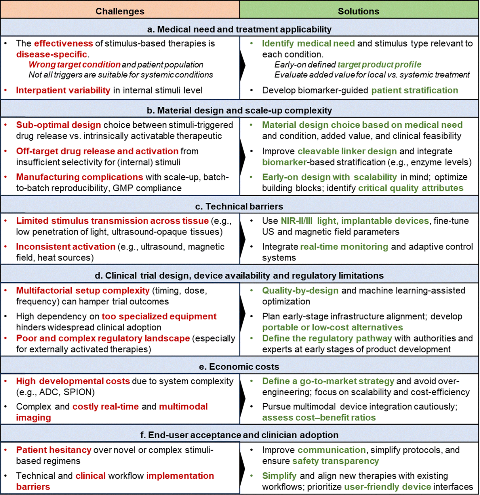 |
To improve the translational success rate in the future, materials research and development should be increasingly guided by clearly defined medical needs and a better understanding of the currently available therapeutic landscape. Together, this will enable a more realistic path toward clinical application.
4.1. Medical need and treatment applicability
Stimuli-responsive therapeutics hold high potential to spatiotemporally control the therapeutic action while minimizing off-target effects. Yet, their treatment performance and applicability are disease- and stimulus-specific. A trigger that is effective for treating one medical condition may be ineffective or impractical in another. Hence, identifying the concrete medical needs to address and the pathophysiological features of the specific indication can help to guide the design of novel stimuli-responsive therapeutics and maximize their eventual clinical relevance.In contrast, internal stimuli-responsive materials, activated by intrinsic biochemical cues such as pH, enzymes or redox gradients, are more amenable to systemic administration and therapies, as they can potentially be engineered so that the activation occurs selectively within the diseased tissues. Yet, the heterogeneity of the endogenous triggers between tissues (both diseased and healthy) and individuals poses a major translational barrier to ensure consistently effective and reproducible responses across patients. Variability in enzyme expression, pH, or redox states can compromise both selectivity and efficacy. A promising strategy to enhance the therapeutic efficacy of internal stimuli-responsive drugs and nanodrugs relies on the identification of biomarkers that correlate with disease states, including acidity, enzyme levels and redox state, and that eventually allows to predict and maximize patient responses to such therapies. Exploring tissue biomarkers and personalized medicine approaches, including the use of companion diagnostics, could help determine patient-specific biochemical features, allowing for tailored treatment strategies.22 Companion diagnostics, relying on imaging and enzyme level assays, may serve as gatekeepers for pre-treatment patient selection, helping identify those most likely to benefit from specific stimuli-responsive treatments.236
4.2. Material design and scale-up complexity
Once the need for stimuli-responsive therapy has been identified, the next step is to design and engineer the corresponding material, which also comes with specific challenges.On the one hand, stimuli-triggered cytotoxic drug delivery aims to employ carrier systems to spatiotemporally control drug delivery, enhancing target-site accumulation and effects, while minimizing off-target damage. This can be exploited for both external (e.g., ThermoDox) and internal stimuli (e.g., ADC), although it has been more clinically advanced for the latter. Employing biocompatible, clinically relevant delivery systems with approved cytotoxic drugs can streamline pharmaceutical development and regulatory trajectories.234 However, because internal stimuli are not always exclusive to diseased tissues, partial off-target release of cytotoxic drugs is often observed, limiting the therapeutic outcomes. Optimization of carrier systems, drugs, and stimuli-sensitive linkers will yield incremental improvements, albeit complete elimination of off-target toxicity using internal stimuli remains challenging. In this regard, exploration of alternative stimuli-sensitive linkers (e.g., carbamate or aminoacrylate for light cleavage in photoactivated chemotherapy,237 or oxanorbornene derivatives for ultrasonic cleavage)238 allows to develop novel materials and open therapeutic opportunities, albeit some of these may not yet align with clinically relevant wavelengths or frequencies.
Combining different approaches,9,239–241 such as ultrasound-assisted drug delivery, chemoradiotherapy or immunotherapy, is also under clinical investigation, aiming to enhance local drug concentration at the target site and potentially reduce off-target toxicity. However, whether these strategies allow for lower, and thereby safer, drug doses remain context-dependent and require further clinical validation. A personalized approach targeting tumor-specific neoantigens offers another promising pathway, with early data suggesting high selectivity and comparable costs to conventional therapies.242,243
In contrast, directly activatable (nano-)materials, as photosensitizers, sonosensitizers, and radioenhancers, are designed to remain biologically inert until the application of an external stimulus, thus offering greater control over the therapeutic action. However, as discussed above, their efficacy is tied to optimal in vivo co-localization of the material with the applied stimulus. Insufficient stimulus application time or intensity (whether light, ultrasound, or magnetic field) can lead to suboptimal therapeutic outcomes, and once the stimulus ceases, its effect often terminates, thus requiring repeated or prolonged stimulation. In addition to these constraints, the engineering complexity of the materials to ensure adequate responses in human tissues can seriously compromise formulation reproducibility, stability and biocompatibility.
Therefore, balancing the advantages and disadvantages of each therapeutic material design strategy crucially requires the consideration of the intended targeted medical indication and the associated pharmaceutical developmental trajectory. Importantly, the choice of the material platform itself already influences the translational potential of the final stimuli-responsive formulation. Therapeutic systems based on approved, clinically tested, and well-characterized nano- and microscale platforms such as liposomes, lipid nanoparticles, certain polymers, or antibodies, generally benefit from more predictable pharmacokinetics and established safety profiles, as well as a clearer regulatory pathway, together facilitating clinical development and translation. In contrast, newly developed or less clinically validated materials (e.g., certain metal–organic frameworks or complex hybrid nanostructures) may face additional challenges related to unknown biocompatibility, long-term safety, large-scale manufacturing, and regulatory acceptance. While some of these emerging systems may offer unique functional advantages, their increased design complexity can delay translation and clinical impact. Hence, selecting a material platform that balances functional performance with translational feasibility can help address the targeted medical need while accelerating clinical implementation.
4.3. Technical barriers
When the therapeutic agent is designed, it should maintain its efficacy not only in in vitro setups, but also in in vivo (clinical) settings. The core technical challenge lies in the limited and variable transmission of external stimuli through biological tissues, which hampers the precise activation of stimuli-responsive therapies at the intended site of action.4.4. Clinical trial design, device availability and regulatory limitations
Following the preclinical development of the new therapeutic agent, this must be assessed clinically, where planning the optimal clinical trial design is crucial to achieve successful outcomes.4.5. Economic costs
A major challenge is that both the high manufacturing and infrastructure costs of stimuli-responsive therapies, especially complex nanomaterials and device-dependent modalities, can make it difficult to attract investors and reach widespread clinical adoption.000, and USD 100000–300000, respectively), other devices, such as magnetic resonance imaging-guided field generators and medical linear accelerators, are significantly costly (both can exceed USD 1 million, with additional maintenance costs). This obviously creates an additional financial barrier for healthcare facilities. The magnetic-activatable SPION formulation NanoTherm is a relevant example collecting numerous translational barriers, since, despite its EMA approval as a medical device in 2010,35 it has never advanced to phase III trials, with limited clinical data. Key barriers include the need for specialized infrastructure and equipment for precise intratumoral SPION injection into brain tumors, dedicated alternating magnetic field generators for hyperthermia activation, and trained personnel to administer the therapy. Therefore, only a small number of hospitals and research centers are able to offer NanoTherm therapy, which, in addition, limits the number of treated patients, and consequently, the volume of clinical data available from the trial.Future research should therefore prioritize the development of simpler and more cost-effective alternatives, including portable ultrasound devices and low-cost magnetic field generators, to make stimuli-responsive therapies more financially viable for routine clinical use. For broad clinical adoption, comprehensive cost-benefit analysis will be essential to demonstrate that the clinical advantages of stimuli-responsive therapies justify the added economic burden when compared to more conventional treatments.
4.6. End-user acceptance and clinician adoption
The success of stimuli-responsive therapies depends not only on the scientific and technical advances, but also on ensuring patient acceptance and broad clinical adoption.Overall, improving end-user compliance requires more effective science communication, increasing the general knowledge for both patients and clinicians, transparent dissemination of safety and efficacy data, and the development of streamlined, user-friendly treatment protocols that minimize the burden on both patients and healthcare providers.
5. Outlook
Stimuli-responsive therapeutics have already achieved considerable clinical impact, with several products used in the treatment of cancer, dental conditions, vision disorders, and infectious diseases. While small-molecule fluorescent dyes remain relevant, the advances in nano- and microtechnology have driven the clinical adoption of novel materials such as SPION, ADC, Hensify nanoparticles, and SonoVue or Definity microbubbles. These materials paved the way for new therapeutic modalities, including sonopermeation, radiotherapy-enhancement, magnetically induced hyperthermia, and improved drug delivery and targeting control. A comparative summary of major stimuli, including advantages, limitations, trade-offs, and translational status, is provided to contextualize the clinical translation landscape of different stimuli and stimuli-responsive nanomedicines and microscale therapeutics (Table 3).| Stimuli | Advantages | Challenges | Main trade-offs | Common indications | Translational status/potential | Clinically relevant examples |
|---|---|---|---|---|---|---|
| Light | Non-invasive, cost-effective, high spatiotemporal control, simple implementation | Limited tissue penetration, oxygen-dependent | High precision vs. low penetration depth | Superficial cancers, dental and skin conditions, vision disorders, intraoperative | Clinically established/High ✓✓✓ | 5-ALA, porphyrins (e.g., liposomal Visudyne), methylene blue (approved) |
| Ultrasound | Non-invasive, deep tissue penetration, high spatiotemporal control, potential for blood–brain barrier opening | Understanding and fine-tuning of ultrasound parameters to maximize response; artifacts and low/no penetration through lungs, bone, and gas filled bowel | High penetration vs. parameter optimization | Brain diseases, Tumor vasculature perfusion | Clinical trials/High ✓✓✓ | Microbubbles (approved for imaging and in trials for therapy) |
| Radiation | Non-invasive, deep tissue penetration, clinically well established | Mostly metal-based materials, administration routes | Physical dose amplification vs. delivery and distribution limitations | Urogenital, lung, and brain cancers | Clinically established/Medium ✓✓ | Hafnium oxide nanoparticles (EMA approved) |
| Heat | Minimally invasive, cost-effective | Poor spatiotemporal and temperature control, side effects | Simplicity vs. complex stimulus control | Liver and breast cancers | Clinical trials/Medium ✓✓ | ThermoDox, UGN-103 hydrogel (both in trials) |
| Magnetic field | Non-invasive, deep tissue penetration | Metal-based materials, side effects, difficulty to locally apply in deep tissues | High penetration vs. high device and running costs | Brain, prostate, pancreatic cancers | Clinically established but limited usage/Medium ✓✓ | SPION (e.g., NanoTherm; EMA approved) |
| Internal stimuli | Versatile, modular design, simple implementation, no external device required | Off-target activation and side effects, limited specificity for heterogenous tumors | Efficacy vs. toxicity | Liquid and solid cancers | Clinically established/High ✓✓✓ | Antibody–drug conjugates (>10 approved), prodrugs |
The successful translation and implementation of stimuli-responsive therapeutics depend on strong cross-disciplinary collaboration to align therapeutic material design with concrete medical needs, infrastructure availability, and patient expectations, while ensuring that the conceptualization of novel stimuli-responsive therapeutics, especially for more complex nano- or microscale materials, is driven by the medical needs, disease features and clinical implementation challenges, striving for material simplicity while ensuring adequate therapeutic performance.
From the materials research perspective, emerging concepts and future directions are focused on (a) optimizing material designs and structural engineering, improving stimulus-responsiveness control in biological settings; (b) exploring materials and technologies responsive to underexplored stimuli such as electrical pulses or shear forces, opening new opportunities to treat indications such as stroke or epilepsy; (c) triggering therapeutic activation and (multi)drug release via combined stimuli, including bidirectional on–off responsive structures; (d) evaluating their integration into implantable devices and vascular grafts for on-demand treatment control in regenerative medicine applications and incorporation into robotic systems to develop remote and self-activatable therapeutics (Fig. 7).
 | ||
| Fig. 7 Design and engineering opportunities for stimuli-responsive therapeutic materials. Several engineering aspects can open new avenues for stimuli-responsive therapeutics, with their design and development driven by clear medical, disease, and patient needs. (A) Optimizing stimulus-responsiveness control in biologically relevant settings through precise material design, engineering, and tuning of physicochemical properties. (B) Exploring alternative physiological stimuli, including electrical pulses, shear forces, and cytokine/chemokine cues, enabling new therapeutic opportunities for neurological or cardiovascular diseases. (C) Developing multi-stimuli and multi-drug-containing systems, where single or combined triggers (e.g., electrical, magnetic, pH) enable on–off drug release and therapy control. (D) Integration of stimuli-responsive therapeutics into implantable devices, incorporating magnetic, mechanical, and electrical responsiveness to support image-guided, on-demand, and closed-loop drug delivery. | ||
5.1. Material design and stimulus-responsiveness control
Promoting quality-by-design approaches of (novel) materials and improved control over the constituting building blocks and their properties (including underexplored engineering aspects such as microbubble shell thickness and porosity, or hydrogel compartmentalization)53 will open new avenues to more accurately construct nano- and microscale materials with enhanced control over specific stimuli (e.g., narrow ultrasound frequencies). Advances in electroactive polymers and semiconductive membranes are already promoting better spatiotemporal control over stimuli activation due to the conversion of the electrical stimuli directly into mechanical motion, enabling fast, reversible, and spatially precise actuation.264,265 Shape memory hydrogels also offer enhanced responsiveness control due to their ability to undergo reversible, stimulus-specific deformation and then recover their original shape in response to environmental triggers.2665.2. Electrical- and shear forces-mediated activation
While traditionally underexplored, electrical stimuli- and shear forces-responsive systems are gaining traction,267 particularly for neuromodulation (e.g., epilepsy)268 or cardiovascular conditions (e.g., clot-targeted therapies).269 Magneto-/electro-responsive polymers270 and mechano-sensitive hydrogels271 offer novel opportunities for precise, non-invasive control of therapeutic release due to shape-morphing capabilities under remote activation.5.3. Multi-stimuli and multi-drug targeted materials
Drug delivery systems can be engineered to be triggered by different, combined stimuli (e.g., pH and redox,272 photo and magnetic,273 or pH and temperature216) to improve drug targeting, and treatment specificity and control. Multi-drug nanomaterials can potentially achieve combined therapeutic effects and overcome drug resistance.274 Besides triggering co-delivery of synergistic drug combinations,275 responsiveness to multiple stimuli enables reversible on–off behavior governed by logic-gate-like mechanisms, where drug release occurs only when specific combinations of triggers are simultaneously present.2765.4. Implantable devices integration and (self-)automation
The integration of stimuli-responsive materials into implantable systems allows for localized, sustained, and on-demand therapeutic delivery directly at the disease site, improving both efficacy and patient compliance. Implantable devices incorporating pH-, redox-, or enzyme-responsive hydrogels have been developed for post-surgical cancer therapy, offering controlled drug release in response to the tumor microenvironment's biochemical cues.277 Active implantable drug delivery systems, including soft robotics, represent a new generation of smart, stimuli-responsive implants capable of remotely controlled and autonomous therapeutic release, integrating powered actuators, control logic, and communication interfaces.278,279 For instance, soft implantable devices have recently been designed for seizure treatment, continuously monitoring electroencephalogram signals and using wireless power transmission to trigger drug release during emergencies.280 Photodynamic therapy can also benefit from implantable devices that use real-time monitoring and wireless power to intelligently modulate treatment based on optical or thermal feedback.248,281,282 Additionally, such systems can be combined with imaging platforms (e.g., SPION),283 enabling real-time visualization of the implant or payload and its therapeutic activity via MRI, PET, or fluorescence imaging to guide and personalize treatments.Progress across these areas will continue to expand the clinical impact of stimuli-responsive therapeutics and enhance their relevance in the treatment of other indications like neurological and cardiovascular diseases, wound healing, and regenerative medicine.
While hurdles remain, the field is entering a critical phase where carefully elaborated material design needs to be ensured, primarily motivated by a disease- and medical need-driven mindset, and supported by robust translational frameworks, to increase the translational success rate of stimuli-responsive (nano- and macroscale) therapeutics and broaden their clinical impact.
Conflicts of interest
The authors declare that they have no known competing financial interests or personal relationships that could have appeared to influence the work reported in this paper.Data availability
The data supporting this article have been included as part of the supplementary information (SI). Keyword search scheme is presented in SI. See DOI: https://doi.org/10.1039/d6cs00165c.Acknowledgements
This research was supported by the Medical Scientist Program of the Faculty of Medicine RWTH Aachen University (MSP-Main 2025). The support of ERC (CoG, 864121; PoC PRIME, 101138100; PoC Open-SESAME, 101247604), DFG (GRK/RTG2735, 331065168), BMBF (PP-TNBC, 16GW0319K), RWTH-Start (MET-Immuno), and the National Academy of Sciences of Ukraine (project no. 0125U000603) is acknowledged. BioRender.com was used to create structural elements of the figures.References
- R. Baskaran, J. Lee and S.-G. Yang, Biomater. Res., 2018, 22, 25 CrossRef PubMed.
- J. Da Silva, C. Bienassis, P. Schmitt, C. Berjaud, M. Guedj and S. Paris, J. Exp. Clin. Cancer Res., 2024, 43, 11 CrossRef CAS PubMed.
- L. R. H. Gerken, M. E. Gerdes, M. Pruschy and I. K. Herrmann, Mater. Horizons, 2023, 10, 4059–4082 RSC.
- J. H. Correia, J. A. Rodrigues, S. Pimenta, T. Dong and Z. Yang, Pharmaceutics, 2021, 13, 1332 CrossRef CAS PubMed.
- X. Zhao, J. Liu, J. Fan, H. Chao and X. Peng, Chem. Soc. Rev., 2021, 50, 4185–4219 RSC.
- Z. Zhang, Y. Du, X. Shi, K. Wang, Q. Qu, Q. Liang, X. Ma, K. He, C. Chi, J. Tang, B. Liu, J. Ji, J. Wang, J. Dong, Z. Hu and J. Tian, Nat. Rev. Clin. Oncol., 2024, 21, 449–467 CrossRef PubMed.
- M. Ashrafizadeh, M. Delfi, A. Zarrabi, A. Bigham, E. Sharifi, N. Rabiee, A. C. Paiva-Santos, A. P. Kumar, S. C. Tan, K. Hushmandi, J. Ren, E. N. Zare and P. Makvandi, J. Controlled Release, 2022, 351, 50–80 CrossRef CAS PubMed.
- Y. Yu, Y. Cheng, J. Tong, L. Zhang, Y. Wei and M. Tian, J. Mater. Chem. B, 2021, 9, 2979–2992 RSC.
- M. Molinaro, D. Skrodzki and D. Pan, Wiley Interdiscip. Rev.:Nanomed. Nanobiotechnol., 2024, 16, e1984 CAS.
- B. Rezaei, P. Yari, S. M. Sanders, H. Wang, V. K. Chugh, S. Liang, S. Mostufa, K. Xu, J. Wang, J. Gómez-Pastora and K. Wu, Small, 2024, 20, 2304848 CrossRef CAS PubMed.
- A. Włodarczyk, S. Gorgoń, A. Radoń and K. Bajdak-Rusinek, Nanomaterials, 2022, 12, 1807 CrossRef PubMed.
- A. Shakeri-Zadeh and J. W. M. Bulte, Nat. Rev. Bioeng., 2025, 3, 245–260 CrossRef CAS PubMed.
- K. Zhu, J. Wang, Z. Wang, Q. Chen, J. Song and X. Chen, Angew. Chem., Int. Ed., 2025, 64, e202422278 CrossRef CAS PubMed.
- D. Li, Y. Yang, D. Li, J. Pan, C. Chu and G. Liu, Small, 2021, 17, 2101976 CrossRef CAS PubMed.
- H. Ding, P. Tan, S. Fu, X. Tian, H. Zhang, X. Ma, Z. Gu and K. Luo, J. Controlled Release, 2022, 348, 206–238 CrossRef CAS PubMed.
- Z. Fu, S. Li, S. Han, C. Shi and Y. Zhang, Signal Transduction Targeted Ther., 2022, 7, 93 CrossRef CAS PubMed.
- X. Meng, Y. Shen, H. Zhao, X. Lu, Z. Wang and Y. Zhao, J. Nanobiotechnol., 2024, 22, 587 CrossRef PubMed.
- Y. Wei, J. Lv, S. Zhu, S. Wang, J. Su and C. Xu, Drug Discovery Today, 2024, 29, 104014 CrossRef CAS PubMed.
- C. Dumontet, J. M. Reichert, P. D. Senter, J. M. Lambert and A. Beck, Nat. Rev. Drug Discovery, 2023, 22, 641–661 CrossRef CAS PubMed.
- S. N. Bhatia, X. Chen, M. A. Dobrovolskaia and T. Lammers, Nat. Rev. Cancer, 2022, 22, 550–556 CrossRef CAS PubMed.
- S. Mura, J. Nicolas and P. Couvreur, Nat. Mater., 2013, 12, 991–1003 CrossRef CAS PubMed.
- T. Lammers, Adv. Mater., 2024, 36, 2312169 CrossRef CAS PubMed.
- A. Grzybowski and K. Pietrzak, Clin. Dermatol., 2012, 30, 451–455 CrossRef PubMed.
- R.-M. Szeimies, T. Dirschka, M. C. Fargnoli, Y. Gilaberte, M. Hædersdal, R. Chavda and P. Calzavara-Pinton, Dermatol. Ther., 2023, 13, 1409–1421 CrossRef PubMed.
- M. Fabre, L. Mateo, D. Lamaa, S. Baillif, G. Pagès, L. Demange, C. Ronco and R. Benhida, Molecules, 2022, 27, 5089 CrossRef CAS PubMed.
- Photodynamic Therapy Market Size And Forecast, https://www.verifiedmarketresearch.com/product/photodynamic-therapy-market/, (accessed October 2025).
- Photodynamic Therapy Market Size and Forecast 2025 to 2034, https://www.precedenceresearch.com/photodynamic-therapy-market, (accessed October 2025).
- Y. Zhang, S. Zhang, Z. Zhang, L. Ji, J. Zhang, Q. Wang, T. Guo, S. Ni, R. Cai, X. Mu, W. Long and H. Wang, Front. Chem., 2021, 9, 728066 CrossRef CAS PubMed.
- H. Kobayashi, P. L. Choyke and M. Ogawa, Curr. Opin. Chem. Biol., 2023, 74, 102289 CrossRef CAS PubMed.
- Y. Kadria-Vili, J. A. Schwartz, T. J. Polascik, G. P. Goodrich, D. Jorden, D. Pinder, N. J. Halas and A. R. Rastinehad, Nanomaterials, 2024, 14, 1261 CrossRef CAS PubMed.
- M. Chaudhry, P. Lyon, C. Coussios and R. Carlisle, Expert Opin. Drug Delivery, 2022, 19, 899–912 CrossRef CAS PubMed.
- A. Lawrie, A. Brisken, S. Francis, D. Cumberland, D. Crossman and C. Newman, Gene Ther., 2000, 7, 2023–2027 CrossRef CAS PubMed.
- B. Rodriguez, D. Rivera, J. Y. Zhang, C. Brown, T. Young, T. Williams, S. Huq, M. Mattioli, A. Bouras and C. G. Hadjpanayis, Pharmaceuticals, 2024, 17, 300 CrossRef CAS PubMed.
- M. Schwake, M. Müther, A.-K. Bruns, B. Zinnhardt, N. Warneke, M. Holling, S. Schipmann, B. Brokinkel, J. Wölfer, W. Stummer and O. Grauer, Cancers, 2022, 14, 541 CrossRef CAS PubMed.
- F. Soetaert, P. Korangath, D. Serantes, S. Fiering and R. Ivkov, Adv. Drug Delivery Rev., 2020, 163–164, 65–83 CrossRef CAS PubMed.
- N. G. Caculitan, J. dela, C. Chuh, Y. Ma, D. Zhang, K. R. Kozak, Y. Liu, T. H. Pillow, J. Sadowsky, T. K. Cheung, Q. Phung, B. Haley, B. C. Lee, R. W. Akita, M. X. Sliwkowski and A. G. Polson, Cancer Res., 2017, 77, 7027–7037 CrossRef CAS PubMed.
- Antibody Drug Conjugates Market Size, Share and Trends 2026 to 2035, https://www.precedenceresearch.com/antibody–drug-conjugates-market, (accessed October 2025).
- J. Yang and J. Kopeček, Curr. Opin. Colloid Interface Sci., 2017, 31, 30–42 CrossRef CAS PubMed.
- J. Zheng, X. Song, Z. Yang, C. Yin, W. Luo, C. Yin, Y. Ni, Y. Wang and Y. Zhang, J. Controlled Release, 2022, 350, 898–921 CrossRef CAS PubMed.
- Q. Peña, A. Wang, O. Zaremba, Y. Shi, H. W. Scheeren, J. M. Metselaar, F. Kiessling, R. M. Pallares, S. Wuttke and T. Lammers, Chem. Soc. Rev., 2022, 51, 2544–2582 RSC.
- Z. Su, D. Xiao, F. Xie, L. Liu, Y. Wang, S. Fan, X. Zhou and S. Li, Acta Pharm. Sin. B, 2021, 11, 3889–3907 CrossRef CAS PubMed.
- K. Song, C. Jiang, S. Huang and X. Li, Mater. Chem. Front., 2025, 9, 189–203 RSC.
- Y. Zong, Y. Lin, T. Wei and Q. Cheng, Adv. Mater., 2023, 35, e2303261 CrossRef PubMed.
- A. H. Maboudi, M. H. Lotfipour, M. Rasouli, M. H. Azhdari, R. MacLoughlin, S. Bekeschus and M. Doroudian, Nanotechnol. Rev., 2024, 13, 20230218 CrossRef CAS.
- H. Cabral, K. Miyata, K. Osada and K. Kataoka, Chem. Rev., 2018, 118, 6844–6892 CrossRef CAS PubMed.
- A. Javia, J. Vanza, D. Bardoliwala, S. Ghosh, L. A. Misra, M. Patel and H. Thakkar, Int. J. Pharm., 2022, 623, 121863 CrossRef CAS PubMed.
- R. A. Barmin, M. Moosavifar, A. Dasgupta, A. Herrmann, F. Kiessling, R. M. Pallares and T. Lammers, Chem. Sci., 2023, 14, 11941–11954 RSC.
- B. Tian, S. Hua and J. Liu, Carbohydr. Polym., 2023, 315, 120972 CrossRef CAS PubMed.
- A. R. Petrovici, M. Pinteala and N. Simionescu, Molecules, 2023, 28, 1086 CrossRef CAS PubMed.
- S. M. Hosseini, J. Mohammadnejad, R. Najafi-Taher, Z. B. Zadeh, M. Tanhaei and S. Ramakrishna, ACS Appl. Bio Mater., 2023, 6, 1323–1338 CrossRef CAS PubMed.
- M. Koohi Moftakhari Esfahani, S. E. Alavi, P. J. Cabot, N. Islam and E. L. Izake, Pharmaceutics, 2022, 14, 1579 CrossRef PubMed.
- S. Devi, M. Kumar, A. Tiwari, V. Tiwari, D. Kaushik, R. Verma, S. Bhatt, B. M. Sahoo, T. Bhattacharya, S. Alshehri, M. M. Ghoneim, A. O. Babalghith and G. E.-S. Batiha, Front. Mater., 2022, 8, 2021 Search PubMed.
- M. Moosavifar, R. A. Barmin, E. Rama, A. Rix, R. A. Gumerov, T. Lisson, C. Bastard, S. Rütten, N. Avraham-Radermacher, J. Koehler, M. Pohl, V. Kulkarni, J. Baier, S. Koletnik, R. Zhang, A. Dasgupta, A. Motta, M. Weiler, I. I. Potemkin, G. Schmitz, F. Kiessling, T. Lammers and R. M. Pallares, Adv. Sci., 2024, 11, 2404385 CrossRef CAS PubMed.
- A. Dasgupta, T. Sun, R. Palomba, E. Rama, Y. Zhang, C. Power, D. Moeckel, M. Liu, A. Sarode, M. Weiler, A. Motta, C. Porte, Z. Magnuska, A. Said Elshafei, R. Barmin, A. Graham, A. McClelland, D. Rommel, E. Stickeler, F. Kiessling, R. M. Pallares, L. De Laporte, P. Decuzzi, N. McDannold, S. Mitragotri and T. Lammers, Proc. Natl. Acad. Sci. U. S. A., 2023, 120, e2218847120 CrossRef CAS PubMed.
- J. C. Hsu, Z. Tang, O. E. Eremina, A. M. Sofias, T. Lammers, J. F. Lovell, C. Zavaleta, W. Cai and D. P. Cormode, Nat. Rev. Methods Primers, 2023, 3, 30 CrossRef CAS PubMed.
- R. Zhang, F. Kiessling, T. Lammers and R. M. Pallares, Drug Delivery Transl. Res., 2023, 13, 378–385 CrossRef PubMed.
- S. Yu, G. Xia, N. Yang, L. Yuan, J. Li, Q. Wang, D. Li, L. Ding, Z. Fan and J. Li, Int. J. Mol. Sci., 2024, 25, 5632 CrossRef CAS PubMed.
- S. M. Dadfar, K. Roemhild, N. I. Drude, S. von Stillfried, R. Knüchel, F. Kiessling and T. Lammers, Adv. Drug Delivery Rev., 2019, 138, 302–325 CrossRef CAS PubMed.
- F. Lux, V. L. Tran, E. Thomas, S. Dufort, F. Rossetti, M. Martini, C. Truillet, T. Doussineau, G. Bort, F. Denat, F. Boschetti, G. Angelovski, A. Detappe, Y. Crémillieux, N. Mignet, B.-T. Doan, B. Larrat, S. Meriaux, E. Barbier, S. Roux, P. Fries, A. Müller, M.-C. Abadjian, C. Anderson, E. Canet-Soulas, P. Bouziotis, M. Barberi-Heyob, C. Frochot, C. Verry, J. Balosso, M. Evans, J. Sidi-Boumedine, M. Janier, K. Butterworth, S. McMahon, K. Prise, M.-T. Aloy, D. Ardail, C. Rodriguez-Lafrasse, E. Porcel, S. Lacombe, R. Berbeco, A. Allouch, J.-L. Perfettini, C. Chargari, E. Deutsch, G. Le Duc and O. Tillement, Br. J. Radiol., 2018, 92, 20180365 CrossRef PubMed.
- R. F. Barth, P. Mi and W. Yang, Cancer Commun., 2018, 38, 1–15 Search PubMed.
- L. Xue, A. S. Thatte, D. Mai, R. M. Haley, N. Gong, X. Han, K. Wang, N. C. Sheppard, C. H. June and M. J. Mitchell, Nat. Rev. Mater., 2024, 9, 100–118 CrossRef CAS.
- L. Gong, Y. Zhang, C. Liu, M. Zhang and S. Han, Int. J. Nanomed., 2021, 16, 1083–1102 CrossRef PubMed.
- K. K. K. Sharma, S. G. Swarts and W. A. Bernhard, J. Phys. Chem. B, 2011, 115, 4843–4855 CrossRef CAS PubMed.
- Y.-F. Zhang and M. Lu, Front. Bioeng. Biotechnol., 2024, 12, 2024 Search PubMed.
- S. Anand, T. A. Chan, T. Hasan and E. V. Maytin, Pharmaceuticals, 2021, 14, 447 CrossRef CAS PubMed.
- B. Parshad, S. Arora, B. Singh, Y. Pan, J. Tang, Z. Hu and H. K. Patra, Commun. Chem., 2025, 8, 100 CrossRef CAS PubMed.
- S. Zahnreich, A. Bhatti, B. Ahmad, S. Drabke, J. Kaufmann and H. Schmidberger, Cells, 2025, 14, 682 CrossRef CAS PubMed.
- Y. Dou, K. Hynynen and C. Allen, J. Controlled Release, 2017, 249, 63–73 CrossRef CAS PubMed.
- Celsion Corporation Receives Recommendation from Independent Data Monitoring Committee to Consider Stopping the Phase III OPTIMA Study, https://investor.celsion.com/news-releases/news-release-details/celsion-corporation-receives-recommendation-independent-data, (accessed December 2025).
- Z. Fralish, A. Chen, S. Khan, P. Zhou and D. Reker, Nat. Rev. Drug Discovery, 2024, 23, 365–380 CrossRef CAS PubMed.
- J. Rautio, N. A. Meanwell, L. Di and M. J. Hageman, Nat. Rev. Drug Discovery, 2018, 17, 559–587 CrossRef CAS PubMed.
- H. Kobayashi, A. Furusawa, A. Rosenberg and P. L. Choyke, Int. Immunol., 2021, 33, 7–15 CrossRef CAS PubMed.
- T. Kato, R. Okada, A. Furusawa, F. Inagaki, H. Wakiyama, H. Furumoto, S. Okuyama, H. Fukushima, P. L. Choyke and H. Kobayashi, Mol. Cancer Ther., 2021, 20, 2262–2273 CrossRef CAS PubMed.
- D. Kobzev, O. Semenova, A. Tatarets, A. Bazylevich, G. Gellerman and L. Patsenker, Dyes Pigm., 2023, 212, 111101 CrossRef CAS.
- B. Worley, V. Harikumar, K. Reynolds, M. A. Dirr, R. E. Christensen, N. Anvery, M. D. Yi, E. Poon and M. Alam, Arch. Dermatol. Res., 2023, 315, 1099–1108 CrossRef CAS PubMed.
- J. Karges, Angew. Chem., Int. Ed., 2022, 61, e202112236 CrossRef CAS PubMed.
- O. Semenova, D. Kobzev, I. Hovor, M. Atrash, F. Nakonechny, O. Kulyk, A. Bazylevich, G. Gellerman and L. Patsenker, Pharmaceutics, 2023, 15, 247 CrossRef CAS PubMed.
- X. Hu, Z. Fang, C. Zhu, Y. Yang, Z. Yang and W. Huang, Adv. Funct. Mater., 2024, 34, 2401325 CrossRef CAS.
- P. Chowdhury and Y.-H. Chan, Mol. Syst. Des. Eng., 2022, 7, 702–719 RSC.
- Y. Zhang, S. Li, X. Fang, B. Miao, Y. Wang, J. Liu, G. Nie and B. Zhang, Nanophotonics, 2022, 11, 5189–5204 CrossRef CAS PubMed.
- H. Yukawa, K. Sato and Y. Baba, Adv. Drug Delivery Rev., 2023, 200, 114863 CrossRef CAS PubMed.
- R. M. Pallares, F. Kiessling and T. Lammers, Nanomedicine, 2024, 19, 2433–2435 CrossRef CAS PubMed.
- Y. Yang, S. Jiang, S. G. Stanciu, H. Peng, A. Wu and F. Yang, Mater. Horizons, 2024, 11, 5815–5842 RSC.
- A. Lopez-Beltran, M. S. Cookson, B. J. Guercio and L. Cheng, BMJ, 2024, 384, e076743 CrossRef CAS PubMed.
- S. Mitragotri, Nat. Rev. Drug Discovery, 2005, 4, 255–260 CrossRef CAS PubMed.
- Y. Meng, K. Hynynen and N. Lipsman, Nat. Rev. Neurol., 2021, 17, 7–22 CrossRef PubMed.
- E. Stride and C. Coussios, Nat. Rev. Phys., 2019, 1, 495–509 CrossRef CAS.
- E. Stride, T. Segers, G. Lajoinie, S. Cherkaoui, T. Bettinger, M. Versluis and M. Borden, Ultrasound Med. Biol., 2020, 46, 1326–1343 CrossRef PubMed.
- I. Lentacker, I. De Cock, R. Deckers, S. C. De Smedt and C. T. W. Moonen, Adv. Drug Delivery Rev., 2014, 72, 49–64 CrossRef CAS PubMed.
- M. Cattaneo, G. Guerriero, G. Shakya, L. A. Krattiger, L. G. Paganella, M. L. Narciso and O. Supponen, Nat. Phys., 2025, 21, 590–598 Search PubMed.
- A. Carpentier, M. Canney, A. Vignot, V. Reina, K. Beccaria, C. Horodyckid, C. Karachi, D. Leclercq, C. Lafon, J.-Y. Chapelon, L. Capelle, P. Cornu, M. Sanson, K. Hoang-Xuan, J.-Y. Delattre and A. Idbaih, Sci. Transl. Med., 2016, 8, 343re2 Search PubMed.
- M. Ahluwalia, M. McDermott, T. Burns, J. de Groot, A. Mogilner, A. Achrol, B. Shah, C. Bettegowda, A. Ozair, A. Khosla, T. Schwartz, A. Sahgal, M. Mishra, R. Everson, J. Weinberg, N. Amankulor, J. Sporrer, C. Cifarelli, A. Rezai, N. Lipsman and G. F. Woodworth, Neuro-Oncol., 2023, 25(Suppl 5), v88 Search PubMed.
- G. Dimcevski, S. Kotopoulis, T. Bjånes, D. Hoem, J. Schjøtt, B. T. Gjertsen, M. Biermann, A. Molven, H. Sorbye, E. McCormack, M. Postema and O. H. Gilja, J. Controlled Release, 2016, 243, 172–181 CrossRef CAS PubMed.
- N. Lipsman, Y. Meng, A. J. Bethune, Y. Huang, B. Lam, M. Masellis, N. Herrmann, C. Heyn, I. Aubert, A. Boutet, G. S. Smith, K. Hynynen and S. E. Black, Nat. Commun., 2018, 9, 2336 CrossRef PubMed.
- F. Kiessling, S. Fokong, P. Koczera, W. Lederle and T. Lammers, J. Nucl. Med., 2012, 53, 345–348 CrossRef CAS PubMed.
- S. Fokong, B. Theek, Z. Wu, P. Koczera, L. Appold, S. Jorge, U. Resch-Genger, M. van Zandvoort, G. Storm, F. Kiessling and T. Lammers, J. Controlled Release, 2012, 163, 75–81 CrossRef CAS PubMed.
- P. Koczera, L. Appold, Y. Shi, M. Liu, A. Dasgupta, V. Pathak, T. Ojha, S. Fokong, Z. Wu, M. van Zandvoort, O. Iranzo, A. J. C. Kuehne, A. Pich, F. Kiessling and T. Lammers, J. Controlled Release, 2017, 259, 128–135 CrossRef CAS PubMed.
- P. Nittayacharn, E. Abenojar, M. B. Cooley, F. M. Berg, C. Counil, A. J. Sojahrood, M. S. Khan, C. Yang, E. Berndl, M. Golczak, M. C. Kolios and A. A. Exner, J. Controlled Release, 2024, 367, 135–147 CrossRef CAS PubMed.
- Y. Cheng, H. Cheng, C. Jiang, X. Qiu, K. Wang, W. Huan, A. Yuan, J. Wu and Y. Hu, Nat. Commun., 2015, 6, 8785 CrossRef CAS PubMed.
- S. L. Huang, Methods Mol. Biol., 2010, 605, 113–128 CrossRef CAS PubMed.
- M. P. Purohit, B. J. Yu, K. S. Roy, Y. Xiang, S. N. Ewbank, M. M. Azadian, A. R. Hart, G. P. B. Muwanga, P. J. Martinez, J. B. Wang, A. K. Taoube, E. Markarian, N. Macedo, A. K. Kwan, D. G. Lopez and R. D. Airan, Nat. Nanotechnol., 2025, 20, 1688–1699 CrossRef CAS PubMed.
- D. Kobzev, O. Semenova, S. Aviel-Ronen, O. Kulyk, R. Carmieli, T. Mirzabekov, G. Gellerman and L. Patsenker, Int. J. Mol. Sci., 2024, 25, 10137 CrossRef CAS PubMed.
- I. Rosenthal, J. Z. Sostaric and P. Riesz, Ultrason. Sonochem., 2004, 11, 349–363 CrossRef CAS PubMed.
- S. J. McMahon, W. B. Hyland, M. F. Muir, J. A. Coulter, S. Jain, K. T. Butterworth, G. Schettino, G. R. Dickson, A. R. Hounsell, J. M. O’Sullivan, K. M. Prise, D. G. Hirst and F. J. Currell, Radiother. Oncol., 2011, 100, 412–416 CrossRef CAS PubMed.
- J. Schuemann, R. Berbeco, D. B. Chithrani, S. H. Cho, R. Kumar, S. J. McMahon, S. Sridhar and S. Krishnan, Int. J. Radiat. Oncol., Biol., Phys., 2016, 94, 189–205 CrossRef PubMed.
- A. Taheri, M. U. Khandaker, F. Moradi and D. A. Bradley, Radiat. Phys. Chem., 2023, 212, 111146 CrossRef CAS.
- N. Jackson, D. Cecchi, W. Beckham and D. B. Chithrani, Molecules, 2024, 29, 2438 CrossRef CAS PubMed.
- P. Qi, Q. Chen, D. Tu, S. Yao, Y. Zhang, J. Wang, C. Xie, C. Pan and H. Peng, Biomater. Sci., 2020, 8, 2778–2785 RSC.
- L. Li, M. Wang, Q. Zhao, P. Bai, H. Hao, Z. Zhang, T. Liu, Y. Yang, K. Pu and R. Zhang, Angew. Chem., Int. Ed., 2025, 64, e202413232 CrossRef CAS PubMed.
- Performance and Safety of thermotherapy with nanoparticles (NanoTherm® Therapy System) as an adjuvant therapy to the Standard of Care treatment of patients with recurrent glioblastoma, https://drks.de/search/en/trial/DRKS00023339/details, (accessed December 2025).
- M. Rezaee, E. Alizadeh, P. Cloutier, D. J. Hunting and L. Sanche, ChemMedChem, 2014, 9, 1145–1149 CrossRef CAS PubMed.
- P. C. N. Teixeira, Cytologic and radiosensibilizer action of the methylene blue, Biophysics Institute Carlos Chagas Filho, Universidade Federal, Rio de Janeiro (Brazil), 1989 Search PubMed.
- X. Yu, B. Liu, N. Zhang, Q. Wang and G. Cheng, Front. Cell Dev. Biol., 2021, 9, 646981 CrossRef PubMed.
- Y. You, R. Wen, R. Pathak, A. Li, W. Li, D. St Clair, M. Hauer-Jensen, D. Zhou and Y. Liang, Cell Death Dis., 2014, 5, e1493 CrossRef CAS PubMed.
- H. K. Rachamala, V. S. Madamsetty, R. S. Angom, N. M. Nakka, S. K. Dutta, E. Wang, D. Mukhopadhyay and K. Pal, J. Exp. Clin. Cancer Res., 2024, 43, 1–17 Search PubMed.
- S. Leo, N. M. C. Gutierrez, A.-L. Bulin, J. L. Coll, L. Sancey, B. Habermeyer and M. Broekgaarden, Eur. J. Med. Chem., 2025, 296, 117861 CrossRef CAS PubMed.
- S. R. Adams, H. C. Yang, E. N. Savariar, J. Aguilera, J. L. Crisp, K. A. Jones, M. A. Whitney, S. M. Lippman, E. E. W. Cohen, R. Y. Tsien and S. J. Advani, Nat. Commun., 2016, 7, 13019 CrossRef CAS PubMed.
- K. Ni, G. Lan, C. Chan, B. Quigley, K. Lu, T. Aung, N. Guo, P. La Riviere, R. R. Weichselbaum and W. Lin, Nat. Commun., 2018, 9, 2351 CrossRef PubMed.
- Z. Chen, F. Han, Y. Du, H. Shi and W. Zhou, Signal Transduction Targeted Ther., 2023, 8, 70 CrossRef PubMed.
- X. Li, H. Wang, Z. Li, F. Tao, J. Wu, W. Guan and S. Liu, Front. Oncol., 2023, 12, 2022 Search PubMed.
- Q. Li, C. W. Kartikowati, S. Horie, T. Ogi, T. Iwaki and K. Okuyama, Sci. Rep., 2017, 7, 9894 CrossRef PubMed.
- Y. Li, R. Zhang, R. Barmin, E. Rama, M. Schoenen, F. Schrank, V. Schulz, I. Slabu, F. Kiessling, T. Lammers and R. M. Pallares, Nanoscale Adv., 2024, 6, 4352–4359 RSC.
- P. Sahoo, P. Choudhary, S. S. Laha, A. Dixit and O. T. Mefford, Chem. Commun., 2023, 59, 12065–12090 RSC.
- A. Maier, Q. Jia, K. Shukla, A. I. Dugulan, P.-L. Hagedoorn, R. van Oossanen, G. van Rhoon, A. G. Denkova and K. Djanashvili, ACS Appl. Nano Mater., 2024, 7, 27465–27475 CrossRef CAS PubMed.
- P. Singh, K. Duraisamy, C. Raitmayr, K. S. Sharma, T. Korzun, K. Singh, A. S. Moses, K. Yamada, V. Grigoriev, A. A. Demessie, Y. Park, Y. T. Goo, B. Mamnoon, A. P. M. Souza, K. Michimoto, K. Farsad, A. Jaiswal, O. R. Taratula and O. Taratula, Adv. Funct. Mater., 2025, 35, 2414719 CrossRef CAS PubMed.
- E. V. Araújo, S. V. Carneiro, D. M. A. Neto, T. M. Freire, V. M. Costa, R. M. Freire, L. M. U. D. Fechine, C. S. Clemente, J. C. Denardin, J. C. S. dos Santos, R. Santos-Oliveira, J. S. Rocha and P. B. A. Fechine, Adv. Colloid Interface Sci., 2024, 328, 103166 CrossRef PubMed.
- S. Patri, N. T. K. Thanh and N. Kamaly, Nanoscale, 2024, 16, 15446–15464 RSC.
- M. Talelli, C. J. F. Rijcken, T. Lammers, P. R. Seevinck, G. Storm, C. F. van Nostrum and W. E. Hennink, Langmuir, 2009, 25, 2060–2067 CrossRef CAS PubMed.
- K. Y. Vlasova, A. Piroyan, I. M. Le-Deygen, H. M. Vishwasrao, J. D. Ramsey, N. L. Klyachko, Y. I. Golovin, P. G. Rudakovskaya, I. I. Kireev, A. V. Kabanov and M. Sokolsky-Papkov, J. Colloid Interface Sci., 2019, 552, 689–700 CrossRef CAS PubMed.
- J. C. Rose, M. Cámara-Torres, K. Rahimi, J. Köhler, M. Möller and L. De Laporte, Nano Lett., 2017, 17, 3782–3791 CrossRef CAS PubMed.
- B. Qian, A. Shen, S. Huang, H. Shi, Q. Long, Y. Zhong, Z. Qi, X. He, Y. Zhang, W. Hai, X. Wang, Y. Cui, Z. Chen, H. Xuan, Q. Zhao, Z. You and X. Ye, Adv. Sci., 2023, 10, 2303033 CrossRef CAS PubMed.
- M. Karimi, P. Sahandi Zangabad, A. Ghasemi, M. Amiri, M. Bahrami, H. Malekzad, H. Ghahramanzadeh Asl, Z. Mahdieh, M. Bozorgomid, A. Ghasemi, M. R. Rahmani Taji Boyuk and M. R. Hamblin, ACS Appl. Mater. Interfaces, 2016, 8, 21107–21133 CrossRef CAS PubMed.
- B. Khan, A. Arbab, S. Khan, H. Fatima, I. Bibi, N. P. Chowdhry, A. Q. Ansari, A. A. Ursani, S. Kumar, J. Hussain and S. Abdullah, MedComm: Biomater. Appl., 2023, 2, e55 Search PubMed.
- W. H. Abuwatfa, N. S. Awad, W. G. Pitt and G. A. Husseini, Polymers, 2022, 14, 925 CrossRef CAS PubMed.
- B. Shriky, A. Kelly, M. Isreb, M. Babenko, N. Mahmoudi, S. Rogers, O. Shebanova, T. Snow and T. Gough, J. Colloid Interface Sci., 2020, 565, 119–130 CrossRef CAS PubMed.
- F. Giugliano, C. Corti, P. Tarantino, F. Michelini and G. Curigliano, Curr. Oncol. Rep., 2022, 24, 809–817 CrossRef CAS PubMed.
- D. Kobzev, C. Prasad, D. Walunj, H. Gotman, O. Semenova, A. Bazylevich, L. Patsenker and G. Gellerman, Eur. J. Med. Chem., 2023, 252, 115298 CrossRef CAS PubMed.
- D. Zhou, X. Zhai, L. Zhang, Z. Xie, Y. Wang, Y. Zhen, R. Gao and Q. Miao, npj Precis. Oncol., 2024, 8, 94 CrossRef CAS PubMed.
- K. Tsuchikama, Y. Anami, S. Y. Y. Ha and C. M. Yamazaki, Nat. Rev. Clin. Oncol., 2024, 21, 203–223 CrossRef CAS PubMed.
- A. Mullard, Nat. Rev. Drug Discovery, 2025, 24, 573–576 CrossRef CAS PubMed.
- W. Ma, X. Wang, D. Zhang and X. Mu, Int. J. Nanomed., 2024, 19, 7547–7566 CrossRef CAS PubMed.
- J. J. Sonju, A. Dahal, S. S. Singh, X. Gu, W. D. Johnson, C. M. R. Muthumula, S. A. Meyer and S. D. Jois, Int. J. Pharm., 2022, 612, 121364 CrossRef CAS PubMed.
- Y. Suzuki and H. Ishihara, Drug Metab. Pharmacokinet., 2021, 41, 100424 CrossRef CAS PubMed.
- A. Klipp, M. Burger and J.-C. Leroux, Adv. Drug Delivery Rev., 2023, 200, 115047 CrossRef CAS PubMed.
- S. Aryal, C.-M. J. Hu and L. Zhang, ACS Nano, 2010, 4, 251–258 CrossRef CAS PubMed.
- T. Chida, Y. Miura, H. Cabral, T. Nomoto, K. Kataoka and N. Nishiyama, J. Controlled Release, 2018, 292, 130–140 CrossRef CAS PubMed.
- X.-Q. Yi, Q. Zhang, D. Zhao, J.-Q. Xu, Z.-L. Zhong, R.-X. Zhuo and F. Li, Polym. Chem., 2016, 7, 1719–1729 RSC.
- V. S. Egorova, E. P. Kolesova, M. Lopus, N. Yan, A. Parodi and A. A. Zamyatnin, Pharmaceutics, 2023, 15, 1848 CrossRef CAS PubMed.
- J. Son, S. Parveen, D. MacPherson, Y. Marciano, R. H. Huang and R. V. Ulijn, Biomater. Sci., 2023, 11, 6457–6479 RSC.
- B. Parshad, A. G. Baker, I. Ahmed, A. Estepa-Fernández, D. Muñoz-Espín and L. Fruk, Small, 2025, 21, 2405732 CrossRef CAS PubMed.
- R. M. Pallares, R. A. Barmin, A. Wang, F. Kiessling and T. Lammers, J. Controlled Release, 2025, 385, 113991 CrossRef CAS PubMed.
- G. Manenti, T. Perretta, M. Nezzo, F. R. Fraioli, B. Carreri, P. E. Gigliotti, A. Micillo, A. Malizia, D. Di Giovanni, C. P. Ryan and F. G. Garaci, Cancers, 2024, 16, 1404 CrossRef CAS PubMed.
- H. S. Jung and H. J. Kim, World J. Surg. Oncol., 2022, 20, 265 CrossRef PubMed.
- Y. Li, Y. Li, Y. Song and S. Liu, Oncol. Rep., 2024, 51, 53 CrossRef CAS PubMed.
- F. Gibault, F. Bailly, M. Corvaisier, M. Coevoet, G. Huet, P. Melnyk and P. Cotelle, ChemMedChem, 2017, 12, 954–961 CrossRef CAS PubMed.
- Z. Alnaieb, E. Osman and S. Medani, Urol. Ann., 2025, 17, 173–178 CrossRef CAS PubMed.
- M. Dias, R. Zhang, T. Lammers and R. M. Pallares, Drug Delivery Transl. Res., 2025, 15, 789–797 CrossRef PubMed.
- D. M. Cognetti, J. M. Johnson, J. M. Curry, S. T. Kochuparambil, D. McDonald, F. Mott, M. J. Fidler, K. Stenson, N. R. Vasan, M. A. Razaq, J. Campana, P. Ha, G. Mann, K. Ishida, M. Garcia-Guzman, M. Biel and A. M. Gillenwater, Head Neck, 2021, 43, 3875–3887 CrossRef PubMed.
- L. C. Gomes-da-Silva, O. Kepp and G. Kroemer, Oncoimmunology, 2020, 9, 1841393 CrossRef PubMed.
- N. L. Miyazaki, A. Furusawa, P. L. Choyke and H. Kobayashi, Cancers, 2023, 15, 5117 CrossRef CAS PubMed.
- Y. Guo, M. Shi, A. Yang, J. Feng, X. Zhu, Y. Choi, G. Hu, J. Pan, C. Hu, R. Luo, Y. Zhang, L. Zhou, Y. Cheng, C. Lüpfert, J. Cai and Y. Shi, Head Neck, 2015, 37, 1081–1087 CrossRef PubMed.
- Theralase(R) Provides Update on Phase II Bladder Cancer Clinical Study, https://www.biospace.com/theralase-r-provides-update-on-phase-ii-bladder-cancer-clinical-study, (accessed November 2025).
- A. V. Balar, A. M. Kamat, G. S. Kulkarni, E. M. Uchio, J. L. Boormans, M. Roumiguié, L. E. M. Krieger, E. A. Singer, D. F. Bajorin, P. Grivas, H. K. Seo, H. Nishiyama, B. R. Konety, H. Li, K. Nam, E. Kapadia, T. Frenkl and R. de Wit, Lancet Oncol., 2021, 22, 919–930 CrossRef CAS PubMed.
- Ruvidar Demonstrates Potential for Durable Responses in Non–Muscle Invasive Bladder Cancer, https://www.onclive.com/view/ruvidar-demonstrates-potential-for-durable-responses-in-non-muscle-invasive-bladder-cancer, (accessed November 2025).
- A. Y. Lin, E. Yang, J. S. Rink, D. Xu, S. Miller and L. I. Gordon, Blood, 2023, 142, 2825 CrossRef.
- V. Paefgen, D. Doleschel and F. Kiessling, Front. Pharmacol., 2015, 6, 197 Search PubMed.
- Y. Guo, H. Lee, C. Kim, C. Park, A. Yamamichi, P. Chuntova, M. Gallus, M. O. Bernabeu, H. Okada, H. Jo and C. Arvanitis, Nat. Commun., 2024, 15, 8021 CrossRef CAS PubMed.
- M. A. Borden, D. E. Kruse, C. F. Caskey, S. Zhao, P. A. Dayton and K. W. Ferrara, IEEE Trans. Ultrason. Ferroelectr. Freq. Control, 2005, 52, 1992–2002 Search PubMed.
- M. A. Rajora, A. Dhaliwal, M. Zheng, V. Choi, M. Overchuk, J. W. H. Lou, C. Pellow, D. Goertz, J. Chen and G. Zheng, Adv. Sci., 2024, 11, 2304453 CrossRef CAS PubMed.
- J. Shin, C. Kong, J. S. Cho, J. Lee, C. S. Koh, M.-S. Yoon, Y. C. Na, W. S. Chang and J. W. Chang, Neurosurg. Focus, 2018, 44, E15 Search PubMed.
- A. Dasgupta, M. Liu, T. Ojha, G. Storm, F. Kiessling and T. Lammers, Drug Discovery Today Technol., 2016, 20, 41–48 CrossRef PubMed.
- C.-C. Wu, L. Szalontay, A. N. Pouliopoulos, S. Bae, X. Berg, H.-J. Wei, A. Webster Carrion, D. Kokossis, C. Sethi, J. Fino, H. Shatravka, J. Lipina, R. Ji, K. Liu, O. Yousefian, M. Gallitto, N. Yoh, Z. Englander, N. McQuillan, M. Tazhibi, G. De Los Santos, P. Canoll, Z. Jin, J. Garvin, R. D. Gartrell, J. Pavisic, A. Maddocks, A. Lignelli, N. Feldstein, E. E. Konofagou and S. Zacharoulis, Sci. Transl. Med., 2025, 17, eadq6645 CrossRef CAS PubMed.
- D. Moore-Palhares, A. Dasgupta, M. Saifuddin, M. L. Anzola Pena, S. Prasla, L. Ho, L. Lu, J. Kung, E. McNabb, L. Sannachi, D. Vesprini, H. Chen, I. Karam, H. Soliman, E. Szumacher, E. Chow, S. Gandhi, M. Trudeau, B. Curpen, G. J. Stanisz, M. Kolios and G. J. Czarnota, PLoS Med., 2024, 21, e1004408 CrossRef PubMed.
- D. Moore-Palhares, M. Saifuddin, A. Dasgupta, M. L. Anzola Pena, S. Prasla, L. Ho, L. Lu, J. Kung, I. Karam, I. Poon, A. Bayley, E. McNabb, G. Stanisz, M. Kolios and G. J. Czarnota, Radiother. Oncol., 2024, 198, 110380 CrossRef PubMed.
- W. Stummer, A. Novotny, H. Stepp, C. Goetz, K. Bise and H. J. Reulen, J. Neurosurg., 2000, 93, 1003–1013 CAS.
- C. G. Hadjipanayis and W. Stummer, J. Neurooncol., 2019, 141, 479–486 CrossRef CAS PubMed.
- A. M. Sonabend, A. Gould, C. Amidei, R. Ward, K. A. Schmidt, D. Y. Zhang, C. Gomez, J. F. Bebawy, B. P. Liu, G. Bouchoux, C. Desseaux, I. B. Helenowski, R. V. Lukas, K. Dixit, P. Kumthekar, V. A. Arrieta, M. S. Lesniak, A. Carpentier, H. Zhang, M. Muzzio, M. Canney and R. Stupp, Lancet Oncol., 2023, 24, 509–522 CrossRef CAS PubMed.
- A. Carpentier, R. Stupp, A. M. Sonabend, H. Dufour, O. Chinot, B. Mathon, F. Ducray, J. Guyotat, N. Baize, P. Menei, J. de Groot, J. S. Weinberg, B. P. Liu, E. Guemas, C. Desseaux, C. Schmitt, G. Bouchoux, M. Canney and A. Idbaih, Nat. Commun., 2024, 15, 1650 CrossRef CAS PubMed.
- M. Haram, R. Hansen, O. F. Myhre, S. Solberg, N. Amini, B. A. Angelsen, C. de, L. Davies and E. Hofsli, WFUMB Ultrasound Open, 2025, 3, 100080 CrossRef.
- F. Han, Y. Wang, X. Dong, Q. Lin, Y. Wang, W. Gao, M. Yun, Y. Li, S. Gao, H. Huang, N. Li, T. Luo, X. Luo, M. Qiu, D. Zhang, K. Yan, A. Li and Z. Liu, Eur. Radiol., 2023, 34, 1481–1492 CrossRef PubMed.
- A. Rix, M. Piepenbrock, B. Flege, S. von Stillfried, P. Koczera, T. Opacic, N. Simons, P. Boor, S. Thoröe-Boveleth, R. Deckers, J.-N. May, T. Lammers, G. Schmitz, E. Stickeler and F. Kiessling, Theranostics, 2021, 11, 9557–9570 CrossRef CAS PubMed.
- X. Sun, S. Guo, J. Yao, H. Wang, C. Peng, B. Li, Y. Wang, Y. Jiang, T. Wang, Y. Yang, J. Cheng, W. Wang, Z. Cao, X. Zhao, X. Li, J. Sun, J. Yang, F. Tian, X. Chen, Q. Li, W. Gao, J. Shen, Q. Zhou, P. Wang, Z. Li, Z. Tian, Z. Zhang, W. Cao, M. Li and Y. Tian, Cardiovasc. Res., 2019, 115, 190–203 CrossRef CAS PubMed.
- Y. Jiang, J. Fan, Y. Li, G. Wu, Y. Wang, J. Yang, M. Wang, Z. Cao, Q. Li, H. Wang, Z. Zhang, Y. Wang, B. Li, F. Sun, H. Zhang, Z. Zhang, K. Li and Y. Tian, Int. J. Cardiol., 2021, 325, 132–139 CrossRef PubMed.
- F. De Felice, L. Belgioia, D. Alterio, P. Bonomo, M. Maddalo, F. Paiar, N. Denaro, R. Corvò, A. Merlotti, P. Bossi, G. L. Pappagallo, R. M. D'Angelillo, S. M. Magrini and S. Arcangeli, Crit. Rev. Oncol. Hematol., 2021, 162, 103345 CrossRef PubMed.
- M. Łazar-Poniatowska, A. Bandura, R. Dziadziuszko and J. Jassem, Transl. Lung Cancer Res., 2021, 10, 2018–2031 CrossRef PubMed.
- Y. Liu, Y. Bao, X. Yang, S. Sun, M. Yuan, Z. Ma, W. Zhang, Y. Zhai, Y. Wang, Y. Men, J. Qin, L. Xue, J. Wang and Z. Hui, Front. Immunol., 2023, 14, 1117448 CrossRef CAS PubMed.
- S. Liu, T. Jiang, L. Xiao, S. Yang, Q. Liu, Y. Gao, G. Chen and W. Xiao, Oncologist, 2021, 26, e1555–e1566 CrossRef CAS PubMed.
- M. Kurdi, A. Alkhotani, T. Alsinani, S. Alkhayyat, Y. Katib, Z. Jastaniah, A. J. Sabbagh, N. S. Butt, F. A. Toonsi, M. Alharbi and S. Baeesa, Clin. Oncol., 2025, 38, 103741 CrossRef CAS PubMed.
- C. Pesce, G. Rodella, A. Fragassi, M. Garofalo, S. Salmaso, P. Caliceti, B. Gallez and A. Malfanti, Nanomedicine, 2025, 20, 2571–2599 CrossRef CAS PubMed.
- X. Liu, Y. Zhang, K.-Y. Yang, N. Zhang, F. Jin, G.-R. Zou, X.-D. Zhu, F.-Y. Xie, X.-Y. Liang, W.-F. Li, Z.-Y. He, N.-Y. Chen, W.-H. Hu, H.-J. Wu, M. Shi, G.-Q. Zhou, Y.-P. Mao, R. Guo, R. Sun, J. Huang, S.-Q. Liang, W.-L. Wu, Z. Su, L. Li, P. Ai, Y.-X. He, J. Zang, L. Chen, L. Lin, S. H. Huang, C. Xu, J.-W. Lv, Y.-Q. Li, S.-B. Hong, Y.-S. Jie, H. Li, S.-W. Huang, Y.-L. Liang, Y.-Q. Wang, Y.-L. Peng, J.-H. Zhu, S.-B. Zang, S.-R. Liu, Q.-G. Lin, H.-J. Li, L. Tian, L.-Z. Liu, H.-Y. Zhao, A.-H. Lin, J.-B. Li, N. Liu, L.-L. Tang, Y.-P. Chen, Y. Sun and J. Ma, Lancet, 2024, 403, 2720–2731 CrossRef CAS PubMed.
- J.-P. Machiels, Y. Tao, L. Licitra, B. Burtness, M. Tahara, D. Rischin, G. Alves, I. P. F. Lima, B. G. M. Hughes, Y. Pointreau, S. Aksoy, S. Laban, R. Greil, M. Burian, M. Hetnał, J.-P. Delord, R. Mesía, M. Taberna, J. N. Waldron, C. Simon, V. Grégoire, K. J. Harrington, R. F. Swaby, Y. Zhang, B. Gumuscu, B. Bidadi, L. L. Siu, D. Rischin, B. G. Hughes, B. Gao, M. McGrath, R. Greil, D. Thurnher, T. Fuereder, M. Burian, S. Rottey, J.-P. Machiels, P. M. Clement, S. Henry, S. Deheneffe, G. Vasconcelos Alves, I. P. F. Lima, J. Mourão Dias, P. R. M. De Marchi, M. P. Mak, A. J. Pereira de Santana Gomes, D. Oliveira de Castro Junior, T. C. Motta, M. L. Agostinho Padoan, A. P. Victorina, S. J. de Azevedo, L. L. Siu, S. Brule, J. Hilton, C. S. Wang, N. Bouganim, M. Webster, J. Walker, N. Chua, A. R. Zambrano, A. Quiroga Echeverri, O. M. Niño Gomez, C. A. Ortiz, L. Rojas, A. Cardona Zorilla, O. M. Urrego Meléndez, P. B. Holečková, B. Melichar, J. Cvek, J. Prausová, M. Vošmik, J.-P. Delord, X. Zasadny, L. Geoffrois, Y. Tao, Y. Pointreau, R. Fietkau, M. Haderlein, A. H. Mueller, U. Schroeder, B. Wollenberg, S. Laban, P. Ivanyi, V. Gruenwald, P. Schafhausen, O. Gutfeld, I. Gluck, A. Popovtzer, A. Meirovitz, S. Billan, B. Brenner, A. Popovtzer, D. Limon, L. Licitra, F. Perri, F. Caponigro, M. Violati, D. Ferrari, F. Nole, F. Bertolini, L. Livi, M. G. Ghi, I. Imarisio, M. Tahara, A. Homma, T. Ueda, Y. Asada, T. Yamazaki, K. Matsumoto, T. Fujii, S. Ikeda, S. Takahashi, T. Kinoshita, K. Sasaki, A. Tsuji, M.-J. Ahn, B. C. Cho, K.-W. Lee, K. H. Lee, M. K. Choi, H. J. Yun, M. P. Hendriks, S. F. Oosting, J. Buter, E. Van Meerten, J. Graham, A. Kawecki, I. Debicka, A. Maciejczyk, M. Pysz, D. Filarska, M. Hetnał, P. Koralewski, A. Wygoda, K. Składowski, M. Talerczyk, A. Berrocal Jaime, P. Pérez Segura, I. Braña García, N. Basté Rotllan, R. Mesía Nin, M. Taberna Sanz, L. Iglesias Docampo, A. Soria Rivas, A. Rueda Domínguez, J. M. Trigo Pérez, R.-L. Hong, S.-H. Li, H.-M. Wang, C.-J. Yen, M.-H. Yang, Y.-F. Chang, Y.-C. Liu, J.-C. Lin, M. Ekenel, H. Harputluoğlu, S. Aksoy, Ö. Özyilkan, A. Bılıcı, M. A. N. Şendur, C. Arslan, K. Harrington, S. Ramkumar, D. Gujral, S. Stewart, M. Powell, A. Sibtain, T. Roques, K. Yip, A. Mirza, M. Sivaramalingam, N. D. Belman, S. Agarwala, I. Anderson, A. Patel, R. Maggiore, M. Baumgart, B. Burtness, M. J. Fidler, V. Kaur, E. Gaughan, F. Worden, C. P. Rodriguez, A. Sukari, D. Wong, S. Yom, W. V. Walsh, J. A. Fiorillo, J. T. Yorio and G. S. Obara, Lancet Oncol., 2024, 25, 572–587 CrossRef CAS PubMed.
- Y. Shiravand, F. Khodadadi, S. M. A. Kashani, S. R. Hosseini-Fard, S. Hosseini, H. Sadeghirad, R. Ladwa, K. O’Byrne and A. Kulasinghe, Curr. Oncol., 2022, 29, 3044–3060 CrossRef PubMed.
- A. G. Robertson and L. M. Rendina, Chem. Soc. Rev., 2021, 50, 4231–4244 RSC.
- Nanobiotix announces first ever radioenhancer to receive european market approval, https://ml-eu.globenewswire.com/Resource/Download/63f3c5b2-c59b-4e44-abb9-168ef2a5df31, (accessed December 2025).
- S. Bonvalot, P. L. Rutkowski, J. Thariat, S. Carrère, A. Ducassou, M.-P. Sunyach, P. Agoston, A. Hong, A. Mervoyer, M. Rastrelli, V. Moreno, R. K. Li, B. Tiangco, A. C. Herraez, A. Gronchi, L. Mangel, T. Sy-Ortin, P. Hohenberger, T. de Baère, A. Le Cesne, S. Helfre, E. Saada-Bouzid, A. Borkowska, R. Anghel, A. Co, M. Gebhart, G. Kantor, A. Montero, H. H. Loong, R. Vergés, L. Lapeire, S. Dema, G. Kacso, L. Austen, L. Moureau-Zabotto, V. Servois, E. Wardelmann, P. Terrier, A. J. Lazar, J. V. M. G. Bovée, C. Le Péchoux and Z. Papai, Lancet Oncol., 2019, 20, 1148–1159 CrossRef CAS PubMed.
- C. Le Tourneau, C. Hoffmann, Z. Takacsi-Nagy, X. Liem, S. Salas, A. Debard, L. Finzi, L. A. Farber, M. Gogishvili, G. Kristesashvili, T. Makharadze and S. S. Yom, J. Clin. Oncol., 2022, 40, TPS6110 CrossRef.
- Y. Hu, S. Paris, H. Barsoumian, C. O. Abana, K. He, M. Wasley, A. I. Younes, F. Masrorpour, D. Chen, L. Yang, J. D. Dunn, J. Zhang, S. Gandhi, Q. N. Nguyen, M. A. Cortez and J. Welsh, Int. J. Radiat. Oncol., Biol., Phys., 2021, 111, 647–657 CrossRef PubMed.
- C. Verry, S. Dufort, J. Villa, M. Gavard, C. Iriart, S. Grand, J. Charles, B. Chovelon, J. L. Cracowski, J. L. Quesada, C. Mendoza, L. Sancey, A. Lehmann, F. Jover, J. Y. Giraud, F. Lux, Y. Crémillieux, S. McMahon, P. J. Pauwels, D. Cagney, R. Berbeco, A. Aizer, E. Deutsch, M. Loeffler, G. Le Duc, O. Tillement and J. Balosso, Radiother. Oncol., 2021, 160, 159–165 CrossRef CAS PubMed.
- NH TherAguix company liquidation on May 2025, https://www.linkedin.com/posts/g%C3%A9raldine-le-duc-801837b1_dear-shareholders-colleagues-collaborators-activity-7333532400267759616-0iq4/, (accessed December 2025).
- M. Koshy, M. Spiotto, L. E. Feldman, J. J. Luke, G. F. Fleming, D. Olson, J. W. Moroney, R. Nanda, A. Rosenberg, A. T. Pearson, A. Juloori, F. Weinberg, C. Ray, R. C. Gaba, P. J. Chang, L. A. Janisch, Z.-Q. Xu, W. Lin, R. R. Weichselbaum and S. J. Chmura, J. Clin. Oncol., 2023, 41, 2527 CrossRef.
- J. D. Lickliter, J. Ruben, G. Kichenadasse, R. Jennens, C. Gzell, R. P. Mason, H. Zhou, J. Becker, E. Unger and B. Stea, Cancer Res. Commun., 2023, 3, 1607–1614 CrossRef CAS PubMed.
- S. Nimalasena, L. Gothard, S. Anbalagan, S. Allen, V. Sinnett, K. Mohammed, G. Kothari, A. Musallam, C. Lucy, S. Yu, G. Nayamundanda, A. Kirby, G. Ross, E. Sawyer, F. Castell, S. Cleator, I. Locke, D. Tait, C. Westbury, V. Wolstenholme, C. Box, S. P. Robinson, J. Yarnold and N. Somaiah, Int. J. Radiat. Oncol., Biol., Phys., 2020, 108, 1019–1029 CrossRef PubMed.
- Y. Ogawa, K. Kubota, N. Aoyama, T. Yamanishi, S. Kariya, N. Hamada, M. Nogami, A. Nishioka, M. Onogawa and M. Miyamura, Cancers, 2015, 7, 2277–2289 CrossRef CAS PubMed.
- S. Obata, Y. Ishimaru, S. Miyagi, M. Nakatake, A. Kuroiwa, Y. Ohta, T. Kan, S. Kanegae, Y. Inoue, R. Nishizato and K. Miyazaki, Mol. Clin. Oncol., 2022, 16, 68 CrossRef PubMed.
- Y. Li, R. A. Barmin, R. Zhang, F. Kiessling, T. Lammers and R. M. Pallares, Adv. Drug Delivery Rev., 2026, 229, 115756 CrossRef CAS PubMed.
- R. Stupp, W. P. Mason, M. J. van den Bent, M. Weller, B. Fisher, M. J. B. Taphoorn, K. Belanger, A. A. Brandes, C. Marosi, U. Bogdahn, J. Curschmann, R. C. Janzer, S. K. Ludwin, T. Gorlia, A. Allgeier, D. Lacombe, J. G. Cairncross, E. Eisenhauer and R. O. Mirimanoff, N. Engl. J. Med., 2005, 352, 987–996 CrossRef CAS PubMed.
- M. Johannsen, B. Thiesen, P. Wust and A. Jordan, Int. J. Hyperthermia, 2010, 26, 790–795 CrossRef PubMed.
- Nanomedicine upscaling for early clinical phases of multimodal cancer therapy, https://cordis.europa.eu/project/id/685795, (accessed December 2025).
- Vall d'Hebron proposes hyperthermic therapy with magnetic nanoparticles to improve treatment of pancreatic cancer with adenocarcinoma, https://vhir.vallhebron.com/en/society/news/vall-dhebron-proposes-hyperthermic-therapy-magnetic-nanoparticles-improve-treatment-pancreatic-cancer-adenocarcinoma, (accessed December 2025).
- Vall d’Hebron enrolls the first patient in a clinical trial designed to treat locally advanced pancreatic cancer with nanoparticles, https://www.vallhebron.com/en/news/news/vall-dhebron-enrolls-first-patient-clinical-trial-designed-treat-locally-advanced-pancreatic-cancer-nanoparticles, (accessed December 2025).
- Z. V. Díaz-Riascos, M. Llaguno-Munive, N. Lafuente-Gómez, Y. Luengo, S. Holmes, J. Volatron, O. Ibarrola, S. Mancilla, F. Sarno, J. J. Aguirre, S. Razafindrakoto, P. Southern, F. J. Terán, A. Keogh, G. Salas, A. Prina-Mello, J. C. Lacal, A. del Pozo, Q. A. Pankhurst, M. Hidalgo, F. Gazeau, Á. Somoza, S. Schwartz and I. Abasolo, ACS Appl. Mater. Interfaces, 2025, 17, 2924–2939 CrossRef PubMed.
- C. Al Sabbagh, J. Seguin, E. Agapova, D. Kramerich, V. Boudy and N. Mignet, Eur. J. Pharm. Biopharm., 2020, 157, 154–164 CrossRef CAS PubMed.
- M. Regenold, P. Bannigan, J. C. Evans, A. Waspe, M. J. Temple and C. Allen, Nanomedicine, 2022, 40, 102484 CrossRef CAS PubMed.
- M. Hossann, Z. Syunyaeva, R. Schmidt, A. Zengerle, H. Eibl, R. D. Issels and L. H. Lindner, J. Controlled Release, 2012, 162, 400–406 CrossRef CAS PubMed.
- UroGen Pharma, https://investors.urogen.com/, (accessed December 2025).
- R. Solanki and D. Bhatia, Gels, 2024, 10, 440 CrossRef CAS PubMed.
- S. M. Prasad, D. Shishkov, N. V. Mihaylov, A. Khuskivadze, P. Genov, V. Terzi, M. Kates, W. C. Huang, M. J. Louie, S. Raju, B. Burger, A. Meads and M. Schoenberg, J. Urol., 2023, 210, 619–629 CrossRef PubMed.
- S. M. Prasad, W. C. Huang, N. D. Shore, B. Hu, M. Bjurlin, G. Brown, P. Genov, D. Shishkov, A. Khuskivadze, T. Ganev, D. Marchev, I. Orlov, E. Kopyltsov, V. Zubarev, A. Nosov, D. Komlev, B. Burger, S. Raju, A. Meads and M. Schoenberg, J. Urol., 2025, 213, 205–216 CrossRef PubMed.
- L. Zhang, Y. Yan, Y. Gao, Y. Chen, J. Yu, N. Ren and L. Sun, Sci. Rep., 2024, 14, 22357 CrossRef CAS PubMed.
- Y. Lv, X. Cui, T. Li, C. Liu, A. Wang, T. Wang, X. Zhou, R. Li, F. Zhang, Y. Hu, T. Zhang and Z. Liu, Clin. Exp. Med., 2025, 25, 139 CrossRef PubMed.
- Q. Wei, P. Li, T. Yang, J. Zhu, L. Sun, Z. Zhang, L. Wang, X. Tian, J. Chen, C. Hu, J. Xue, L. Ma, T. Shimura, J. Fang, J. Ying, P. Guo and X. Cheng, J. Hematol. Oncol., 2024, 17, 1 CrossRef CAS PubMed.
- Y. Ma, Y. Huang, Y. Zhao, S. Zhao, J. Xue, Y. Yang, W. Fang, Y. Guo, Y. Han, K. Yang, Y. Li, J. Yang, Z. Fu, G. Chen, L. Chen, N. Zhou, T. Zhou, Y. Zhang, H. Zhou, Q. Liu, Y. Zhu, H. Zhu, S. Xiao, L. Zhang and H. Zhao, Lancet Oncol., 2024, 25, 901–911 CrossRef CAS PubMed.
- A. L. B. Seynhaeve, B. M. Dicheva, S. Hoving, G. A. Koning and T. L. M. ten Hagen, J. Controlled Release, 2013, 172, 330–340 CrossRef CAS PubMed.
- E. Gonzalez-Ochoa, A. C. Veneziani and A. M. Oza, Clin. Med. Insights: Oncol., 2023, 17, 11795549231187264 CrossRef PubMed.
- U. A. Matulonis, D. Lorusso, A. Oaknin, S. Pignata, A. Dean, H. Denys, N. Colombo, T. Van Gorp, J. A. Konner, M. R. Marin, P. Harter, C. G. Murphy, J. Wang, E. Noble, B. Esteves, M. Method and R. L. Coleman, J. Clin. Oncol., 2023, 41, 2436–2445 CrossRef CAS PubMed.
- K. N. Moore, A. Angelergues, G. E. Konecny, S. N. Banerjee, S. Pignata, N. Colombo, J. W. Moroney, C. Cosgrove, J.-Y. Lee, A. Roszak, S. Breuer, J. M. Tromp, D. Bello-Roufai, L. Gilbert, R. Miller, T. K. N. Myers, Y. Wang, A. Berkenblit, D. Lorusso and T. Van Gorp, J. Clin. Oncol., 2023, 41, LBA5507 CrossRef.
- US Food and Drug Administration (FDA), https://www.fda.gov/drugs/resources-information-approved-drugs/fda-approves-mirvetuximab-soravtansine-gynx-fra-positive-platinum-resistant-epithelial-ovarian, (accessed December 2025).
- M. Johnson, A. El-Khoueiry, N. Hafez, N. Lakhani, H. Mamdani, J. Rodon, R. E. Sanborn, J. Garcia-Corbacho, V. Boni, M. Stroh, A. L. Hannah, S. Wang, H. Castro and A. Spira, Clin. Cancer Res., 2021, 27, 4521–4530 CrossRef CAS PubMed.
- R. F. Riedel, V. Chua, A. Moradkhani, N. Krkyan, A. Ahari, A. Osada and S. P. Chawla, Oncologist, 2022, 27, 809–e765 CrossRef PubMed.
- I. Judson, J. Verweij, H. Gelderblom, J. T. Hartmann, P. Schöffski, J.-Y. Blay, J. M. Kerst, J. Sufliarsky, J. Whelan, P. Hohenberger, A. Krarup-Hansen, T. Alcindor, S. Marreaud, S. Litière, C. Hermans, C. Fisher, P. C. W. Hogendoorn, A. P. dei Tos and W. T. A. van der Graaf, Lancet Oncol., 2014, 15, 415–423 CrossRef CAS PubMed.
- E. Tchaparian, H.-Y. Lin, Y. Chen, J. N. Hunter, S. Yin, H. Ng and A. Wu, Front. Pharmacol., 2024, 15, 2024 Search PubMed.
- E. Tchaparian and D. Chu, Drug Metab. Pharmacokinet., 2017, 32, S45 CrossRef.
- S. Pant, T. Dragovich, C. Lieu, A. Jimeno, M. Kundranda, D. Menter, E. Tchaparian, Y. C. Chen and S. Kopetz, Invest. New Drugs, 2023, 41, 25–34 CrossRef CAS PubMed.
- J. M. Metselaar and T. Lammers, Drug Delivery Transl. Res., 2020, 10, 721–725 CrossRef PubMed.
- D. J. Craig, N. S. Nanavaty, M. Devanaboyina, L. Stanbery, D. Hamouda, G. Edelman, L. Dworkin and J. J. Nemunaitis, Future Oncol., 2021, 17, 1683–1694 CrossRef CAS PubMed.
- J. T. Jørgensen, Transl. Oncol., 2021, 14, 101063 CrossRef PubMed.
- K. M. Kuznetsov, K. Cariou and G. Gasser, Chem. Sci., 2024, 15, 17760–17780 RSC.
- J. Fan, R. Lennarz, K. Zhang, A. Mourran, J. Meisner, M. Xuan, R. Göstl and A. Herrmann, Nat. Commun., 2025, 16, 1–13 Search PubMed.
- P. Cruz-Nova, A. Ancira-Cortez, G. Ferro-Flores, B. Ocampo-García and B. Gibbens-Bandala, Pharmaceutics, 2022, 14, 1095 CrossRef CAS PubMed.
- J. Chen, B. Wang, Y. Wang, H. Radermacher, J. Qi, J. Momoh, T. Lammers, Y. Shi, A. Rix and F. Kiessling, Adv. Sci., 2024, 11, 2306139 CrossRef CAS PubMed.
- F. Meric-Bernstam, J. Larkin, J. Tabernero and C. Bonini, Lancet, 2021, 397, 1010–1022 CrossRef CAS PubMed.
- Z. Zhang, M. Lu, Y. Qin, W. Gao, L. Tao, W. Su and J. Zhong, Front. Immunol., 2021, 12, 2021 Search PubMed.
- A. Novakova, S. A. Morris, L. Vaiarelli and S. Frank, Vaccines, 2025, 13, 144 CrossRef CAS PubMed.
- D. M. Patel, N. N. Patel and J. K. Patel, in Emerging Technologies for Nanoparticle Manufacturing, ed. J. K. Patel and Y. V. Pathak, Springer International Publishing, Cham, 2021, pp. 511–539 Search PubMed.
- C. Domingues, A. Santos, C. Alvarez-Lorenzo, A. Concheiro, I. Jarak, F. Veiga, I. Barbosa, M. Dourado and A. Figueiras, ACS Nano, 2022, 16, 9994–10041 CrossRef CAS PubMed.
- D. A. Dri, F. Rinaldi, M. Carafa and C. Marianecci, Drug Delivery Transl. Res., 2023, 13, 757–769 CrossRef CAS PubMed.
- F. Pisanello, M. De Vittorio and F. Pisano, Neurophotonics, 2024, 11, S11512 CAS.
- K. Kim, I. S. Min, T. H. Kim, D. H. Kim, S. Hwang, K. Kang, K. Kim, S. Park, J. Lee, Y. U. Cho, J. W. Lee, W. H. Yeo, Y. M. Song, Y. Jung and K. J. Yu, npj Flexible Electron., 2023, 7, 1–12 Search PubMed.
- L. Demi, T. Egan and M. Muller, Appl. Sci., 2020, 10, 462 CrossRef.
- M. Vicentini, R. Ferrero and A. Manzin, Int. J. Therm. Sci., 2024, 203, 109151 CrossRef CAS.
- C. Le Tourneau, J. J. Lee and L. L. Siu, J. Natl. Cancer Inst., 2009, 101, 708–720 CrossRef CAS PubMed.
- J. Antony, Design of Experiments for Engineers and Scientists, Elsevier, 2023 Search PubMed.
- R. Rampado and D. Peer, J. Controlled Release, 2023, 358, 398–419 CrossRef CAS PubMed.
- L. Rao, Y. Yuan, X. Shen, G. Yu and X. Chen, Nat. Nanotechnol., 2024, 19, 1769–1781 CrossRef CAS PubMed.
- A. Boutet, R. Madhavan, G. J. B. Elias, S. E. Joel, R. Gramer, M. Ranjan, V. Paramanandam, D. Xu, J. Germann, A. Loh, S. K. Kalia, M. Hodaie, B. Li, S. Prasad, A. Coblentz, R. P. Munhoz, J. Ashe, W. Kucharczyk, A. Fasano and A. M. Lozano, Nat. Commun., 2021, 12, 1–13 Search PubMed.
- M. E. Reis, A. Bettencourt and H. M. Ribeiro, Front. Med., 2022, 9, 821094 CrossRef PubMed.
- A. Bhushan and P. Misra, Curr. Oncol. Rep., 2024, 26, 1224–1235 CrossRef PubMed.
- C. Xue, S. Hu, Z.-H. Gao, L. Wang, M.-X. Luo, X. Yu, B.-F. Li, Z. Shen and Z.-S. Wu, Nat. Commun., 2021, 12, 2928 CrossRef CAS PubMed.
- H. W. Choi, J. H. Lim, T. Kang and B. G. Chung, Antioxidants, 2022, 11, 2137 CrossRef CAS PubMed.
- M. Liang, S. Hu, Y. Han, Z. Liu, C.-P. Li, J. Hao and P. Xue, ACS Appl. Mater. Interfaces, 2023, 15, 37855–37866 CrossRef CAS PubMed.
- H. Li, Y. Feng, Q. Luo, Z. Li, X. Li, H. Gan, Z. Gu, Q. Gong and K. Luo, Theranostics, 2023, 13, 5386–5417 CrossRef CAS PubMed.
- J. Bischof, G. Fletcher, P. Verkade, C. Kuntner, J. Fernandez-Rodriguez, L. Chaabane, L. A. Rose, A. Walter, M. Vandenbosch, M. A. M. J. van Zandvoort, A. Zaritsky, A. Keppler and M. Parsons, npj Imaging, 2024, 2, 1–6 CrossRef PubMed.
- M. L. Daud and G. G. De Simone, Ecancermedicalscience, 2024, 18, 1821 CrossRef PubMed.
- M. E. Alkahtani, M. Elbadawi, C. A. R. Chapman, R. A. Green, S. Gaisford, M. Orlu and A. W. Basit, Adv. Healthcare Mater., 2024, 13, 2301759 CrossRef CAS PubMed.
- N. Di Trani, A. Silvestri, A. Sizovs, Y. Wang, D. R. Erm, D. Demarchi, X. Liu and A. Grattoni, Lab Chip, 2020, 20, 1562–1576 RSC.
- T. Song, K. Li, J. Wang, X. Sun, S. Li, C. Yang and P. Li, J. Mater. Chem. B, 2025, 13, 8975–9000 RSC.
- J. Yi, G. Zou, J. Huang, X. Ren, Q. Tian, Q. Yu, P. Wang, Y. Yuan, W. Tang, C. Wang, L. Liang, Z. Cao, Y. Li, M. Yu, Y. Jiang, F. Zhang, X. Yang, W. Li, X. Wang, Y. Luo, X. J. Loh, G. Li, B. Hu, Z. Liu, H. Gao and X. Chen, Nature, 2023, 624, 295–302 CrossRef CAS PubMed.
- T. J. Foutz and M. Wong, Biomed. J., 2022, 45, 27–37 CrossRef CAS PubMed.
- S. Zhou, W. Zhao, J. Hu, C. Mao and M. Zhou, Adv. Healthcare Mater., 2023, 12, 2202578 CrossRef CAS PubMed.
- E. Yarali, M. Baniasadi, A. Zolfagharian, M. Chavoshi, F. Arefi, M. Hossain, A. Bastola, M. Ansari, A. Foyouzat, A. Dabbagh, M. Ebrahimi, M. J. Mirzaali and M. Bodaghi, Appl. Mater. Today, 2022, 26, 101306 CrossRef.
- K. Zhang, Y. Zhou, J. Zhang, Q. Liu, C. Hanenberg, A. Mourran, X. Wang, X. Gao, Y. Cao, A. Herrmann and L. Zheng, Nat. Commun., 2024, 15, 1–11 Search PubMed.
- M. Tang, Y. H. Liu, X. M. Xu, Y. M. Zhang and Y. Liu, Bioorg. Med. Chem., 2022, 57, 116649 CrossRef CAS PubMed.
- E. S. Abu Serea, I. Orue, J. Á. García, S. Lanceros-Méndez and J. Reguera, ACS Appl. Nano Mater., 2023, 6, 18466–18479 CrossRef CAS.
- J. Gong, T. Shi, J. Liu, Z. Pei, J. Liu, X. Ren, F. Li and F. Qiu, Biomed. Pharmacother., 2023, 161, 114505 CrossRef CAS PubMed.
- G. Kuang, J. Ding, W. Xie, Z. Ye and Q. Zhang, Int. J. Nanomedicine, 2025, 20, 5181–5192 CrossRef PubMed.
- B. Arsuffi, G. Siqueira, G. Nyström, S. Titotto, T. Magrini and C. Daraio, Adv. Funct. Mater., 2024, 34, 2409864 CrossRef CAS.
- Z. Mazidi, S. Javanmardi, S. M. Naghib and Z. Mohammadpour, Chem. Eng. J., 2022, 433, 134569 CrossRef CAS.
- F. Del Bono, N. Di Trani, D. Demarchi, A. Grattoni and P. Motto Ros, Lab Chip, 2025, 25, 3608–3629 RSC.
- Z. Chen, J. Chen, S. Jung, H. Y. Kim, M. Lo Preti, C. Laschi, Z. Ren, M. Sitti, R. J. Full and G. Z. Yang, Matter, 2025, 8, 102045 CrossRef.
- N. Di Trani, A. Silvestri, G. Bruno, T. Geninatti, C. Y. X. Chua, A. Gilbert, G. Rizzo, C. S. Filgueira, D. Demarchi and A. Grattoni, Lab Chip, 2019, 19, 2192–2204 RSC.
- W. S. Kim, M. I. Khot, H. M. Woo, S. Hong, D. H. Baek, T. Maisey, B. Daniels, L. P. Coletta, B. J. Yoon, D. G. Jayne and S. I. Park, Nat. Commun., 2022, 13(1), 2178 CrossRef CAS PubMed.
- B. J. Liang, S. Pang, R. Perttila, C.-H. Ma, P. Srivastava, B. Gaitan, A. J. Sorrin, N. Fadul, I. Rahman, Z. Ylniemi, D. M. Roque, T. Hasan, P. Uusimaa and H.-C. Huang, Sci. Adv., 2023, 9, eadi3441 CrossRef CAS PubMed.
- E. Rama, S. R. Mohapatra, Y. Sugimura, T. Suzuki, S. Siebert, R. Barmin, J. Hermann, J. Baier, A. Rix, T. Lemainque, S. Koletnik, A. S. Elshafei, R. M. Pallares, S. M. Dadfar, R. H. Tolba, V. Schulz, J. Jankowski, C. Apel, P. Akhyari, S. Jockenhoevel and F. Kiessling, Biomaterials, 2024, 311, 122669 CrossRef CAS PubMed.
| This journal is © The Royal Society of Chemistry 2026 |