

 Open Access Article
 This Open Access Article is licensed under a
 Creative Commons Attribution 3.0 Unported Licence
The working lives of neuronal 5-HT2A receptors from the outside in
Aneesah Jabar,
Mikayla Pinto and
Terence E. Hébert *
*
Department of Pharmacology and Therapeutics, McGill University, Montreal, Quebec H3G 1Y6, Canada. E-mail: terence.hebert@mcgill.ca
First published on 7th May 2026
Abstract
5-HT2A receptors have been shown to play critical roles in regulating neuronal signalling networks and brain plasticity. They have become important targets for treatment of resistant forms of mental health disorders such as depression, anxiety and post-traumatic stress disorder. Like many G protein-coupled receptors (GPCRs), signalling downstream of 5-HT2A depends on where the receptor is in the cell. Events and signalling pathways modulated by the cell surface pool of receptors may be distinct from those regulated by an internal pool of receptors. In this review, we parse out how the cell organizes and regulates this location bias and explore approaches to target the distinct receptor pools pharmacologically.
1. Introduction
G protein-coupled receptors (GPCRs) are transmembrane receptors that signal primarily through heterotrimeric G proteins and β-arrestin. They are expressed ubiquitously throughout the body and regulate numerous physiological functions, so it is not particularly surprising that GPCRs are targets for more than 30% of FDA-approved drugs.1 The human genome encodes over 800 GPCRs, and approximately 370 are non-sensory GPCRs.1 Most of these non-sensory receptors are present in the brain, including members of the serotonin (5-hydroxytryptamine or 5-HT) receptor family.1,2 Although serotonin is primarily produced in the gastrointestinal tract, its function as a neurotransmitter depends on its synthesis in the brainstem.3 In serotonergic neurons of the raphe nuclei, tryptophan is hydroxylated to 5-hydroxytryptophan and subsequently decarboxylated to synthesize 5-HT.3,4 Following its synthesis, serotonin is stored in these neurons, which send ascending projections to the forebrain and descending projections to the medulla and spinal cord.3 The release of serotonin at synaptic terminals modulates various regions of the central nervous system through 5-HT receptors.3 Due to their widespread distribution and established role in depression, anxiety and schizophrenia, these receptors stand out as promising drug targets.5 The 14 subtypes of 5-HT receptors are grouped into seven classes (5-HT1–7), all of which are GPCRs except for 5-HT3.6 Of relevance here, the 5-HT2A receptor is distributed across several brain regions but is most densely expressed in pyramidal neurons and in γ-aminobutyric acid (GABA) interneurons in layer 4/5 of the pre-frontal cortex.7–10 Currently, this receptor is targeted by several antidepressants (trazodone and mirtazapine) and atypical antipsychotics (aripiprazole, risperidone and olanzapine), which predominantly act as receptor antagonists.11 In contrast, recent research has highlighted 5-HT2A receptor agonism as a mechanism underlying psychedelic-assisted therapies, which has garnered attention for its potential to elicit rapid and sustained clinical benefits for treatment-resistant neuropsychiatric conditions.12–19 This review focuses on the complexities in understanding psychedelic signalling via the 5-HT2A receptor and highlights spatiotemporal signalling as a promising avenue for future research.2. A shift towards psychedelic-assisted therapy
Much of what we understand about the effects of psychedelics on the body and mind originates from their long-standing spiritual and healing use by indigenous peoples throughout the Americas.20–22 Subsequent studies in the 1950s and 1960s suggested that psychedelics were psychomimetic and could be used to study neuropsychiatric diseases; they have since been investigated for their potential to treat several mental illnesses.23–27 Serotonergic psychedelics primarily act through 5-HT2A agonism to alter perception, thought, and mood. These hallucinogenic effects ultimately led to their prohibition in the 1970s. More recently, there has been renewed interest in psychedelic therapy for treatment-resistant neuropsychiatric disorders, as it may address some limitations of the existing treatments. On average, classically prescribed antidepressants take 3–6 weeks before the onset of therapeutic benefits; even then, at least 30% of diagnosed individuals remain treatment resistant.11,28 Furthermore, several early-phase clinical trials of psychedelic-assisted therapy have demonstrated rapid, promising therapeutic outcomes, some of which can last up to 12 months.14,15Psychedelic-assisted therapy involves administering a controlled dose of a psychedelic, typically 25 mg of psilocybin, combined with several hours of psychotherapy before, during and after the session.21,29,30 Observational studies of recreational psychedelic use in the broader population, as well as evidence from small-scale trials, indicate that these compounds exhibit a favourable safety profile with low abuse potential.20–22,31–33 While commonly reported adverse effects such as nausea and headache are typically described as mild symptoms,34 we cannot ignore psychedelic agonism at 5-HT2B receptors, which represents a more serious concern due to its association with cardiac valvulopathy.35,36 Despite the accumulating evidence supporting the development of psychedelic-assisted therapy, its adoption as a standard treatment remains limited due to unresolved concerns about the hallucinations or subjective effects. Amid ongoing debate regarding the necessity of hallucinations for clinical improvement, acute subjective psychedelic experiences appear to be therapeutically meaningful, as mystical experience questionnaire (MEQ) scores can predict psilocybin treatment success.37,38 The MEQ comprises four areas to assess the acute effects of psychedelics: mystical experience, positive mood, transcendence of time and space, and ineffability.37 Likewise, the historic use of psychedelics in community sessions highlights the dependence of context and setting on therapeutic outcomes. It was shown that intersubjective experiences in community psychedelic practice improve psychological well-being and social connectedness.39 The importance of group experience was equally noted in a pilot study involving indigenous participants. In this study, ketamine-associated experiences paired with a community setting was necessary for participants to break harmful patterns and to connect with their inner selves.40 Therefore, a community setting and guided sessions remain crucial factors in therapeutic psychedelic practices, in part, why therapy requires hours of clinical supervision. Regardless of the potential therapeutic relevance, the subjective effects of psychedelics increase costs and reduce accessibility, thereby impeding their widespread clinical development. Moreover, the unpredictable nature of hallucinations among individuals has led to strict exclusion criteria in psychedelic trials, often omitting those with a family history of psychotic disorders—who frequently have comorbidities of depression and other conditions.13,29,41–43 Therefore, a clearer understanding of whether it is possible to separate hallucinogenic from therapeutic effects will depend on mechanistic studies.
2.1 Hallucinations require 5-HT2A receptors
As hallucinations remain a prominent concern in psychedelic therapy, it is important to understand their mechanisms of action to clarify how these compounds alter conscious experiences. Serotonergic psychedelics can broadly be divided into three chemical classes: tryptamines, ergolines and phenethylamines (Table 1). While this structurally diverse group of compounds has affinity for multiple serotonin, dopamine, histamine and adrenergic receptors, it is generally accepted that the subjective effects depend on 5-HT2A receptor activation.36 Radioligand binding experiments support the interpretation that 5-HT2A receptor activation by psychedelics leads to hallucinations.44 Further evidence supporting this includes the loss of hallucinogenic effects in 5-HT2A rodent knockout models45 and the attenuation of subjective effects by 5-HT2A antagonists in both rodents and humans.46–49 That said, rodent studies rely primarily on the head-twitch response (HTR) as a proxy for hallucinogenic effects in humans.50 Nevertheless, as a surrogate for hallucinations, the HTR has several limitations. Compounds from various drug classes that are not known to be hallucinogenic in humans can elicit a rodent HTR. These include select benzodiazepines, the cannabinoid CB1 antagonist SR 141716A, some 5-HT1AR antagonists, [Met]enkephalin, and carbachol.51 Additionally, the activation of 5-HT2A receptors across several brain regions can produce a HTR, obscuring which specific regions are necessary for generating these responses.51 Notably, relying on HTR alone as a measure of hallucinogenic potential overlooks higher-order effects often altered in humans, such as attention and cognition. To track such alterations due to hallucinogens in rodents, the addition of the signal detection task or the 5-choice serial reaction time test may be beneficial.52 Taken together, most studies to date show 5-HT2A as necessary for the hallucinogenic effects of psychedelics, but its precise role in mediating therapeutic effects is less understood.![[thin space (1/6-em)]](https://www.rsc.org/images/entities/char_2009.gif) LogP3) computed using XLogP3 3.0 (PubChem release 2025.04.14) for serotonin, LSD, 2-Br-LSD, lisuride, psilocin, DMT, 5-MeO-DMT, TBG, volinanserin, and bufotenine, (PubChem release 2025.09.15) for ariadne, ketanserin, and pimavanserin, (PubChem release 2024.11.20) for NBO, and (PubChem release 2021.10.14) for DOI. Partition coefficients (XLogP3-AA) computed for NBO, TBG, volinanserin, ketanserin, and pimavanserin. All chemical structures were obtained from PubChem
LogP3) computed using XLogP3 3.0 (PubChem release 2025.04.14) for serotonin, LSD, 2-Br-LSD, lisuride, psilocin, DMT, 5-MeO-DMT, TBG, volinanserin, and bufotenine, (PubChem release 2025.09.15) for ariadne, ketanserin, and pimavanserin, (PubChem release 2024.11.20) for NBO, and (PubChem release 2021.10.14) for DOI. Partition coefficients (XLogP3-AA) computed for NBO, TBG, volinanserin, ketanserin, and pimavanserin. All chemical structures were obtained from PubChem
| Drug | Hallucinogenic potential | Human or rodent study | Partition coefficient (LogP) |
Chemical class | Chemical structure |
|---|---|---|---|---|---|
| Serotonin | No | Endogenous reference ligand | 0.2 | Tryptamine |  |
| Psilocin | Yes | Human181,182 | 2.1 | Tryptamine | 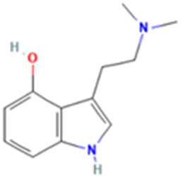 |
| LSD | Yes | Human183 and Rat85 | 3 | Ergoline |  |
| 2-Br-LSD | No | Human,77 and Rat67 | 4 | Ergoline |  |
| Lisuride | No | Human184 and Rat85 | 2.7 | Ergoline |  |
| Ariadne | No | Human and Rat75 | 2.8 | Phenethylamine |  |
| 25CN-NBOH (NBO) | Yes | Mouse185 | 3.1 | Phenethylamine | 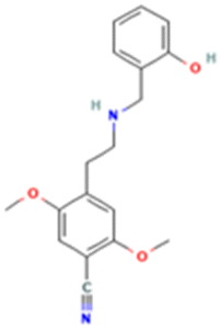 |
| DOI | Yes | Rat186 | 2.5 | Phenethylamine |  |
| N,N-Dimethyltryptamine (DMT) | Yes | Human187 | 2.5 | Tryptamine |  |
| 5-MeO-DMT | Yes | Human188 and Mice189 | 1.5 | Tryptamine |  |
| Bufotenine | Yes | Human and Rat190 | 1.2 | Tryptamine |  |
| Tabernanthalog (TBG) | No | Rat74 | 2.4 | Tryptamine |  |
| Ketanserin | No | Human191 and Rat192 | 2.6 | Quinazoline |  |
| Volinanserin (MDL100907) | No | Rats47 | 3.9 | Piperidine |  |
| Pimavanserin | No | Human193 | 4.5 | Urea |  |
2.2 5-HT2A-mediated therapeutic effects
Psychedelics are under investigation for treating several conditions such as depression, anxiety, obsessive-compulsive disorder, substance use disorder, and post-traumatic stress disorder. Besides the monoamine hypothesis, one underlying theory in depression is the presence of cortical atrophy and reduced neuronal connections.53,54 Given that reduced dendritic growth is a common feature of several neuropsychiatric conditions, enhancing dendritic plasticity may represent a unifying mechanism of psychedelic therapy, consistent with the evidence that their therapeutic effects arise from promoting neuroplasticity and the formation of new neural connections.55 Neuroplastic changes similarly accompany the chronic use of classic antidepressants and the rapid-acting N-methyl-D-aspartate (NMDA) receptor antagonist ketamine.56–61 In vitro, the therapeutic readout of psychedelics is typically measured by quantifying dendritic growth using Sholl analysis,62 while in vivo, it is evaluated using behavioural paradigms such as a forced swim test, sucrose preference test and tail suspension test.63 Much like our knowledge of the implications of hallucinogenic effects and 5-HT2A, the association between 5-HT2A and therapeutic outcomes has also been demonstrated by antagonist and knockout studies.64–69While psychedelics can induce neuroplasticity in brain regions that lack 5-HT2A,66 overwhelming evidence indicates that 5-HT2A-mediated plasticity in the prefrontal cortex (PFC) is critical for therapeutic effects. Still, a direct causal link between 5-HT2A receptor signalling, structural plasticity, and behavioural outcomes remains unconfirmed. To address this issue, Shao and colleagues demonstrated that psilocybin increased spine density in both pyramidal tract and intratelencephalic neurons within the medial frontal cortex; however, only plasticity of pyramidal tract neurons was necessary for improvements in stress-related behaviours.68 To establish a direct connection between structural plasticity and 5-HT2A signalling, it was shown that a selective 5-HT2A knockout in pyramidal tract neurons abolished psilocybin-induced spine growth.68 Furthermore, in mice, the localized deletion of 5-HT2A in the medial frontal cortex prevented psilocybin from reducing stress-driven phenotypes.68 In another study, tabernanthalog (TBG), a non-hallucinogenic analogue of 5-MeO-DMT, was shown to rely on 5-HT2A for both dendritic spine growth in layer 5 pyramidal neurons and antidepressant-like effects in the tail suspension test.64 Consequently, the selective photoablation of TBG-induced dendritic spines in the prefrontal cortex abolished antidepressant-like responses.64 These approaches help to demonstrate the relation between 5-HT2A-mediated cortical spinogenesis and the behavioural effects of these psychoplastogens.
Generally, psychedelics are thought to exert their therapeutic effects by driving structural and functional neuroplasticity. Neuroplasticity is a broad term which can encompass neuritogenesis, dendritogenesis, spinogenesis and synaptogenesis. To understand whether these changes are consistent across studies and compounds, it may be beneficial to report several elements of structural and functional neuroplasticity in response to serotonergic psychedelics.70 Similarly, quantifying overall dendritic spine density alone overlooks changes in finer dendritic spine structural features, commonly grouped into four morphological categories: filopodia, thin, stubby, and mushroom. Due to their large size and stability, mushroom spines are often considered more mature and long-lasting compared to the rest.71 Although stubby spines are the second-largest subtype, they are still regarded as immature structures and believed to transition into more mature spine types.65,71,72 In contrast, thin spines form immature, highly plastic synapses that can strengthen with activity and are thought to participate in learning processes.65,72 The presence of filopodia-like structures peaks early in development, and they represent the smallest and most dynamic protrusions, with lifetimes lasting only up to a few hours.72 Hence, the characteristics and dynamics of dendritic spines represent unique stages of plasticity and may be functionally distinct regarding behavioural outcomes in psychedelic therapy. One study demonstrated that in a 5-HT2A-dependent manner, DOI-treated mice displayed increased cortical dendritic density by upregulating stubby and thin dendritic spines but not mushroom spines.65 In a separate experiment yielding similar results, DMT, LSD and DOI also favoured the growth of thin and filopodia spines over mushroom spines.70 Additional long-term assessments are required to understand whether the duration of structural neuroplasticity is necessary for the rapid or long-term behavioural changes. For instance, psilocybin and 25CN-NBOH give rise to persistent functional plasticity and antidepressant-like effects 12 weeks post-administration, without sustained structural plasticity at the same timepoint.73 Currently, these subtle details of spinogenesis are less understood with respect to 5-HT2A-mediated structural plasticity. Therefore, it may be more informative to examine changes in dendritic spine populations and their distribution, along with their respective rates of elimination and formation. Perhaps this approach will provide a more granular understanding of long-term behavioural changes than the total spine number alone. While neuroplasticity is a common readout for predicting therapeutic effects of 5-HT2A agonists, it is ultimately an indirect surrogate, often assessed outside of the relevant circuit-level context. Integrating long-term measures of neuroplasticity in combination with in vivo behavioural outcomes may strengthen the correlation to therapeutic outcomes. Overall, the causal chain of events from receptor activation, precise signalling mechanisms, dendritic spine growth and therapeutic benefit must be further understood and likely to differ depending on the specific psychedelic examined.
3. The diversity of 5-HT2A agonism: partial agonism versus biased signalling
An abiding mystery is that serotonin, among other 5-HT2A agonists, is not perceived as hallucinogenic in humans. In fact, several studies have shown that non-hallucinogenic 5-HT2A agonists, with varying selectivity, such as lisuride, ariadne, and 2-bromo-LSD can mediate several therapeutic effects, either through neuroplastic changes or behavioural paradigms in pre-clinical trials.67,74–78 This distinction has prompted several groups to explore whether it is possible to develop 5-HT2A agonists that retain therapeutic effects with minimal hallucinogenic activity. Characterizing psychedelic signalling may clarify which pathways mediate hallucinogenic versus therapeutic properties.A single GPCR can couple to multiple G proteins and trigger a number of distinct intracellular signalling cascades, ultimately driving gene transcription and translation to shape cellular behaviour and function. When serotonin binds to 5-HT2A, guanosine diphosphate (GDP) is exchanged for guanosine triphosphate (GTP), thus leading to the dissociation of the Gαq/11 subunit from Gβγ subunits. Gαq/11 activates phospholipase C (PLC), which cleaves the membrane phosphatidylinositol 4,5-bisphosphate (PtdIns(4,5)P2 or PIP2) to subsequently produce inositol 1,4,5-triphosphate (IP3) and diacylglycerol (DAG). IP3 binds to receptors on the endoplasmic reticulum, permitting Ca2+ release from the endoplasmic reticulum into the cytoplasm. Ca2+ and DAG are protein kinase C (PKC) activators; both Ca2+ and DAG are required for the activation of α, β and γ PKC isozymes, whereas novel PKC isozymes (δ, ε, η and θ) only depend on DAG. PKC activation leads to extracellular signal-regulated kinases 1/2 (ERK 1/2) through the mitogen-activated protein kinases (MAPK).79 Consequently, active, ligand-bound receptors are phosphorylated by GPCR kinases (GRKs), which subsequently recruit β-arrestin to desensitize the initial wave of signalling.80 Furthermore, β-arrestin can mediate receptor internalization, which can ultimately lead to receptor recycling and degradation or, importantly, elicit a second wave of signalling.80,81
Two major explanations posit why some 5-HT2A agonists are hallucinogenic while others are not: functional selectivity and partial agonism. Functional selectivity, or ligand bias, commonly refers to a ligand's preferential activation of G protein- or β-arrestin-driven pathways.80 This may include a given receptor's ability to couple to G proteins beyond those initially identified as receptor-coupled. In the case of 5-HT2A, psychedelics have been proposed to involve activation of Gαi/o proteins in addition to Gαq/11 alone.45,82–84 If some psychedelics are indeed biased, it may be possible to design drugs that selectively activate or avoid pathways differentially associated with certain physiological outcomes. In vivo, biased agonism may underlie some psychedelic-like behaviours observed with LSD and not with lisuride.45,67,85–87 Various studies describe LSD as a β-arrestin-biased ligand,87–89 whereas lisuride, a structurally similar non-hallucinogenic ligand, is classified as G protein biased.90 Although this distinction does not apply to all psychedelic and non-psychedelic analogues, LSD and lisuride are frequently used as benchmark comparators.85 β-arrestin 2 knockout mice treated with LSD showed reduced or no behaviours associated with psychedelic action, including measures of HTR, pre-pulse inhibition and motor activities, indicating that these behaviours at least partially depend on β-arrestin 2.87 These findings have been challenged by other attempts to synthesize biased 5-HT2A agonists.
Based on a second binding mode observed in serotonin and psilocin, one group synthesized β-arrestin-biased 5-HT2A ligands, with IHCH-7079 and IHCH-7086 being the two most potent ones.91 Interestingly, these compounds did not trigger a HTR but retained efficacy in alleviating depressive-like behaviours, as shown by tail suspension and forced swim tests.91 These findings align with recent studies demonstrating that β-arrestin-biased 5-HT2A agonists fail to elicit HTR; instead, HTR positively correlates with 5-HT2A-driven, Gq signalling efficacy.92 Another structure-based screen of a virtual chemical library identified (R)-69 and (R)-70, agonists with greater 5-HT2A selectivity compared to several classical psychedelics. While these G protein-biased compounds produced minimal HTR and did not disrupt prepulse inhibition compared to LSD, they nevertheless elicited antidepressant-like phenotypes in mice.76 Regardless, translating signalling bias measured in heterologous systems should be done with caution, as inconsistencies can arise from assays measuring proximal versus distal signalling events, and in the distinct stoichiometry of receptors, G proteins, and effectors noted between immortalized cell lines (e.g., HEK 293 cells or SH-SY5Y cells), primary neurons, and in vivo models.80 Moreover, psychedelics are chemically diverse and exhibit a rich polypharmacology; thus, the ligand bias observed for one agonist, such as LSD, cannot be extrapolated to others without validation. Indeed, psychedelics belonging to separate chemical classes from LSD, such as psilocin or DMT, would stabilize slightly different 5-HT2A receptor conformations, which may contribute to differences in signalling.93 The vast differences between psychedelics complicate decisions about whether prioritizing biased ligands is the most effective strategy for psychedelic drug discovery.
In contrast to functional selectivity, an alternative hypothesis suggests that non-psychedelic 5-HT2A agonists simply behave as weak partial agonists.67,75,91,92,94,95 Biosensor-based studies indicate that bias toward Gαq/11 or β-arrestin does not consistently distinguish psychedelic from non-psychedelic agonists.92,94 Instead, non-hallucinogenic ligands are generally less efficacious than psychedelics in both Gαq/11-driven pathways (Gαq/11 dissociation from Gβγsubunits, IP1 accumulation, and increases in cytosolic Ca2+) and β-arrestin recruitment.94 Thus, partial agonism may explain why some 5-HT2A agonists are non-hallucinogenic, but still, several psychedelics exist that are weak agonists. Some evidence suggests there is a threshold of Gαq/11 signalling needed to induce hallucinogenic effects, a higher threshold than what is required for therapeutic effects.92
4. 5-HT2A receptors and location bias
Neither functional selectivity nor partial agonism provides a consistent overarching explanation for observed differences between hallucinogenic and non-hallucinogenic 5-HT2A ligands. A common property among psychedelics, regardless of the chemical structure, is their ability to cross cell membranes. These properties differ drastically from the endogenous 5-HT2A ligand serotonin (Table 1 and Fig. 1). The ability of a ligand to access distinct intracellular receptor pools highlights an important nuance regarding functional selectivity called location bias. This phenomenon is noted where the same receptor can initiate distinct signalling cascades when activated in different subcellular locations.96 While GPCRs are widely studied as plasma membrane-bound receptors, their localization to various subcellular compartments is gaining attention, including in the context of psychedelic signalling at 5-HT2A receptors.69 | ||
| Fig. 1 5-HT2A localization and signalling. (A) In pyramidal neurons, 5-HT2A receptors localize to distal dendritic regions and the soma while they are largely absent in the axon terminal. Experimentally known intracellular 5-HT2A receptor pools exist in early and late endosomes, the Golgi apparatus, and mitochondria indicated by black boxes surrounding these locations. The localization in the nucleus and endoplasmic reticulum remains speculative. It is unknown whether intracellular receptor pools are a result of internalization from the cell surface or due to the biosynthetic pathway. Ligand internalization may be due to transporters or passive diffusion across the cell membrane. (B) Schematic of 5-HT2A receptor signalling partners at the cell surface. These may differ across intracellular compartments possibly causing distinct signalling events such as recruitment of Gαi and Gα12/13 by the 5-HT2A receptor. Created with https://BioRender.com.69 | ||
4.1 Subcellular distribution of neuronal 5-HT2A receptors
To study location bias, it is essential to understand the subcellular location of 5-HT2A in a given cellular model, determine whether it is active at those sites, and assess how its downstream signalling cascades and gene expression profiles may vary based on location. However, many of these parameters are poorly characterized for 5-HT2A. Early studies using light and electron microscopy combined with immunocytochemistry revealed that across rat brain regions, most 5-HT2A receptors could be detected in intracellular compartments.7,8 In several regions, including cortical neurons, 5-HT2A receptors were localized to somato-dendritic and axonal areas.7,8 Within neurons, receptor expression tends to increase in distal dendritic regions compared to the soma but is largely absent in axon terminals and dendritic spines.7,8 While these studies did not identify receptor association with the Golgi apparatus, mitochondria, lysosome or endosome-like vesicles, 5-HT2A was found to co-localize with microtubule-associated protein 1A, suggesting potential roles in neuronal plasticity and dendritic growth.7,8 While such techniques provide high spatial resolution, they may fail to capture transient or dynamic receptor distribution within specific subcellular compartments.Considering that it is common to study GPCR signalling in immortalized cell lines, it is important to understand if location bias translates across experimental models. Immunocytochemistry and co-localization assays of fluorescently tagged proteins have revealed differences in the subcellular location between 5-HT2A in primary neurons and HEK 293 T cells.69 Both endogenous and overexpressed 5-HT2A in neurons colocalized with the Golgi apparatus to a greater extent than 5-HT2A in HEK 293 T cells, see Fig. 1.69 Furthermore, 5-HT2A also co-localized, to some extent, with mitochondria, early endosomes and late endosomes in both cell types.69 Understanding that 5-HT2A receptors are not confined to the plasma membrane is only one piece of the puzzle; the overall contribution of internal receptors to cellular signalling remains largely unexplored.
4.2 Lipophilicity of serotonergic psychedelics and other 5-HT2A ligands
Location bias has been thoroughly studied for several GPCRs, but it is a more recent addition in psychedelic research. Vargas and colleagues first showed that serotonergic psychedelics induce structural plasticity and antidepressant-like effects through internal 5-HT2A receptor pools in cortical neurons.69 In that study, a series of approaches were implemented to reveal the contribution of intracellular receptors to neuroplasticity, including the use of membrane-impermeable 5-HT2A antagonists to selectively block cell-surface receptors. Similarly, DMT and psilocybin lose their plasticity-inducing abilities when chemically modified with highly charged groups that prevent membrane permeation.69 Intriguingly, when serotonin is permitted to cross the plasma membrane through electroporation or SERT overexpression, it gains the ability to induce spinogenesis in vitro, and, in vivo, produces antidepressant-like effects.69 To date, this remains the only study to directly examine 5-HT2A location bias. Their findings, as well as broader GPCR literature, emphasize the potential importance of ligand permeability in potentially therapeutic outcomes. The following sections explore possible implications for 5-HT2A location bias in our understanding of psychedelic mechanisms.Ligand permeability is generally described by the logarithm of the partition coefficient (LogP), which is determined by the ratio of the non-ionized ligand concentration in octanol to its concentration in water;97 several computational methods to estimate LogP values also exist.98 Alternatively, the distribution coefficient, LogDpHX, is a more informative measure as it accounts for both the pH of the intracellular compartment and the ionized and unionized forms of the compound.97 Among well-characterized 5-HT2A agonists and antagonists, there is a spectrum of LogP values (Table 1). By correlating signalling profiles with permeability data, we can begin to disentangle contributions from internal versus surface receptor pools.
Molecular dynamics (MD) simulations provide an efficient computational approach to predict how alterations in a ligand's chemical groups affect permeability. Expectedly, MD simulations predicted that the protonation of tryptamine-based compounds DMT, bufotenine, 5-Meo-DMT and psilocin hinders their membrane partitioning.98,99 Such studies suggest that during membrane partitioning, psychedelics can alter membrane properties such as thickness, stiffness, and lipid order.99–101 These changes create thinner, loosely packed bilayers that may affect receptor dynamics, conformation, and ligand binding.102,103 Even among structurally similar ligands, subtle chemical differences influence how ligands prime the local receptor environment. For example, although bufotenine did not spontaneously partition the membrane, it was predicted to disrupt lipid membrane order to a greater extent than DMT and 5-MeO-DMT.99 Considering the structural diversity of psychedelics, ergolines and phenethylamines may perturb the membrane in distinct ways from tryptamines, as their chemical groups can differentially engage with lipid and phosphate groups.101 Several researchers have used MD simulations and artificial lipid membranes to explore how subtle chemical modifications can alter the permeability of tryptamine-class psychedelics. While serotonin is not generally permeable, simulations in artificial membranes indicate that it can partially insert into the bilayer and slightly reduce membrane stiffness and thickness.100,101 In contrast, DOI more strongly decreases lipid tail order and bilayer rigidity, promoting vesicular fusion.101 Therefore, psychedelics, or other lipophilic drugs, might reshape the membrane environment, tuning receptor behaviour simultaneously or prior to ligand binding. These tools not only strengthen our understanding of location bias but also provide valuable strategies for designing new chemical entities.99,101,104
It is likely that certain ligand–membrane interactions play a role in modulating GPCR signalling. Although phosphoinositides represent only a minor component of cell membranes, they are highly dynamic and essential for maintaining organelle identity, as well as GPCR signalling and trafficking.103,105,106 One of the earliest signalling events of 5-HT2A after Gαq/11 subunit dissociation is the hydrolysis of PIP2 by PLC. Differences in membrane permeability among 5-HT2A agonists may cause them to partition at different rates which in turn can alter membrane composition, possibly reflected by variations in PIP2 hydrolysis.106 In a study of PIP2 depletion in HEK 293 cells, both DOI (LogP = 2.5) and lisuride (LogP = 2.7) achieved similar sustained depletions of PIP2, but lisuride did so more rapidly.106 However, in primary cortical neurons, the magnitude and kinetics of the response were different; DOI induced a faster and greater PIP2 depletion than lisuride.106 Nevertheless, in both cell types, lisuride and DOI produced a greater PIP2 depletion than serotonin (LogP = 0.2), not accounting for PIP2 replenishment.106 Considering that lisuride and DOI are more lipophilic than serotonin, the previous results may reflect their similarities in membrane permeability. Understanding how psychedelics partition into membranes and influence PIP2 hydrolysis may advance our understanding of their signalling patterns. PIP2 has been shown to bind certain GPCRs, further stabilizing Gα protein interactions with the receptor.103 Additionally, PIP2 can facilitate interactions between β-arrestin and GPCR complexes; specifically, PIP2 influences β-arrestin orientation and interactions with the cell membrane.107 Therefore, it is reasonable to assume that a PIP2-mediated regulation of β-arrestin might differ in response to more lipophilic ligands, consequently influencing receptor internalization, desensitization or second-wave signalling. Testing both permeable and impermeable versions of the same ligand or using antagonists that vary in permeability (Table 1) to selectively block certain receptor pools may deepen our understanding.
Additional structural studies have demonstrated the role of plasma membrane lipids in modulating 5-HT1A activity.108 Phosphatidylinositol 4-phosphate (PtdIns4P), the precursor of PIP2, binds between the receptor–G protein complex and promotes GTP hydrolysis, with PIP2 showing a similar but weaker effect.108 In parallel, membrane cholesterols enhance PtdIns4P binding and modulates aripiprazole–5-HT1A interactions by stabilizing TM1 and TM7 of the receptor.108 Whether lipid-mediated interactions observed with other receptors also occur with psychedelics at 5-HT2A receptors remains unknown, partly due to the complexity introduced by different cellular models. In human cells, lipid composition varies based on the developmental stage, disease state, and among different brain regions.109,110 Even within specified regions such as the cortex, lipid profiles are highly heterogeneous.109 Furthermore, membrane compositions in immortalized cell lines can significantly differ from those in primary cell cultures, further complicating our understanding of location bias.111–113 Therefore, more work is needed to determine how 5-HT2A ligands prime the membrane and how the bidirectional relationship between the receptor and the phospholipid environment can shape signalling outcomes by different psychedelics.114
Ligand permeability can be advantageous in understanding location bias, however, designing experimental ligands based on permeability alone is unlikely to yield a physiologically relevant candidate. Previous studies highlight an important question about whether there is a favourable range of permeability or if specific ligand–membrane interactions are required for a given signalling outcome. Permeability beyond a certain LogP value might not meaningfully enhance a compound's pharmacological performance. Highly lipophilic ligands can become trapped within the plasma membrane, whereas less permeable ligands, when given enough time, may also cross the membrane and reach equilibrium.115 Likewise, the kinetic profile by which psychedelics and their analogues permeate the cell may alter membrane organization in a way that influences 5-HT2A signalling. Additionally, since several non-hallucinogenic ligands display comparable LogP values to those of classical psychedelics (Table 1), these differences in hallucinogenic potential are unlikely to reflect a simple intracellular versus extracellular dichotomy. Instead, these processes are likely multifactorial, depending on the nature of compartment-specific signalling pathways; however, the extent to which these compounds retain canonical Gαq/11 and β-arrestin coupling versus alternate signalling partners remains unknown.
5. Avenues worth exploring to understand the 5-HT2A location bias
While a more general distinction has been made between surface and internal receptor contributions to psychedelic outcomes, the precise subcellular contributions of 5-HT2A signalling remain, to our knowledge, incompletely understood.69 Endosomal signalling from internalized agonist-activated receptors at the plasma membrane represents one facet of location bias, but several other GPCRs can also signal in the nucleus, mitochondria, and Golgi.116 Here, we use endosomal signalling as an example of how to approach signalling from internalized receptors. To study location bias, it is necessary to (1) determine if 5-HT2A receptors in different compartments can reach active conformations and (2) understand the location-specific contribution to signalling.5.1 Endosomal receptor signalling
As described earlier, upon receptor activation, GRKs phosphorylate the C-terminal tail of the receptor, promoting β-arrestin recruitment, which can both desensitize a response and facilitate clathrin-dependent receptor internalization.80 Receptor internalization does not always halt signalling; many G proteins (e.g., Gαq/11, Gαs, and Gαi/o) and receptors (e.g., β2-adrenergic receptor, dopamine D1 receptor and opioid receptors) continue to signal in endosomes, sometimes in ways distinct from their plasma membrane-bound versions.117–124 In several studies, GPCR endocytosis has resulted in sustained signalling of cAMP, ERK1/2, PKC and mTOR.125,126 This spatial shift in signalling may alter functionally relevant distal outcomes, as seen in the case of endosomal β2-adrenergic receptors, which can generate distinct transcriptional123 and translational120 profiles. In the case of Gαs-coupled receptors, there are several proposed mechanisms as to why endosomal signalling differs. First, endosomal receptors are increasingly distant from phosphodiesterases near the cell surface, while also in closer proximity to the nucleus, increasing the likelihood of cAMP signalling-dependent gene expression.123 Second, β-arrestin-mediated internalization can scaffold components of the MAPK pathway in proximity to endosomal receptors, thereby prolonging some signalling pathways.125,127 Notably, the conformation in which β-arrestin binds to GPCRs can facilitate different signalling outcomes. The “core” conformation, required for receptor desensitization, is where β-arrestin interacts with both the transmembrane core of the receptor and the phosphorylated C-terminal tail.128 In contrast, interactions through the “tail” conformation permit receptor internalization without desensitization.128 Thus, in some cases of GPCR internalization, β-arrestin remains bound to the C-terminal tail while the receptor core simultaneously remains available for G protein interactions.125 These interactions vary across GPCR classes, offering insight into which receptors are predisposed to prolonged signalling pathways. Class A GPCRs, such as 5-HT2A, are generally thought to form more transient interactions with β-arrestin, whereas class B GPCRs form more stable complexes due to longer C-terminal tails.125 For instance, in class B GPCRs, such as the parathyroid hormone 1 receptor (PTHR), sustained cAMP production from endosomes has been linked to the presence of β-arrestin–receptor complexes.117,129 But for the thyroid-stimulating hormone receptor, a class A GPCR, β-arrestin interactions are transient and, after internalization, are not thought to directly participate in prolonged cAMP signalling at the trans Golgi network.117,130 However, it is clear that the role of β-arrestin varies among receptors even within the same GPCR class;131 therefore, the interplay between 5-HT2A and β-arrestin recruitment will have to be further studied in the context of spatiotemporal signalling from endosomes.Several studies have shown that Gαq/11-coupled receptors can also elicit endosomal signalling, including the angiotensin II type 1 receptor, B2 bradykinin receptor, oxytocin receptor, thromboxane receptor, muscarinic acetylcholine receptor M3, and neurokinin 1 (NK1) receptor.124,132 Among these examples, signalling from specific compartments has resulted in unique physiological outcomes. Activation of the NK1 receptor in endosomes by substance P was shown to sustain spinal neuron firing and nociception.132 Subsequent targeting of endosomal NK1R by cholestanol-conjugated antagonists that facilitate internalization produced more effective maintenance of pain relief than plasma membrane-restricted NK1R inhibition, demonstrated by a reduced von Frey response in mice.132 Similarly, the PTHR can signal from both the plasma membrane and endosomes.133 To investigate the role of PTHR location on biological outcomes, one group used the spatially biased PTHR ligand, PTH7d, to prevent β-arrestin coupling to PTHR and maintain cAMP signalling at the cell surface but not at endosomes.133 The absence of endosomal signalling decreased the formation of the rate-limiting hydroxylase necessary for vitamin D production, highlighting the distinct physiological outcomes of PTHR endosomal signalling.133 Lastly, a recent pre-print highlighted the therapeutic relevance of the constitutively internalized human cytomegalovirus-encoded receptor (US28), which primarily signals through Gαq/11 from endosomal compartments.134 The study demonstrated that confining US28 to the plasma membrane led to the upregulation of oncomodulatory genes, as well as increased neutrophil chemotaxis and cell proliferation in glioblastoma spheroids. These findings suggest that plasma membrane Gαq/11 signalling drives oncogenic processes more strongly than endosomal signalling.134 Based on our knowledge of the previously mentioned Gαq/11-coupled GPCRs, it is worth examining whether 5-HT2A also signals from endosomes or other endomembrane locations as has been reviewed for numerous other GPCRs.116,135–137
While conventional GPCR signalling is initiated at the plasma membrane, we have seen that in some cases, receptor internalization can result in sustained or second wave signalling events. However, for signalling to occur at separate compartments, it requires the presence of appropriate G proteins, effectors, and substrates. Currently, tools exist to detect Gαq protein trafficking to endosomes as well as their activation.124 Regarding the angiotensin II type 1 receptor (AT1R), it was found that although Gαq trafficked to endosomes independently of β-arrestin, endosomal Gαq activation partially relied on the presence of β-arrestin.124 The same study also suggested that endosomal Gαq activity is enhanced by prior activation at the plasma membrane and by β-arrestin-dependent AT1R internalization.124 Whether these findings are generalizable to other Gαq/11-coupled GPCRs like 5-HT2A has yet to be determined.
Although Gαq/11 proteins and Gαq/11-coupled GPCRs are active at endosomes, the mechanisms by which they propagate their signals are unresolved, as some canonical substrates and effectors are missing in endosomes.138 The substrate for PLC, PtdIns(4,5)P2, is present in the plasma membrane but is essentially absent in endosomes. This change in lipid environment occurs during endocytosis when clathrin binds the AP2 adapter complex, creating a depletion in the enzyme that converts PtdIns(4)P to PtdIns(4,5)P2.138 Therefore, it is unlikely that DAG is produced in endosomes, and plasma membrane-derived DAG is not expected to diffuse readily between membranes. This is consistent with the inability of current detection methods to observe DAG in early endosomes.124,138 Although PKC has been identified in endosomes, most isoforms remain inactive without DAG.138,139 Thus, Gαq/11 activation at endosomes may initiate non-conventional pathways, either independently of PLC or via mechanisms that depend on prior activation at the plasma membrane. This could include the direct activation of endosomal adenylyl cyclase isoforms by calmodulin, ERK5 activation, and trafficking of active PKC to endosomes.138,140 Internal 5-HT2A signalling may resemble plasma membrane signalling by activating pathways downstream of Gαq/11, or it may engage in separate pathways by coupling to alternative G proteins such as Gαi/o, or with a greater reliance on Gβγ and β-arrestin signalling in such compartments. In any case, the kinetics of signalling are likely to be altered.
5.2 Rho GTPases: an underexplored pathway in 5-HT2A psychedelic action
An alternative signalling cascade of 5-HT2A receptors may involve the activation of certain Rho family GTPases, which are found both at the plasma membrane and in intracellular compartments including the nucleus, endosomes and the Golgi apparatus.141 Rho GTPases are regulated by guanine nucleotide exchange factors (GEFs), which catalyze the exchange of GDP to GTP, and by GTPase-activating proteins which promote GTP hydrolysis.142 Rho GTPases act as molecular switches to mediate several roles including cytoskeleton organization, cell shape, microtubule dynamics, cell migration, and, notably, dendritic spine formation and synaptic plasticity.142–144 In Xenopus laevis tadpoles, Rac1 and Cdc42 mediate dendritic branch addition and retraction.143 Additionally, transamidation of Rac1 and Cdc42 by transglutaminase has been shown to produce transient dendritic spine enlargements following 5-HT2A/5-HT2C stimulation by DOI in primary rat cortical neurons.145 Specifically, an alternative signalling cascade of 5-HT2A receptors may involve RhoA.146 RhoA is an important part of dendritic branch elongation, with reduced RhoA activity promoting dendritic growth, a process that may be regulated by NMDA receptor activity.143 Furthermore, Rho GEFs can be activated by select G proteins, mainly Gα12/13 and, with a lower potency, Gαq/11, both of which may be coupled to 5-HT2A.92,147,148 This is exemplified by the proposed involvement of Rho in the 5-HT2A receptor G12/13-coupled activation of PLA2.83 Additionally, p63RhoGEF, an effector of Gαq/11 found in the brain, enables Gαq/11 to activate RhoA, RhoB and RhoC.147–150 Evidently, RhoA activation by Gαq/11 can occur in a PLC and Ca2+-independent manner, suggesting a possible signalling pathway in the endosome where PLC is largely absent.138,148 However, p63RhoGEF, is predominantly localized to the plasma membrane and the related cytoplasmic RhoA effector GEFT has minimal activation unless artificially recruited to the plasma membrane.149,151 These observations suggest that Gαq/11-mediated Rho activation is likely influenced by GEF localization. Importantly, psychedelic-induced 5-HT2A activation increases RhoA activity in HEK 293 cells.152 Given the diverse subcellular localizations and distinct physiological roles of Rho GTPases, together with evidence that Gαq/11 can signal from intracellular compartments, it is tempting to speculate that intracellular Gαq/11 engages Rho GTPases to promote dendritic growth and spine formation.124,143 Regardless, a deeper understanding of 5-HT2A-mediated Gαq/11 signalling through p63RhoGEF and Rho GTPases may provide insight into the mechanisms underlying psychedelic-induced plasticity.5.3 Candidate ligands for intracellular 5-HT2A receptors
In cortical neurons, the contribution of compartment-specific 5-HT2A receptors is not fully understood, but collectively, the intracellular receptor pools promote neuroplasticity when activated by exogenous lipophilic psychedelics.69 Considering serotonin is hydrophilic, this raises the question of whether another endogenous ligand might modulate these receptors. Once serotonin is synthesized in serotonergic neurons of the raphe nuclei, it is packaged into vesicles by vesicular monoamine transporter 2 until it is later released into the synaptic cleft. Serotonergic neurons project widely throughout the brain and express active transporters such as serotonin reuptake transporters (SERT) to modulate serotonin levels. Since serotonin is protonated at physiological pH, it does not readily cross cell membranes; instead, it relies on SERT for uptake. SERT is highly expressed on presynaptic serotonergic terminals but less so on postsynaptic neurons, including those in the cortex.4,153,154 Nevertheless, we should not rule out serotonin, as several proposed mechanisms could allow it to reach internal 5-HT2A receptor pools in these neurons.SERT has a high affinity for biogenic amines, but serotonin uptake can still occur passively through low-affinity but high-capacity transporters, including organic cation transporters (OCT1–3) and plasma membrane monoamine transporters (PMAT).4,155 The broad expression of these transporters across the brain suggests they may permit serotonin entry into cortical pyramidal neurons, possibly even at low serotonin concentrations.4,156 Evidently, electron microscopy has revealed that OCT3 exists in the plasma membrane, mitochondrial, and nuclear pools of cortical neurons thereby providing an opportunity for serotonin to cross several membranes.4 In fact, the role of transporters in spatially compartmentalized GPCR signalling is not uncommon. For example, the activation of Golgi-bound dopamine D1 receptors depends on dopamine uptake via OCT2, and likewise, OCT3 may help permit norepinephrine access to the nuclear pool of β1-adrenergic receptors in astrocytes, driving nuclear protein kinase A (PKA) activity.157,158 Similarly, neuronal PMAT and OCT3 transport norepinephrine into the lumen of secretory vesicles, enabling activation of intracellular β2-adrenergic receptors, a mechanism proposed to contribute to long-term potentiation and synaptic plasticity.159 Therefore, modulating the expression or function of certain transporters may help clarify whether they permit serotonin to activate intracellular 5-HT2A. Evidently, the fate of serotonin once it enters postsynaptic neurons may be irrelevant to neuroplasticity, as it is likely to exist in low quantities and is subjected to degradation by monoamine oxidases. This is further supported by findings that serotonin-induced dendritic growth is observed only when SERT is overexpressed in specific neurons.69 Nonetheless, experimentally, little is known about whether, and to what extent, serotonin activates internal 5-HT2A receptors in transporter overexpression systems. Instead, intracellular serotonin may function as an antioxidant within the mitochondria and regulate gene expression through the serotonylation of nuclear histones.4
Aside from serotonin, other endogenous ligands may activate intracellular 5-HT2A receptors, including DMT. While DMT is well-recognized as the psychoactive component of ayahuasca, it is also naturally detected in human urine, blood, and cerebrospinal fluid.160,161 Whether DMT is synthesized in the human brain or is produced peripherally and crosses the blood–brain barrier remains uncertain. However, the rat brain can produce DMT at levels comparable to those of classical monoamine neurotransmitters and contains enzymes involved in its biosynthesis, though several synthetic pathways have been proposed.162,163 These findings suggest that DMT could be an endogenous ligand for intracellular 5-HT2A receptors, although its physiological role is still under speculation.164–166
6. Perspectives
There is currently no consensus on whether the hallucinogenic and therapeutic effects of psychedelics can be mechanistically separated. Existing evidence does not clearly establish whether these subjective experiences are required for clinical benefit or represent unnecessary secondary effects. To this end, several frameworks have been proposed for understanding the mechanisms through which psychedelics induce each effect. Of these, partial agonism, biased agonism, and location bias are at the forefront. While generally accepted as non-hallucinogenic, high doses of lisuride, a partial 5-HT2A agonist, have been shown to induce hallucinations in Parkinson's patients.167 This indicates a possible signalling threshold for hallucinations, such that partial agonism prevents hallucinogenic effects while higher doses overcome this threshold. Additionally, the synthesis of several β-arrestin or Gαq/11-biased 5-HT2A agonists has resulted in contradicting physiological outcomes.91,92 Even so, partial agonism and biased agonism do not provide an all-encompassing explanation for the differences in hallucinogenic and non-hallucinogenic 5-HT2A ligands. Therefore, location bias becomes an important nuance in psychedelic signalling profiles. As Vargas et al. have demonstrated, intracellular 5-HT2A signalling mediates the neuroplasticity necessary for psychedelic therapeutic effects.69 Among the ongoing debates on the necessity of hallucinations for the therapeutic benefit, each of these frameworks may be crucial for elucidating how these effects can be separated.Moving forward, we believe that the focus should be shifted towards understanding how location bias shapes psychedelic actions at 5-HT2A. To summarize our current understanding, the only direct evidence for 5-HT2A comes from Vargas et al. (2023) in HEK 293 cells and primary cortical neurons. From colocalization and electron microscopy experiments, we know that 5-HT2A is distributed throughout several compartments, yet precise signalling pathways inside specific subcellular compartments versus the cell surface receptor pool have not been experimentally determined. Although the remaining evidence for 5-HT2A location bias is largely indirect, consistent findings from other GPCRs in heterologous and primary cells suggest that it may play a role in shaping 5-HT2A-mediated effects for psychedelic and non-psychedelic compounds.
As discussed, several intracellular signalling events are thought to mediate physiological responses that are not otherwise achieved by plasma membrane-bound receptors. Therefore, the intracellular pool of 5-HT2A receptors may also regulate distinct physiological outcomes, influencing both therapeutic and hallucinogenic effects, representing an interesting area for future discovery.69 Intriguingly, serotonin appears to elicit the HTR via intracellular 5-HT2A receptors, which merits further testing of other non-hallucinogenic ligands (Table 1) in similar assays to determine whether this subset of receptors is responsible for both therapeutic and hallucinogenic outcomes.69
The use of chemically modified ligands to spatially restrict psychedelics has enabled broad distinctions between internal and external receptors, yet much is unknown about location bias in psychedelic research. Moving forward, we must pinpoint the specific subcellular receptors and the signalling pathways attributed to psychedelic and non-psychedelic 5-HT2A agonists. This includes making a distinction between actively signalling intracellular 5-HT2A receptors versus receptors undergoing post-translational modifications and trafficking to the plasma membrane via the secretory pathway. Currently, several tools exist to delineate location-specific signalling events in vitro, including genetically encoded fluorescent nanobodies and mini-G proteins that recognize the active state of a GPCR.119,158,168 Biosensors confined to select subcellular compartments permit the recording of site-specific signalling events, as demonstrated using a CAAX motif or a FYVE domain that target the plasma membrane and endosomes, respectively.169,170 Similarly, we can leverage endocytosis inhibitors and chemically inducible dimerization strategies to probe the contribution of endosomal receptors to signalling outcomes.123,168 In a related approach, exploiting nuclear export and nuclear localization signals in biosensor constructs allows for a compartment-specific readout of several kinases, including ERK and PKA.171,172 Receptors present in the Golgi apparatus are markedly more difficult to study, given that they can house both internalized receptors from the plasma membrane and newly synthesized GPCRs. Additionally, the use of cycloheximide can block the 5-HT2A biosynthetic pathway, which makes it possible to separate Golgi-localized receptor signalling.173 A combination of these tools has improved our knowledge about active GPCRs in endosomes (e.g., β2-adrenergic receptor), the Golgi (e.g., dopamine 1 receptor, opioid receptors, and thyrotropin receptor (trans-Golgi network)) and the nucleus (e.g., angiotensin type II 1 and 2 receptors, metabotropic glutamate receptor, and β1- and β2-adrenergic receptors), to name a few.116,119,122,158,168,169 While these techniques have been extensively studied in immortalized cell lines, further investigation is warranted in primary neurons, cortical neuronal stem cells, and in vivo, particularly for 5-HT2A receptors. Overexpression systems can result in altered receptor trafficking and distribution due to transfection conditions (e.g., DNA concentrations, transfection efficiency, and translation rates) or the absence of scaffolding proteins such as postsynaptic density protein 95,174 leading to effects that cannot be recapitulated in physiologically relevant cell types.69,130 Furthermore, despite the high sequence homology between rat and human 5-HT2A receptors, they still display marked differences attributed to their C-terminus.173 For instance, only the human 5-HT2A receptor requires β-arrestin and GRK-2 for internalization, also taking roughly twice as long as the rat 5-HT2A receptor to recycle post-internalization.152,173,175 This reveals an important caveat when comparing receptor signalling and trafficking data in different cellular systems in the literature.
Understanding the relationship between proximal 5-HT2A signalling events, distal signalling, and subsequent gene expression also remains an important area of investigation. Very few studies have examined transcriptional profiles between hallucinogenic and non-hallucinogenic 5-HT2A agonists,45,176 but whether the activation of 5-HT2A in certain locations drives distinct transcriptome or translatome profiles is not known. Moreover, 5-HT2A can form heterodimers with several receptors, including the dopamine D2 receptor and the metabotropic glutamate receptor 2 (mGluR2), further altering its signalling properties.177–179 5-HT2A dimerization represents a relevant aspect of psychedelic mechanisms, especially since these complexes alter receptor trafficking properties.180 Therefore, signalling profiles between hallucinogenic and non-hallucinogenic agonists, driven from different subcellular compartments, should be followed by functional assays to determine their impact on behavioural outcomes in vivo. This task becomes complicated because such effects likely occur at a circuit level, involving multiple receptors, genes and brain regions. From our current knowledge, the 5-HT2A receptor is critical in mediating the therapeutic benefits of psychedelics in neuropsychiatric diseases. Several tools are at our disposal to discern between 5-HT2A signalling biases, revealing that the diverse signalling at the cell surface and intracellular compartments represents a vast pool of knowledge waiting to be uncovered.
Author contributions
A. J. wrote the primary draft; M. P. added to this draft and developed the figure and table. T. E. H. edited the draft and contributed to the writing of the final version.Conflicts of interest
There are no conflicts to declare.Data availability
Since this is an invited review, there are no data to share.Acknowledgements
This work was supported by funding from the Consortium Québécois sur la Découverte du Médicament (CQDM) and Brain Canada.References
- J. S. Lorente, A. V. Sokolov, G. Ferguson, H. B. Schiöth, A. S. Hauser and D. E. Gloriam, Nat. Rev. Drug Discovery, 2025, 24, 458–479 CrossRef CAS PubMed.
- D. K. Vassilatis, J. G. Hohmann, H. Zeng, F. Li, J. E. Ranchalis, M. T. Mortrud, A. Brown, S. S. Rodriguez, J. R. Weller, A. C. Wright, J. E. Bergmann and G. A. Gaitanaris, Proc. Natl. Acad. Sci. U. S. A., 2003, 100, 4903–4908 CrossRef CAS PubMed.
- A. Bakshi and P. Tadi, StatPearls, StatPearls Publishing Copyright © 2025, StatPearls Publishing LLC., Treasure Island (FL), 2025 Search PubMed.
- P. W. Andrews, C. Bosyj, L. Brenton, L. Green, P. J. Gasser, C. A. Lowry and V. M. Pickel, Proc. R. Soc. B, 2022, 289, 20221565 CrossRef CAS PubMed.
- M. Pourhamzeh, F. G. Moravej, M. Arabi, E. Shahriari, S. Mehrabi, R. Ward, R. Ahadi and M. T. Joghataei, Cell. Mol. Neurobiol., 2022, 42, 1671–1692 CrossRef CAS PubMed.
- J. D. McCorvy and B. L. Roth, Pharmacol. Ther., 2015, 150, 129–142 CrossRef CAS PubMed.
- V. Cornea-Hébert, M. Riad, C. Wu, S. K. Singh and L. Descarries, J. Comp. Neurol., 1999, 409, 187–209 CrossRef.
- V. Cornea-Hébert, K. C. Watkins, B. L. Roth, W. K. Kroeze, P. Gaudreau, N. Leclerc and L. Descarries, Neuroscience, 2002, 113, 23–35 CrossRef PubMed.
- R. L. Jakab and P. S. Goldman-Rakic, Proc. Natl. Acad. Sci. U. S. A., 1998, 95, 735–740 CrossRef CAS PubMed.
- D. L. Willins, A. Y. Deutch and B. L. Roth, Synapse, 1997, 27, 79–82 CrossRef CAS PubMed.
- G. Ślifirski, M. Król and J. Turło, Int. J. Mol. Sci., 2021, 22, 9015 CrossRef PubMed.
- M. P. Bogenschutz, S. Ross, S. Bhatt, T. Baron, A. A. Forcehimes, E. Laska, S. E. Mennenga, K. O’Donnell, L. T. Owens, S. Podrebarac, J. Rotrosen, J. S. Tonigan and L. Worth, JAMA Psychiatry, 2022, 79, 953–962 CrossRef PubMed.
- A. K. Davis, F. S. Barrett, D. G. May, M. P. Cosimano, N. D. Sepeda, M. W. Johnson, P. H. Finan and R. R. Griffiths, JAMA Psychiatry, 2021, 78, 481–489 CrossRef PubMed.
- S. Ellis, C. Bostian, A. Donnelly, W. Feng, K. Eisen, M. Lean, E. Conlan, M. Ostacher, S. Aaronson and T. Suppes, J. Affective Disord., 2025, 389, 119655 CrossRef CAS PubMed.
- S. Ellis, C. Bostian, W. Feng, E. Fischer, G. Schwartz, K. Eisen, M. Lean, E. Conlan, M. Ostacher, S. Aaronson and T. Suppes, J. Affective Disord., 2025, 369, 381–389 CrossRef CAS PubMed.
- J. J. Fuentes, F. Fonseca, M. Elices, M. Farré and M. Torrens, Front. Psychiatry, 2019, 10, 943 CrossRef PubMed.
- G. M. Goodwin, S. T. Aaronson, O. Alvarez, P. C. Arden, A. Baker, J. C. Bennett, C. Bird, R. E. Blom, C. Brennan, D. Brusch, L. Burke, K. Campbell-Coker, R. Carhart-Harris, J. Cattell, A. Daniel, C. DeBattista, B. W. Dunlop, K. Eisen, D. Feifel, M. Forbes, H. M. Haumann, D. J. Hellerstein, A. I. Hoppe, M. I. Husain, L. A. Jelen, J. Kamphuis, J. Kawasaki, J. R. Kelly, R. E. Key, R. Kishon, S. K. Peck, G. Knight, M. H. B. Koolen, M. Lean, R. W. Licht, J. L. Maples-Keller, J. Mars, L. Marwood, M. C. McElhiney, T. L. Miller, A. Mirow, S. Mistry, T. Mletzko-Crowe, L. N. Modlin, R. E. Nielsen, E. M. Nielson, S. R. Offerhaus, V. O’Keane, T. Páleníček, D. Printz, M. C. Rademaker, A. V. Reemst, F. Reinholdt, D. Repantis, J. Rucker, S. Rudow, S. Ruffell, A. J. Rush, R. A. Schoevers, M. Seynaeve, S. Shao, J. C. Soares, M. Somers, S. C. Stansfield, D. Sterling, A. Strockis, J. Tsai, L. Visser, M. Wahba, S. Williams, A. H. Young, P. Ywema, S. Zisook and E. Malievskaia, N. Engl. J. Med., 2022, 387, 1637–1648 CrossRef CAS PubMed.
- M. W. Johnson, A. Garcia-Romeu and R. R. Griffiths, Am. J. Drug Alcohol Abuse, 2017, 43, 55–60 CrossRef PubMed.
- S. Ross, A. Bossis, J. Guss, G. Agin-Liebes, T. Malone, B. Cohen, S. E. Mennenga, A. Belser, K. Kalliontzi, J. Babb, Z. Su, P. Corby and B. L. Schmidt, J. Psychopharmacol., 2016, 30, 1165–1180 CrossRef CAS PubMed.
- F. J. Carod-Artal, Neurologia, 2015, 30, 42–49 CrossRef CAS PubMed.
- M. Johnson, W. Richards and R. Griffiths, J. Psychopharmacol., 2008, 22, 603–620 CrossRef CAS PubMed.
- D. E. Nichols, Pharmacol. Rev., 2016, 68, 264–355 CrossRef CAS PubMed.
- J. O. Cole and M. M. Katz, JAMA, 1964, 187, 758–761 CAS.
- B. G. Eisner and S. Cohen, J. Nerv. Ment. Dis., 1958, 127, 528–539 CrossRef CAS PubMed.
- D. E. Nichols and C. D. Nichols, in International Review of Neurobiology, ed. D. J. Nutt and H. Thurgur, Academic Press, 2025, vol. 181, pp. 3–43 Search PubMed.
- H. Osmond, Ann. N. Y. Acad. Sci., 1957, 66, 418–434 CrossRef CAS PubMed.
- D. W. Woolley and E. Shaw, Proc. Natl. Acad. Sci. U. S. A., 1954, 40, 228–231 CrossRef CAS PubMed.
- R. S. McIntyre, M. Alsuwaidan, B. T. Baune, M. Berk, K. Demyttenaere, J. F. Goldberg, P. Gorwood, R. Ho, S. Kasper, S. H. Kennedy, J. Ly-Uson, R. B. Mansur, R. H. McAllister-Williams, J. W. Murrough, C. B. Nemeroff, A. A. Nierenberg, J. D. Rosenblat, G. Sanacora, A. F. Schatzberg, R. Shelton, S. M. Stahl, M. H. Trivedi, E. Vieta, M. Vinberg, N. Williams, A. H. Young and M. Maj, World Psychiatry, 2023, 22, 394–412 CrossRef PubMed.
- C. A. MacCallum, L. A. Lo, C. A. Pistawka and J. K. Deol, Front. Psychiatry, 2022, 13, 1040217 CrossRef PubMed.
- S. M. Wang, S. Kim, W. S. Choi, H. K. Lim, Y. S. Woo, C. U. Pae and W. M. Bahk, Clin. Psychopharmacol. Neurosci., 2024, 22, 222–231 CrossRef CAS PubMed.
- D. E. Nichols, Pharmacol. Ther., 2004, 101, 131–181 CrossRef CAS PubMed.
- D. E. Nichols and C. S. Grob, Forensic Sci. Int., 2018, 284, 141–145 CrossRef CAS PubMed.
- M. V. Uthaug, A. K. Davis, T. F. Haas, D. Davis, S. B. Dolan, R. Lancelotta, C. Timmermann and J. G. Ramaekers, J. Psychopharmacol., 2022, 36, 309–320 CrossRef PubMed.
- M. W. Johnson, R. Andrew Sewell and R. R. Griffiths, Drug Alcohol Depend., 2012, 123, 132–140 CrossRef CAS PubMed.
- D. P. Effinger, S. S. Schalk, J. L. King, J. R. Wallingford, C. K. O’Connell, J. R. Calderon, B. J. Kopecky, J. D. McCorvy and S. M. Thompson, ACS Pharmacol. Transl. Sci., 2025, 8, 3923–3931 CrossRef CAS PubMed.
- M. K. Jain, R. H. Gumpper, S. T. Slocum, G. P. Schmitz, J. S. Madsen, T. A. Tummino, C.-M. Suomivuori, X.-P. Huang, L. Shub, J. F. DiBerto, K. Kim, C. DeLeon, B. E. Krumm, J. F. Fay, M. Keiser, A. S. Hauser, R. O. Dror, B. Shoichet, D. E. Gloriam, D. E. Nichols and B. L. Roth, Neuron, 2025, 113, 3129–3142.e3129 CrossRef CAS PubMed.
- F. S. Barrett, M. W. Johnson and R. R. Griffiths, J. Psychopharmacol., 2015, 29, 1182–1190 CrossRef CAS PubMed.
- D. B. Yaden and R. R. Griffiths, ACS Pharmacol. Transl. Sci., 2021, 4, 568–572 CrossRef CAS PubMed.
- H. Kettner, F. E. Rosas, C. Timmermann, L. Kärtner, R. L. Carhart-Harris and L. Roseman, Front. Pharmacol., 2021, 12, 623985 CrossRef CAS PubMed.
- E. Manson, E. Ryding, W. Taylor, G. Peekeekoot, S. G. Gloeckler, P. Allard, C. Johnny, K. T. Greenway and S. Dames, J. Psychoact. Drugs, 2023, 55, 539–548 CrossRef PubMed.
- G. B. Cassano, S. Pini, M. Saettoni, P. Rucci and L. Dell'Osso, J. Clin. Psychiatry, 1998, 59, 60–68 CrossRef CAS PubMed.
- D. Etchecopar-Etchart, T. Korchia, A. Loundou, P.-M. Llorca, P. Auquier, C. Lançon, L. Boyer and G. Fond, Schizophr. Bull., 2020, 47, 298–308 CrossRef PubMed.
- M. V. Vargas, R. Meyer, A. A. Avanes, M. Rus and D. E. Olson, Front. Psychiatry, 2021, 12, 727117 CrossRef PubMed.
- M. Titeler, R. A. Lyon and R. A. Glennon, Psychopharmacology, 1988, 94, 213–216 CrossRef CAS PubMed.
- J. González-Maeso, N. V. Weisstaub, M. Zhou, P. Chan, L. Ivic, R. Ang, A. Lira, M. Bradley-Moore, Y. Ge, Q. Zhou, S. C. Sealfon and J. A. Gingrich, Neuron, 2007, 53, 439–452 CrossRef PubMed.
- A. M. Becker, A. Klaiber, F. Holze, I. Istampoulouoglou, U. Duthaler, N. Varghese, A. Eckert and M. E. Liechti, Int. J. Neuropsychopharmacol., 2022, 26, 97–106 CrossRef PubMed.
- A. M. Jaster, H. Elder, S. A. Marsh, M. de la Fuente Revenga, S. S. Negus and J. González-Maeso, Psychopharmacology, 2022, 239, 1665–1677 CrossRef CAS PubMed.
- K. H. Preller, J. B. Burt, J. L. Ji, C. H. Schleifer, B. D. Adkinson, P. Stämpfli, E. Seifritz, G. Repovs, J. H. Krystal, J. D. Murray, F. X. Vollenweider and A. Anticevic, eLife, 2018, 7, e35082 CrossRef PubMed.
- K. H. Preller, A. Razi, P. Zeidman, P. Stämpfli, K. J. Friston and F. X. Vollenweider, Proc. Natl. Acad. Sci. U. S. A., 2019, 116, 2743–2748 CrossRef PubMed.
- S. J. Corne and R. W. Pickering, Psychopharmacologia, 1967, 11, 65–78 CrossRef CAS PubMed.
- A. L. Halberstadt and M. A. Geyer, in Encyclopedia of Behavioral Neuroscience, ed. G. F. Koob, M. L. Moal and R. F. Thompson, Academic Press, Oxford, 2010, pp. 12–20 Search PubMed.
- K. M. Turner, J. Peak and T. H. J. Burne, Front. Behav. Neurosci., 2016, 9, 370 Search PubMed.
- H. K. Manji, G. J. Moore, G. Rajkowska and G. Chen, Mol. Psychiatry, 2000, 5, 578–593 CrossRef CAS PubMed.
- L. Schmaal, D. P. Hibar, P. G. Sämann, G. B. Hall, B. T. Baune, N. Jahanshad, J. W. Cheung, T. G. M. van Erp, D. Bos, M. A. Ikram, M. W. Vernooij, W. J. Niessen, H. Tiemeier, A. Hofman, K. Wittfeld, H. J. Grabe, D. Janowitz, R. Bülow, M. Selonke, H. Völzke, D. Grotegerd, U. Dannlowski, V. Arolt, N. Opel, W. Heindel, H. Kugel, D. Hoehn, M. Czisch, B. Couvy-Duchesne, M. E. Rentería, L. T. Strike, M. J. Wright, N. T. Mills, G. I. de Zubicaray, K. L. McMahon, S. E. Medland, N. G. Martin, N. A. Gillespie, R. Goya-Maldonado, O. Gruber, B. Krämer, S. N. Hatton, J. Lagopoulos, I. B. Hickie, T. Frodl, A. Carballedo, E. M. Frey, L. S. van Velzen, B. W. J. H. Penninx, M. J. van Tol, N. J. van der Wee, C. G. Davey, B. J. Harrison, B. Mwangi, B. Cao, J. C. Soares, I. M. Veer, H. Walter, D. Schoepf, B. Zurowski, C. Konrad, E. Schramm, C. Normann, K. Schnell, M. D. Sacchet, I. H. Gotlib, G. M. MacQueen, B. R. Godlewska, T. Nickson, A. M. McIntosh, M. Papmeyer, H. C. Whalley, J. Hall, J. E. Sussmann, M. Li, M. Walter, L. Aftanas, I. Brack, N. A. Bokhan, P. M. Thompson and D. J. Veltman, Mol. Psychiatry, 2017, 22, 900–909 CrossRef CAS PubMed.
- S. Sonda, D. Pendin, S. Comai, S. De Martin, P. Manfredi and A. Mattarei, Trends Pharmacol. Sci., 2025, 46, 1130–1143 CrossRef CAS PubMed.
- C. Agnorelli, M. Spriggs, K. Godfrey, G. Sawicka, B. Bohl, H. Douglass, A. Fagiolini, H. Parastoo, R. Carhart-Harris, D. Nutt and D. Erritzoe, Neurosci. Biobehav. Rev., 2025, 172, 106132 CrossRef CAS PubMed.
- A. Johansen, S. Armand, P. Plavén-Sigray, A. Nasser, B. Ozenne, I. N. Petersen, S. H. Keller, J. Madsen, V. Beliveau, K. Møller, A. Vassilieva, C. Langley, C. Svarer, D. S. Stenbæk, B. J. Sahakian and G. M. Knudsen, Mol. Psychiatry, 2023, 28, 4272–4279 CrossRef CAS PubMed.
- M. Morais, P. A. Santos, A. Mateus-Pinheiro, P. Patrício, L. Pinto, N. Sousa, P. Pedroso, S. Almeida, A. Filipe and J. M. Bessa, J. Psychopharmacol., 2014, 28, 1178–1183 CrossRef PubMed.
- V. Phoumthipphavong, F. Barthas, S. Hassett and A. C. Kwan, eNeuro, 2016, 3, ENEURO.0133-0115.2016 CrossRef PubMed.
- N. K. Savalia, L.-X. Shao and A. C. Kwan, Trends Neurosci., 2021, 44, 260–275 CrossRef CAS PubMed.
- J. F. M. Vetencourt, A. Sale, A. Viegi, L. Baroncelli, R. De Pasquale, O. F. O'Leary, E. Castrén and L. Maffei, Science, 2008, 320, 385–388 CrossRef PubMed.
- J. D. López-Cabrera, L. A. Hernández-Pérez, R. Orozco-Morales and J. V. Lorenzo-Ginori, J. Neurosci. Methods, 2020, 343, 108835 CrossRef PubMed.
- V. Krishnan and E. J. Nestler, in Molecular and Functional Models in Neuropsychiatry, ed. J. J. Hagan, Springer Berlin Heidelberg, Berlin, Heidelberg, 2011, pp. 121–147 Search PubMed.
- I. K. Aarrestad, L. P. Cameron, E. M. Fenton, A. B. Casey, D. R. Rijsketic, S. D. Patel, R. Sambyal, S. B. Johnson, C. Ly, J. Viswanathan, E. V. Barragan, S. A. Lozano, N. Seban, H. Hu, N. A. Powell, M. Chytil, R. Meyer, D. Rose, C. Hempel, E. Olson, H. D. Hansen, C. A. Madsen, G. M. Knudsen, C. Redd, D. G. Wheeler, N. Guanzon, J. Muir, J. J. Hennessey, G. Quon, J. D. McCorvy, S. P. Gandhi, K. Rasmussen, C. Liston, J. A. Gray, B. D. Heifets, A. S. Nord, C. K. Kim and D. E. Olson, Nat. Neurosci., 2025, 28, 1919–1931 CrossRef CAS PubMed.
- M. de la Fuente Revenga, B. Zhu, C. A. Guevara, L. B. Naler, J. M. Saunders, Z. Zhou, R. Toneatti, S. Sierra, J. T. Wolstenholme, P. M. Beardsley, G. W. Huntley, C. Lu and J. González-Maeso, Cell Rep., 2021, 37, 109836 CrossRef CAS PubMed.
- T. G. Ekins, C. Rybicki-Kler, T. Deng, I. A. W. Brooks, I. Jedrasiak-Cape, E. Donoho and O. J. Ahmed, Mol. Psychiatry, 2025, 31, 1431–1442 CrossRef PubMed.
- V. Lewis, E. M. Bonniwell, J. K. Lanham, A. Ghaffari, H. Sheshbaradaran, A. B. Cao, M. M. Calkins, M. A. Bautista-Carro, E. Arsenault, A. Telfer, F.-F. Taghavi-Abkuh, N. J. Malcolm, F. El Sayegh, A. Abizaid, Y. Schmid, K. Morton, A. L. Halberstadt, A. Aguilar-Valles and J. D. McCorvy, Cell Rep., 2023, 42, 112203 CrossRef CAS PubMed.
- L.-X. Shao, C. Liao, P. A. Davoudian, N. K. Savalia, Q. Jiang, C. Wojtasiewicz, D. Tan, J. D. Nothnagel, R.-J. Liu, S. C. Woodburn, O. M. Bilash, H. Kim, A. Che and A. C. Kwan, Nature, 2025, 642, 411–420 CrossRef CAS PubMed.
- M. V. Vargas, L. E. Dunlap, C. Dong, S. J. Carter, R. J. Tombari, S. A. Jami, L. P. Cameron, S. D. Patel, J. J. Hennessey, H. N. Saeger, J. D. McCorvy, J. A. Gray, L. Tian and D. E. Olson, Science, 2023, 379, 700–706 CrossRef CAS PubMed.
- C. Ly, A. C. Greb, L. P. Cameron, J. M. Wong, E. V. Barragan, P. C. Wilson, K. F. Burbach, S. Soltanzadeh Zarandi, A. Sood, M. R. Paddy, W. C. Duim, M. Y. Dennis, A. K. McAllister, K. M. Ori-McKenney, J. A. Gray and D. E. Olson, Cell Rep., 2018, 23, 3170–3182 CrossRef CAS PubMed.
- E. Pchitskaya and I. Bezprozvanny, Front. Synaptic Neurosci., 2020, 12, 31 CrossRef PubMed.
- K. P. Berry and E. Nedivi, Neuron, 2017, 96, 43–55 CrossRef CAS PubMed.
- H. M. Kramer, M. Hibicke, J. Middleton, A. M. Jaster, J. L. Kristensen and C. D. Nichols, Neuropsychopharmacology, 2025, 51, 641–649 CrossRef PubMed.
- L. P. Cameron, R. J. Tombari, J. Lu, A. J. Pell, Z. Q. Hurley, Y. Ehinger, M. V. Vargas, M. N. McCarroll, J. C. Taylor, D. Myers-Turnbull, T. Liu, B. Yaghoobi, L. J. Laskowski, E. I. Anderson, G. Zhang, J. Viswanathan, B. M. Brown, M. Tjia, L. E. Dunlap and D. E. Olson, Nature, 2021, 589, 474–479 CrossRef CAS PubMed.
- M. J. Cunningham, H. A. Bock, I. C. Serrano, B. Bechand, D. J. Vidyadhara, E. M. Bonniwell, D. Lankri, P. Duggan, A. L. Nazarova, A. B. Cao, M. M. Calkins, P. Khirsariya, C. Hwu, V. Katritch, S. S. Chandra, J. D. McCorvy and D. Sames, ACS Chem. Neurosci., 2023, 14, 119–135 CrossRef CAS PubMed.
- A. L. Kaplan, D. N. Confair, K. Kim, X. Barros-Álvarez, R. M. Rodriguiz, Y. Yang, O. S. Kweon, T. Che, J. D. McCorvy, D. N. Kamber, J. P. Phelan, L. C. Martins, V. M. Pogorelov, J. F. DiBerto, S. T. Slocum, X.-P. Huang, J. M. Kumar, M. J. Robertson, O. Panova and J. A. Ellman, Nature, 2022, 610, 582–591 CrossRef CAS PubMed.
- M. Karst, J. H. Halpern, M. Bernateck and T. Passie, Cephalalgia, 2010, 30, 1140–1144 CrossRef PubMed.
- Y. Qu, L. Chang, L. Ma, X. Wan and K. Hashimoto, Pharmacol., Biochem. Behav., 2023, 222, 173500 CrossRef CAS PubMed.
- D. Kamato, L. Thach, R. Bernard, V. Chan, W. Zheng, H. Kaur, M. Brimble, N. Osman and P. J. Little, Front. Cardiovasc. Med., 2015, 2, 14 Search PubMed.
- P. Kolb, T. Kenakin, S. P. H. Alexander, M. Bermudez, L. M. Bohn, C. S. Breinholt, M. Bouvier, S. J. Hill, E. Kostenis, K. A. Martemyanov, R. R. Neubig, H. O. Onaran, S. Rajagopal, B. L. Roth, J. Selent, A. K. Shukla, M. E. Sommer and D. E. Gloriam, Br. J. Pharmacol., 2022, 179, 3651–3674 CrossRef CAS PubMed.
- O. Takenouchi, H. Yoshimura and T. Ozawa, Sci. Rep., 2018, 8, 677 CrossRef PubMed.
- S. Karaki, C. Becamel, S. Murat, C. Mannoury la Cour, M. J. Millan, L. Prézeau, J. Bockaert, P. Marin and F. Vandermoere, Mol. Cell. Proteomics, 2014, 13, 1273–1285 CrossRef CAS PubMed.
- D. M. Kurrasch-Orbaugh, J. C. Parrish, V. J. Watts and D. E. Nichols, J. Neurochem., 2003, 86, 980–991 CrossRef CAS PubMed.
- Z. Xu, H. Wang, J. Yu, Y. Deng, X. Tian, R. Ni, F. Xia, L. Yang, C. Xu, L. Zhang, R. Luo, P. Chen, X. Zhang, Y. Liu, J. Hou, M. Zhang, S. Chen, L. Su, H. Sun and Z. Shao, Nature, 2026, 651, 829–837 CrossRef CAS PubMed.
- G. C. Glatfelter, E. Pottie, J. S. Partilla, C. P. Stove and M. H. Baumann, ACS Pharmacol. Transl. Sci., 2024, 7, 641–653 CrossRef CAS PubMed.
- T. Páleníček, Z. Hliňák, V. Bubeníková-Valešová, T. Novák and J. Horáček, Prog. Neuro-Psychopharmacol. Biol. Psychiatry, 2010, 34, 588–596 CrossRef PubMed.
- R. M. Rodriguiz, V. Nadkarni, C. R. Means, V. M. Pogorelov, Y.-T. Chiu, B. L. Roth and W. C. Wetsel, Sci. Rep., 2021, 11, 17690 CrossRef CAS PubMed.
- K. Kim, T. Che, O. Panova, J. F. DiBerto, J. Lyu, B. E. Krumm, D. Wacker, M. J. Robertson, A. B. Seven, D. E. Nichols, B. K. Shoichet, G. Skiniotis and B. L. Roth, Cell, 2020, 182, 1574–1588.e1519 CrossRef CAS PubMed.
- D. Wacker, C. Wang, V. Katritch, G. W. Han, X. P. Huang, E. Vardy, J. D. McCorvy, Y. Jiang, M. Chu, F. Y. Siu, W. Liu, H. E. Xu, V. Cherezov, B. L. Roth and R. C. Stevens, Science, 2013, 340, 615–619 CrossRef CAS PubMed.
- V. M. Pogorelov, R. M. Rodriguiz, B. L. Roth and W. C. Wetsel, Front. Mol. Biosci., 2023, 10, 1233743 CrossRef CAS PubMed.
- D. Cao, J. Yu, H. Wang, Z. Luo, X. Liu, L. He, J. Qi, L. Fan, L. Tang, Z. Chen, J. Li, J. Cheng and S. Wang, Science, 2022, 375, 403–411 CrossRef CAS PubMed.
- J. Wallach, A. B. Cao, M. M. Calkins, A. J. Heim, J. K. Lanham, E. M. Bonniwell, J. J. Hennessey, H. A. Bock, E. I. Anderson, A. M. Sherwood, H. Morris, R. de Klein, A. K. Klein, B. Cuccurazzu, J. Gamrat, T. Fannana, R. Zauhar, A. L. Halberstadt and J. D. McCorvy, Nat. Commun., 2023, 14, 8221 CrossRef CAS PubMed.
- S. Ali, X. Tian, K. A. Cunningham and J. Zhou, J. Med. Chem., 2025, 68, 9831–9835 CrossRef CAS PubMed.
- A. Ippolito, S. Vasudevan, S. Hurley, G. Gilmour, F. Westhorpe, G. Churchill and T. Sharp, Br. J. Pharmacol., 2025 DOI:10.1111/bph.70109.
- R. Li, H. Yan, Y. Chen, Y. Liu, L. Tang, J. Yu, J. Liu, H. Wang, S. Wang and J. Cheng, J. Med. Chem., 2025, 68, 21683–21700 CrossRef CAS PubMed.
- D. S. Eiger, C. Hicks, J. Gardner, U. Pham and S. Rajagopal, BioEssays, 2023, 45, e2300123 CrossRef PubMed.
- Z. Blumenfeld, K. Bera, E. Castrén and H. A. Lester, Neuropsychopharmacology, 2024, 49, 246–261 CrossRef CAS PubMed.
- T. Cheng, Y. Zhao, X. Li, F. Lin, Y. Xu, X. Zhang, Y. Li, R. Wang and L. Lai, J. Chem. Inf. Model., 2007, 47, 2140–2148 CrossRef CAS PubMed.
- F. Zohairi, H. Khandelia and A. A. Hakami Zanjani, Chem. Phys. Lipids, 2023, 251, 105279 CrossRef CAS PubMed.
- T. Q. T. Nguyen, F. W. Lund, A. A. H. Zanjani and H. Khandelia, Biochim. Biophys. Acta, Biomembr., 2022, 1864, 183957 CrossRef CAS PubMed.
- D. Saha Roy, A. Singh, V. A. Vaidya, D. Huster, K. R. Mote and S. Maiti, ACS Chem. Neurosci., 2024, 15, 4066–4074 CrossRef CAS PubMed.
- R. S. Cantor, Biochemistry, 2003, 42, 11891–11897 CrossRef CAS PubMed.
- H.-Y. Yen, K. K. Hoi, I. Liko, G. Hedger, M. R. Horrell, W. Song, D. Wu, P. Heine, T. Warne, Y. Lee, B. Carpenter, A. Plückthun, C. G. Tate, M. S. P. Sansom and C. V. Robinson, Nature, 2018, 559, 423–427 CrossRef CAS PubMed.
- V. F. Palmisano, C. Agnorelli, A. Fagiolini, D. Erritzoe, D. Nutt, S. Faraji and J. J. Nogueira, Biochemistry, 2024, 63, 419–428 CAS.
- G. Di Paolo and P. De Camilli, Nature, 2006, 443, 651–657 CrossRef CAS PubMed.
- R. Kundu, S. Mondal, A. Kapadia, A. A. Banerjee, O. A. Kucherak, A. S. Klymchenko, S. P. Koushika, R. Venkatramani, V. A. Vaidya and A. Datta, Chem. Sci., 2025, 16, 10970–10982 RSC.
- A. A. S. Gomes, M. Di Michele, R. A. Roessner, M. Damian, P. M. Bisch, N. Sibille, M. Louet, J.-L. Banères and N. Floquet, Nat. Commun., 2025, 16, 4982 CrossRef CAS PubMed.
- P. Xu, S. Huang, H. Zhang, C. Mao, X. E. Zhou, X. Cheng, I. A. Simon, D.-D. Shen, H.-Y. Yen, C. V. Robinson, K. Harpsøe, B. Svensson, J. Guo, H. Jiang, D. E. Gloriam, K. Melcher, Y. Jiang, Y. Zhang and H. E. Xu, Nature, 2021, 592, 469–473 CrossRef CAS PubMed.
- A. Bhaduri, E. K. Neumann, A. R. Kriegstein and J. V. Sweedler, JACS Au, 2021, 1, 2261–2270 CrossRef CAS PubMed.
- T. Harayama and H. Riezman, Nat. Rev. Mol. Cell Biol., 2018, 19, 281–296 CrossRef CAS PubMed.
- F. R. Marlet, S. S. Muñoz, N. Sotiraki, J. N. Eliasen, J. Woessmann, J. Weicher, J. E. Dreier, E. M. Schoof, K. A. Kohlmeier, K. Maeda and C. Galvagnion, Biochim. Biophys. Acta, Mol. Basis Dis., 2024, 1870, 167212 CrossRef CAS PubMed.
- J. L. Symons, K. J. Cho, J. T. Chang, G. Du, M. N. Waxham, J. F. Hancock, I. Levental and K. R. Levental, Soft Matter, 2021, 17, 288–297 RSC.
- Y. Zhang, D. Baycin-Hizal, A. Kumar, J. Priola, M. Bahri, K. M. Heffner, M. Wang, X. Han, M. A. Bowen and M. J. Betenbaugh, Anal. Chem., 2017, 89, 1477–1485 CrossRef CAS PubMed.
- R. Dawaliby, C. Trubbia, C. Delporte, M. Masureel, P. Van Antwerpen, B. K. Kobilka and C. Govaerts, Nat. Chem. Biol., 2016, 12, 35–39 CrossRef CAS PubMed.
- C. T. Szlenk, J. B. Gc and S. Natesan, Mol. Pharmacol., 2019, 96, 527–541 CrossRef CAS PubMed.
- B. G. Allen, C. Merlen, A. F. Branco, D. Pétrin and T. E. Hébert, Cell. Signalling, 2024, 123, 111358 CrossRef CAS PubMed.
- D. Calebiro, T. Miljus and S. O’Brien, Trends Biochem. Sci., 2025, 50, 46–60 CrossRef CAS PubMed.
- N. M. Fisher and M. von Zastrow, Proc. Natl. Acad. Sci. U. S. A., 2025, 122, e2420623122 CrossRef CAS PubMed.
- R. Irannejad, J. C. Tomshine, J. R. Tomshine, M. Chevalier, J. P. Mahoney, J. Steyaert, S. G. Rasmussen, R. K. Sunahara, H. El-Samad, B. Huang and M. von Zastrow, Nature, 2013, 495, 534–538 CrossRef CAS PubMed.
- M. J. Klauer, K. L. Hall, C. A. D. Jagla and N. G. Tsvetanova, Proc. Natl. Acad. Sci. U. S. A., 2025, 122, e2414738122 CrossRef CAS PubMed.
- S. J. Kotowski, F. W. Hopf, T. Seif, A. Bonci and M. von Zastrow, Neuron, 2011, 71, 278–290 CrossRef CAS PubMed.
- M. Stoeber, D. Jullié, B. T. Lobingier, T. Laeremans, J. Steyaert, P. W. Schiller, A. Manglik and M. von Zastrow, Neuron, 2018, 98, 963–976.e965 CrossRef CAS PubMed.
- B. K. A. Willette, J.-F. Zhang, J. Zhang and N. G. Tsvetanova, Nat. Chem. Biol., 2024, 20, 151–161 CrossRef CAS PubMed.
- S. C. Wright, V. Lukasheva, C. Le Gouill, H. Kobayashi, B. Breton, S. Mailhot-Larouche, É. Blondel-Tepaz, N. Antunes Vieira, C. Costa-Neto, M. Héroux, N. A. Lambert, E. S. L. T. Parreiras and M. Bouvier, Proc. Natl. Acad. Sci. U. S. A., 2021, 118 Search PubMed.
- L. Chen, C. Mao and Y. Jiang, Eur. J. Pharmacol., 2025, 1008, 178311 CrossRef CAS PubMed.
- V. L. Wehbi, H. P. Stevenson, T. N. Feinstein, G. Calero, G. Romero and J.-P. Vilardaga, Proc. Natl. Acad. Sci. U. S. A., 2013, 110, 1530–1535 CrossRef CAS PubMed.
- L. M. Luttrell, F. L. Roudabush, E. W. Choy, W. E. Miller, M. E. Field, K. L. Pierce and R. J. Lefkowitz, Proc. Natl. Acad. Sci. U. S. A., 2001, 98, 2449–2454 CrossRef CAS PubMed.
- T. J. Cahill, A. R. B. Thomsen, J. T. Tarrasch, B. Plouffe, A. H. Nguyen, F. Yang, L.-Y. Huang, A. W. Kahsai, D. L. Bassoni, B. J. Gavino, J. E. Lamerdin, S. Triest, A. K. Shukla, B. Berger, J. Little, A. Antar, A. Blanc, C.-X. Qu, X. Chen and R. J. Lefkowitz, Proc. Natl. Acad. Sci. U. S. A., 2017, 114, 2562–2567 CrossRef CAS PubMed.
- T. N. Feinstein, V. L. Wehbi, J. A. Ardura, D. S. Wheeler, S. Ferrandon, T. J. Gardella and J.-P. Vilardaga, Nat. Chem. Biol., 2011, 7, 278–284 CrossRef CAS PubMed.
- R. C. Werthmann, S. Volpe, M. J. Lohse and D. Calebiro, FASEB J., 2012, 26, 2043–2048 CrossRef CAS PubMed.
- E. E. Blythe and M. von Zastrow, Nat. Chem. Biol., 2024, 20, 323–332 CrossRef CAS PubMed.
- D. D. Jensen, T. Lieu, M. L. Halls, N. A. Veldhuis, W. L. Imlach, Q. N. Mai, D. P. Poole, T. Quach, L. Aurelio, J. Conner, C. K. Herenbrink, N. Barlow, J. S. Simpson, M. J. Scanlon, B. Graham, A. McCluskey, P. J. Robinson, V. Escriou, R. Nassini and N. W. Bunnett, Sci. Transl. Med., 2017, 9, eaal3447 CrossRef PubMed.
- A. D. White, K. A. Peña, L. J. Clark, C. S. Maria, S. Liu, F. G. Jean-Alphonse, J. Y. Lee, S. Lei, Z. Cheng, C.-L. Tu, F. Fang, N. Szeto, T. J. Gardella, K. Xiao, S. H. Gellman, I. Bahar, I. Sutkeviciute, W. Chang and J.-P. Vilardaga, Sci. Signaling, 2021, 14, eabc5944 CrossRef CAS PubMed.
- C. Daly, A. Wright, R. Heukers, C. M. McKee, R. C. Coll, E. Evergren, M. J. Smit, A. R. B. Thomsen and B. Plouffe, bioRxiv, 2025, preprint DOI:10.1101/2025.06.12.659276.
- I. Fasciani, M. Carli, F. Petragnano, F. Colaianni, G. Aloisi, R. Maggio, M. Scarselli and M. Rossi, Biomolecules, 2022, 12, 1343 CrossRef CAS PubMed.
- F. Liccardo, A. Luini and R. Di Martino, Cells, 2022, 11, 528 CrossRef CAS PubMed.
- U. Pham, A. Chundi and S. Rajagopal, Handb. Exp. Pharmacol., 2026, 290, 87–119 CrossRef PubMed.
- C. Daly and B. Plouffe, Br. J. Pharmacol., 2025, 182, 3068–3089 CrossRef CAS PubMed.
- A. Lladó, F. Tebar, M. Calvo, J. Moretó, A. Sorkin and C. Enrich, Mol. Biol. Cell, 2004, 15, 4877–4891 CrossRef PubMed.
- C. A. Heckman, T. Biswas, D. M. Dimick and M. L. Cayer, Biomolecules, 2020, 10, 1288 CrossRef CAS PubMed.
- S. Phuyal and H. Farhan, Front. Cell Dev. Biol., 2019, 7, 127 CrossRef PubMed.
- S. Etienne-Manneville and A. Hall, Nature, 2002, 420, 629–635 Search PubMed.
- Z. Li, L. Van Aelst and H. T. Cline, Nat. Neurosci., 2000, 3, 217–225 CrossRef CAS PubMed.
- K. McNair, R. Spike, C. Guilding, G. C. Prendergast, T. W. Stone, S. R. Cobb and B. J. Morris, J. Neurosci., 2010, 30, 3508–3517 CrossRef CAS PubMed.
- Z. Mi, T. Si, K. Kapadia, Q. Li and N. A. Muma, Neuropharmacology, 2017, 117, 93–105 CrossRef CAS PubMed.
- R. Mitchell, D. McCulloch, E. Lutz, M. Johnson, C. MacKenzie, M. Fennell, G. Fink, W. Zhou and S. C. Sealfon, Nature, 1998, 392, 411–414 CrossRef CAS PubMed.
- S. Lutz, A. Shankaranarayanan, C. Coco, M. Ridilla, M. R. Nance, C. Vettel, D. Baltus, C. R. Evelyn, R. R. Neubig, T. Wieland and J. J. Tesmer, Science, 2007, 318, 1923–1927 CrossRef CAS PubMed.
- S. Vogt, R. Grosse, G. Schultz and S. Offermanns, J. Biol. Chem., 2003, 278, 28743–28749 CrossRef CAS PubMed.
- J. Goedhart, J. van Unen, M. J. W. Adjobo-Hermans and T. W. J. Gadella, Sci. Rep., 2013, 3, 2284 CrossRef CAS PubMed.
- R. J. Rojas, M. E. Yohe, S. Gershburg, T. Kawano, T. Kozasa and J. Sondek, J. Biol. Chem., 2007, 282, 29201–29210 CrossRef CAS PubMed.
- J. van Unen, N. R. Reinhard, T. Yin, Y. I. Wu, M. Postma, T. W. J. Gadella and J. Goedhart, Sci. Rep., 2015, 5, 14693 CrossRef CAS PubMed.
- J. A. K. John Jayakumar, M. M. Panicker and B. Basu, Sci. Rep., 2020, 10, 21675 CrossRef CAS PubMed.
- C. Lebrand, O. Cases, R. Wehrlé, R. D. Blakely, R. H. Edwards and P. Gaspar, J. Comp. Neurol., 1998, 401, 506–524 CrossRef CAS PubMed.
- F. C. Zhou, Y. Sari and J. K. Zhang, Dev. Brain Res., 2000, 119, 33–45 CrossRef CAS PubMed.
- H. Duan and J. Wang, J. Pharmacol. Exp. Ther., 2010, 335, 743–753 CrossRef CAS PubMed.
- S. L. Lightman and L. L. Iversen, Br. J. Pharmacol., 1969, 37, 638–649 CrossRef CAS PubMed.
- K. C. Benton, D. S. Wheeler, B. Kurtoglu, M. B. Z. Ansari, D. P. Cibich, D. A. Gonzalez, M. R. Herbst, S. Khursheed, R. C. Knorr, D. Lobner, J. G. Maglasang, K. E. Rohr, A. Taylor, R. C. Twining, P. J. Witt and P. J. Gasser, Glia, 2022, 70, 1777–1794 CrossRef CAS PubMed.
- N. M. Puri, G. R. Romano, T.-Y. Lin, Q. N. Mai and R. Irannejad, eLife, 2022, 11, e75468 CrossRef CAS PubMed.
- B. Lee, X. Xing, E. A. Hammes, Z. Zeng, Z. M. Estrada-Tobar, K. Kim, K. E. Ireton, K. N. Mimi Man, A. A. Jacobi, R. A. Berumen, J. C. Weiner, A. Shen, B. Xu, J. Wang, P. J. Gasser, M. F. Navedo, Y. K. Xiang, E. Díaz, C.-Y. Chen and J. W. Hell, Cell Rep., 2025, 44, 116011 Search PubMed.
- S. A. Barker, E. H. McIlhenny and R. Strassman, Drug Test. Anal., 2012, 4, 617–635 CrossRef CAS PubMed.
- J. Kärkkäinen, T. Forsström, J. Tornaeus, K. Wähälä, P. Kiuru, A. Honkanen, U. H. Stenman, U. Turpeinen and A. Hesso, Scand. J. Clin. Lab. Invest., 2005, 65, 189–199 CrossRef PubMed.
- J. G. Dean, T. Liu, S. Huff, B. Sheler, S. A. Barker, R. J. Strassman, M. M. Wang and J. Borjigin, Sci. Rep., 2019, 9, 9333 CrossRef PubMed.
- N. G. Glynos, L. Carter, S. J. Lee, Y. Kim, R. T. Kennedy, G. A. Mashour, M. M. Wang and J. Borjigin, Sci. Rep., 2023, 13, 280 CrossRef CAS PubMed.
- C. Chaves, R. G. Dos Santos, S. M. Dursun, M. Tusconi, M. G. Carta, E. Brietzke and J. E. C. Hallak, Front. Psychiatry, 2024, 15, 1485337 CrossRef PubMed.
- J. Schimmelpfennig and K. Jankowiak-Siuda, Neuropharmacology, 2025, 268, 110314 CrossRef CAS PubMed.
- J. H. Jiménez and J. C. Bouso, J. Psychopharmacol., 2022, 36, 905–919 CrossRef PubMed.
- J. Kehler and M. S. T. Lindskov, J. Psychopharmacol., 2025, 39, 889–895 CrossRef CAS PubMed.
- A. Bock, B. T. Lobingier, M. Stoeber and N. G. Tsvetanova, Nat. Chem. Biol., 2025, 21, 1148–1159 CrossRef CAS PubMed.
- A. Godbole, S. Lyga, M. J. Lohse and D. Calebiro, Nat. Commun., 2017, 8, 443 Search PubMed.
- Y. Namkung, C. Le Gouill, V. Lukashova, H. Kobayashi, M. Hogue, E. Khoury, M. Song, M. Bouvier and S. A. Laporte, Nat. Commun., 2016, 7, 12178 Search PubMed.
- N. Komatsu, K. Aoki, M. Yamada, H. Yukinaga, Y. Fujita, Y. Kamioka and M. Matsuda, Mol. Biol. Cell, 2011, 22, 4647–4656 CrossRef CAS PubMed.
- S. Mehta, Y. Zhang, R. H. Roth, J.-F. Zhang, A. Mo, B. Tenner, R. L. Huganir and J. Zhang, Nat. Cell Biol., 2018, 20, 1215–1225 CrossRef CAS PubMed.
- A. Bhattacharya, S. Sankar and M. M. Panicker, J. Neurochem., 2010, 112, 723–732 Search PubMed.
- A. I. Abbas, P. N. Yadav, W. D. Yao, M. I. Arbuckle, S. G. Grant, M. G. Caron and B. L. Roth, J. Neurosci., 2009, 29, 7124–7136 CrossRef CAS PubMed.
- J. A. Gray, D. J. Sheffler, A. Bhatnagar, J. A. Woods, S. J. Hufeisen, J. L. Benovic and B. L. Roth, Mol. Pharmacol., 2001, 60, 1020–1030 CrossRef CAS PubMed.
- J. González-Maeso, T. Yuen, B. J. Ebersole, E. Wurmbach, A. Lira, M. Zhou, N. Weisstaub, R. Hen, J. A. Gingrich and S. C. Sealfon, J. Neurosci., 2003, 23, 8836–8843 CrossRef PubMed.
- L. Albizu, T. Holloway, J. González-Maeso and S. C. Sealfon, Neuropharmacology, 2011, 61, 770–777 CrossRef CAS PubMed.
- S. Łukasiewicz, A. Polit, S. Kędracka-Krok, K. Wędzony, M. Maćkowiak and M. Dziedzicka-Wasylewska, Biochim. Biophys. Acta, Mol. Cell Res., 2010, 1803, 1347–1358 CrossRef PubMed.
- R. Toneatti, J. M. Shin, U. H. Shah, C. R. Mayer, J. M. Saunders, M. Fribourg, P. T. Arsenovic, W. G. Janssen, S. C. Sealfon, J. F. López-Giménez, D. L. Benson, D. E. Conway and J. González-Maeso, Sci. Signaling, 2020, 13, eaaw3122 CrossRef CAS PubMed.
- J. Liu, H. Tang, C. Xu, S. Zhou, X. Zhu, Y. Li, L. Prézeau, T. Xu, J.-P. Pin, P. Rondard, W. Ji and J. Liu, Nat. Commun., 2022, 13, 6365 CrossRef CAS PubMed.
- F. Hasler, U. Grimberg, M. A. Benz, T. Huber and F. X. Vollenweider, Psychopharmacology, 2004, 172, 145–156 CrossRef CAS PubMed.
- F. X. Vollenweider, M. F. Vollenweider-Scherpenhuyzen, A. Bäbler, H. Vogel and D. Hell, NeuroReport, 1998, 9, 3897–3902 CrossRef CAS PubMed.
- F. Holze, P. Gasser, F. Müller, P. C. Dolder and M. E. Liechti, Biol. Psychiatry, 2023, 93, 215–223 CrossRef CAS PubMed.
- W. M. Herrmann, R. Horowski, K. Dannehl, U. Kramer and K. Lurati, Headache, 1977, 17, 54–60 CrossRef CAS PubMed.
- W. E. Fantegrossi, B. W. Gray, J. M. Bailey, D. A. Smith, M. Hansen and J. L. Kristensen, Psychopharmacology, 2015, 232, 1039–1047 CrossRef CAS PubMed.
- R. Schreiber, M. Brocco, V. Audinot, A. Gobert, S. Veiga and M. J. Millan, J. Pharmacol. Exp. Ther., 1995, 273, 101–112 CrossRef CAS PubMed.
- S. Szára, Experientia, 1956, 12, 441–442 CrossRef PubMed.
- J. Barsuglia, A. K. Davis, R. Palmer, R. Lancelotta, A.-M. Windham-Herman, K. Peterson, M. Polanco, R. Grant and R. R. Griffiths, Front. Psychol., 2018, 9, 2459 CrossRef PubMed.
- M. S. Riga, L. Lladó-Pelfort, F. Artigas and P. Celada, Neuropharmacology, 2018, 142, 219–230 CrossRef CAS PubMed.
- M. C. McBride, J. Psychoact. Drugs, 2000, 32, 321–331 CrossRef CAS PubMed.
- L. Andrén, A. Svensson, B. Dahlöf, R. Eggertsen and L. Hansson, Acta Med. Scand., 1983, 214, 125–130 CrossRef.
- J. E. Leysen, F. Awouters, L. Kennis, P. M. Laduron, J. Vandenberk and P. A. J. Janssen, Life Sci., 1981, 28, 1015–1022 CrossRef CAS PubMed.
- J. Cummings, S. Isaacson, R. Mills, H. Williams, K. Chi-Burris, A. Corbett, R. Dhall and C. Ballard, Lancet, 2014, 383, 533–540 CrossRef CAS PubMed.
| This journal is © The Royal Society of Chemistry 2026 |