
 Open Access Article
 This Open Access Article is licensed under a
 Creative Commons Attribution 3.0 Unported Licence
A cysteine reactive chloroalkane probe enables HaloTag ligation for downstream chemical proteomics analysis
Rubaba R. Abanti ab,
Dongqing Wua and
Pavel Kielkowski*ab
ab,
Dongqing Wua and
Pavel Kielkowski*ab
aLMU Munich, Department of Chemistry, Butenandtstr. 5-13, 81377 Munich, Germany. E-mail: pavel.kielkowski@cup.lmu.de
bCluster for Nucleic Acid Therapeutics Munich (CNATM), Munich, Germany
First published on 9th April 2026
Abstract
Chemical proteomics is a powerful method to track proteins labelled by reactive small molecules in living cells on a proteome-wide scale. The strategy relies on the reactivity and specificity of bioorthogonal ‘click reactions’. Although a variety of bioorthogonal reactions have been developed to facilitate chemical proteomics, their reactivity and specificity might not be comparable with enzymatic reactions. Here, we describe an iodoacetamide chloroalkane cysteine reactive probe that is, upon reaction with the nucleophilic cysteine of thioredoxin (TrxA), efficiently and specifically conjugated with the HaloTag protein. The TrxA–HaloTag conjugate is utilized for downstream chemoproteomics analysis, including in-gel shift assay and mass spectrometry-based proteomics. The TrxA–HaloTag conjugation in whole cell lysate allows fast and efficient pull-down of labelled proteins on anti-HaloTag nanobeads, resulting in low background after mass spectrometric analysis. The main advantage of the system is its high efficiency and complete bioorthogonality due to enzymatic reactivity that is characteristic for HaloTag ligation. This study demonstrates the utility of chloroalkane small compound probes for chemoproteomics applications.
Introduction
Chemical proteomics is a strategy used for the deconvolution of non-covalent and covalent interactions between small molecules and proteins on a proteome-wide scale. This includes activity-based protein profiling of reactive amino acid residues1,2 on proteins using cysteine reactive probes3,4 and interrogation of natural product interactions with proteins.5 The other application field of chemical proteomics is the profiling of protein post-translational modifications, which includes protein glycosylation,6–8 lipidation,9,10 ADP-ribosylation,11–13 AMPylation14,15 and tyrosination.16,17 The analysis and visualization of the interaction between the chemical proteomic probe and protein is usually done by in-gel fluorescence analysis or by mass spectrometry. However, the critical step of chemical proteomic strategies relies on the specificity and reactivity of bioorthogonal reactions to conjugate the probe with the reporter or an affinity tag for downstream detection or enrichment of labelled proteins. Therefore, the size and reactivity of the functional group attached to the probe are the main prerequisites for success and specificity of the results in any chemical proteomic study. There are many bioorthogonal reactions available, including Staudinger ligation,7 Cu(I)-catalyzed azide–alkyne cycloaddition (CuAAC),18,19 strain-promoted azide–alkyne cycloaddition (SPAAC),20 inverse electron-demand Diels–Alder (IEDDA) reaction21 and azomethine imine–isonitrile reaction22 to name a few most common types. They are mainly distinguished by reaction kinetics, size of the functional groups and synthetic accessibility. The chemical methods to establish protein–(probe)–reporter linkage have been complemented by engineered enzymatic systems such as HaloTag,23 SNAP-tag,24,25 Connectase26 and TUB-tag27 as selected examples, which all possess fast and specific reactivity characteristics for enzymatic reactions. In particular, HaloTag has been used for diverse applications because of its fast reaction kinetics, stability and relatively small size of the protein, and versatility of the system.28,29 Initially, the haloalkane dehalogenase protein DhaA from the bacterial species Rhodococcus rhodochrous was engineered in order to result in an irreversible ester bond between a probe and an aspartate's side chain carboxylic acid of the HaloTag active site.23 The HaloTag system was used for many applications, including the imaging of fusion proteins and affinity protein purification.28,30 Recently, the HaloTag system was successfully used for a chemical proteomic strategy using proteolysis targeting chimeras (PROTACs).31 In the PROTAC approach, small compounds equipped with a chloroalkane group were designed to target the protein of interest (POI) for proteasomal degradation after conjugation with a HaloTag-E3 ligase fusion protein. Together, these studies underline the robustness and yet untapped application potential of the HaloTag system in chemical proteomics.In this study, we were inspired by a previously developed shift-assay that is based on CuAAC between the alkyne-containing probe labelled protein and an azide–polyethylene glycol (PEG) linker, in which PEG is a branched polymer of five to ten kilodalton in molecular weight resulting in the shift of the labelled protein towards higher mass after separation by sodium dodecyl sulfate–polyacrylamide gel electrophoresis (SDS-PAGE).32,33 Here, we designed and synthesized cysteine reactive iodoacetamide chloroalkane probe 1, which was reacted with modified thioredoxin containing only one cysteine. The thioredoxin–chloroalkane probe was characterized by intact protein mass spectrometry and then used for the monitoring of the conjugation reaction with HaloTag by an in-gel shift assay. The optimized reaction conditions were then tested in cell lysates and pull-down reactions utilizing anti-HaloTag nanobeads followed by LC-MS/MS analysis. Together, our results demonstrate that small compound probes containing a chloroalkane tag can be utilized for both gel- and mass spectrometry-based chemical proteomic studies, leveraging the high specificity and efficiency of HaloTag ligation.
Results and discussion
To initiate the study, the cysteine reactive iodoacetamide chloroalkane probe 1 was synthesized containing a linker composed of one ethylene glycol unit and 6-chlorohexyl, which resembles the most used HaloTag ligand TAMRA-chloroalkane (Fig. 1A and Fig. S1).23 In order to perform the proof-of-principle experiments showing the utility of HaloTag ligation in chemical proteomics by gel- and mass spectrometry-based analysis (Fig. 1B), we recombinantly expressed and purified Escherichia coli protein TrxA containing a cysteine to serine mutation (TrxAC33S) resulting in a protein with a single cysteine residue that can be reacted with the cysteine reactive probe 1 (Fig. 1C and SI).34 The reaction between TrxAC33S and probe 1 at a ratio of 1![[thin space (1/6-em)]](https://www.rsc.org/images/entities/char_2009.gif) :10 gave about 8–10% of the modified TrxAC33S-chloroalkane as determined by intact protein mass spectrometry (Fig. 1D). The unmodified TrxAC33S and TrxAC33S-chloroalkane were precipitated to remove the excess of probe 1. First, concentration-dependent labelling was performed to examine the reactivity of TrxAC33S-chloroalkane with the HaloTag. On the Coomassie stained SDS-PAGE, we were delighted to observe the TrxAC33S–HaloTag conjugate at about 50 kDa together with HaloTag itself (34 kDa) and TrxAC33S (12 kDa, Fig. 1E). The successful formation of the TrxAC33S–HaloTag conjugate was further verified by western blot using the anti-TrxA and anti-HaloTag antibodies (Fig. 1E). Both confirmed the specific formation of the TrxAC33S–HaloTag conjugate compared to the negative control experiments, including the free TrxAC33S or HaloTag, only labelled TrxAC33S-chloroalkane without HaloTag, non-modified TrxAC33S incubated with HaloTag, and HaloTag treated with probe 1 (Fig. 1E).
:10 gave about 8–10% of the modified TrxAC33S-chloroalkane as determined by intact protein mass spectrometry (Fig. 1D). The unmodified TrxAC33S and TrxAC33S-chloroalkane were precipitated to remove the excess of probe 1. First, concentration-dependent labelling was performed to examine the reactivity of TrxAC33S-chloroalkane with the HaloTag. On the Coomassie stained SDS-PAGE, we were delighted to observe the TrxAC33S–HaloTag conjugate at about 50 kDa together with HaloTag itself (34 kDa) and TrxAC33S (12 kDa, Fig. 1E). The successful formation of the TrxAC33S–HaloTag conjugate was further verified by western blot using the anti-TrxA and anti-HaloTag antibodies (Fig. 1E). Both confirmed the specific formation of the TrxAC33S–HaloTag conjugate compared to the negative control experiments, including the free TrxAC33S or HaloTag, only labelled TrxAC33S-chloroalkane without HaloTag, non-modified TrxAC33S incubated with HaloTag, and HaloTag treated with probe 1 (Fig. 1E).
 | ||
| Fig. 1 Proof-of-concept: HaloTag application with chemical proteomic probes. (A) Structure of the cysteine reactive chloroalkane iodoacetamide probe 1. (B) Concept of HaloTag application with chemical proteomic probes for gel- and mass spectrometry-based analysis. (C) Structure of TrxAC33S labelled with chloroalkane linker. (D) Intact protein mass spectrometry of TrxAC33S and TrxAC33S modified with the chloroalkane probe 1. (E) Shift assay of concentration-dependent TrxA-chloroalkane conjugation with Halo-Tag (500 nM) analyzed by Coomassie stain (left) and western blots using anti-TrxA and anti-HaloTag antibodies (middle). The graph (right) shows the quantification of three independent replicates; the x-axis is in log10 scale; AUC – area under the curve. POI – protein of interest. HT – HaloTag. | ||
Next, to further characterize the conjugation reaction conditions, the TrxAC33S/TrxAC33S-chloroalkane mixture (5 µM) was titrated with HaloTag showing the first observable conjugate on Coomassie stain and on western blots in reaction with 50 nM HaloTag (Fig. 2A). The HaloTag reaction with its ligand shows very fast reaction kinetics reaching the diffusion limit of compounds in solution. Therefore, we have tested the time-dependent labelling of TrxAC33S-chloroalkane (2 µM) with HaloTag (200 nM, Fig. 2B). The conjugation product could be observed already after two minutes, reaching a plateau after ten minutes (Fig. 2B). This fast reaction kinetics might be explained by the polarity and charge of the TrxA protein surface that may play a critical role, similar to that for rhodamine substrates.35 It remains to be tested whether this is a specific property of TrxA or it applies to proteins in general. After this series of initial experiments with purified proteins, we asked whether the conjugation would still be selective and with the same fast kinetics in complex protein mixtures of cell lysates. To test this, three different cell lysates from HeLa, HEK293T and SH-SY5Y cells were used as well as varied concentrations of TrxAC33S-chloroalkane to explore the detection limits. Satisfyingly, in all conditions, it was possible to observe the specific formation of the TrxAC33S–HaloTag conjugate in both Coomassie stained SDS-PAGE and western blot (Fig. 2C). In parallel, the time-dependent reaction between TrxAC33S-chloroalkane and HaloTag in HEK293T cell lysate confirmed the fast reaction kinetics with clearly observable conjugate formation already after one minute of reaction time, suggesting even faster reaction kinetics facilitated by the surrounding protein-rich environment (Fig. 2D). To corroborate the anticipated reaction mechanism involving the reactive aspartate of the HaloTag active site, we performed an additional treatment after the TrxAC33S-chloroalkane conjugation with HaloTag using the commercial TAMRA-chloroalkane. As expected, we observed decreasing fluorescence signal with increasing TrxAC33S-chloroalkane presence in the reaction mixture (Fig. 2E). Next, although the HaloTag system is compatible with a wide range of buffers, we examined the influence of a set of commonly used buffers. The results confirmed the efficient conjugation in most of the buffers, with somewhat decreased reactivity in sodium dodecyl sulfate (SDS) containing buffers (Fig. S2 and Table S1). Similarly to other chemical biology strategies to conjugate or bring two proteins in close proximity, the length of the linker can play a crucial role. The fast reaction kinetics of TAMRA-chloroalkane is dependent on specific interactions between the TAMRA-ligand and HaloTag. Probe 1 uses the same linker length as the TAMRA-ligand to fit well into the hydrophobic pocket of the HaloTag to reach the active site and we were wondering whether extending the linker would significantly change the reactivity. Therefore, we synthesized two additional probes 2 and 3, each with a prolonged linker length by one ethylene glycol unit (Fig. 2F). Interestingly, both probes yielded the anticipated TrxAC33S–HaloTag product in the tested conditions, albeit the buffer seems to have a stronger impact on the overall complex yield in contrast to 1. Taken together, collectively these experiments demonstrate that HaloTag leads to productive, specific and fast conjugation with proteins labelled with a reactive small compound probe as exemplified using the cysteine reactive probe 1 allowing a gel-based shift assay to be run to detect the formation of the complex.
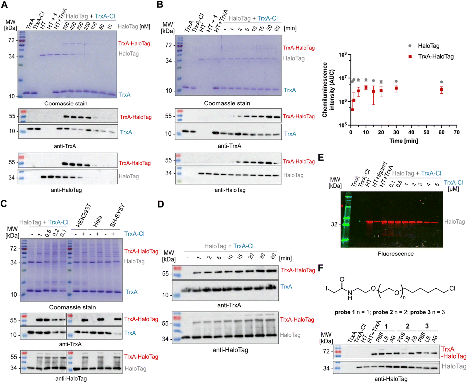 | ||
| Fig. 2 Validation of TrxAC33S-chloroalkane conjugation with the HaloTag. (A) TrxA-chloroalkane (5 µM) conjugation with HaloTag concentration gradient analyzed by Coomassie and western blot; reaction conditions: 15 min at 25 °C. (B) Time-dependent conjugation of TrxAC33S-chloroalkane (2 µM) with HaloTag (200 nM) analyzed by Coomassie and western blot (left). The graph (right) shows the quantification of three independent replicates; the x-axis is in log10 scale; AUC – area under the curve. (C) Conjugation of TrxAC33S-chloroalkane (5 µM) with HaloTag (500 nM) in cell lysates incubated for 15 min at 25 °C and analyzed by Coomassie stain and western blot. (D) Time-dependent conjugation of TrxAC33S-chloroalkane (5 µM) with HaloTag (500 nM) in cell lysates analyzed by western blot. (E) Pulse-chase assay with HaloTag TMR fluorescent ligand: analysis of HaloTag active site occupancy after the reaction with TrxAC33S-chloroalkane using TAMRA-chloroalkane (1 µM) ligand. (F) Evaluation of the linker length impact on conjugation efficiency with HaloTag (200 nM). Each probe was tested in three different commonly used buffers and analyzed by western blot. Reaction conditions: TrxAC33S-chloroalkane (2 µM), 15 min at 25 °C. PBS – phosphate-buffered saline, LB – lysis buffer, AB – activity buffer, and HT – HaloTag. | ||
Typical explorative chemical proteomic experiments involve both gel- and mass spectrometry-based analysis of the probe labelled proteins. To extend the application of the HaloTag system in chemical proteomics beyond the gel-based shift assay, we turned our focus to the possibility of the pull-down of labelled proteins by the utilization of commercially available high-affinity anti-HaloTag magnetic nanobeads. To this end, as described above, the TrxAC33S-chloroalkane (2 µM) was incubated in HEK293T cell lysate with HaloTag (200 nM) for 15 minutes. After the incubation, the reaction mixture was directly transferred onto the magnetic nanobeads and washed three times with a wash buffer containing Tris–HCl, NaCl and EDTA, with removal of the wash buffer after each step by separation on a magnetic rack (Fig. 3A). The efficient pull-down of the TrxAC33S–HaloTag conjugate was confirmed by gel-based analysis (Fig. S3). Finally, the enriched protein(s) were proteolytically digested by trypsin and the resulting peptides subjected directly to LC-MS/MS analysis. In parallel, control experiments including the lysate incubated either with unmodified TrxAC33S or HaloTag or both were processed in the same way. The analysis of the acquired mass spectra and comparison of all conditions showed highly efficient enrichment of the TrxAC33S–HaloTag conjugate with minimal background (Fig. 3B and Fig. S4). Together, this exemplifies the utility of HaloTag conjugates for mass spectrometry-based chemical proteomic pull-down experiments to analyze the small compound probe labelled proteins in complex protein mixtures.
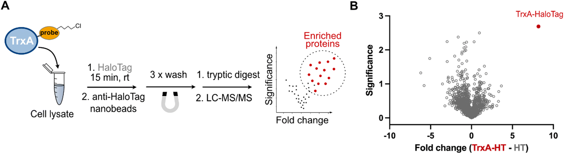 | ||
| Fig. 3 Application of HaloTag for the pull-down of labelled proteins and mass spectrometry. (A) General scheme of the workflow. (B) Volcano plot visualizing pull-down of the TrxAC33S–HaloTag conjugate from HEK293T cell lysate. Lysates containing HaloTag and TrxAC33S were compared to lysates containing HaloTag and TrxAC33S-chloroalkane, and both were incubated for 15 min at 25 °C. Significance is defined by −log10(p-value); p-value was calculated by Student's t-test. Fold change is in log2 scale. The experiment was done in three independent replicates. HT – HaloTag. | ||
In summary, in this study we extend the chemical proteomics toolbox for analysis of proteins labelled with activity-based probes containing the chloroalkane reactive moiety. This strategy leverages the fast and specific reactivity of the HaloTag protein, replacing the conventional ‘click reactions’, enabling downstream analysis by gel-based shift assay and pull-down of the labelled protein followed by mass spectrometry. Principally, the strategy is fully bioorthogonal, fast, specific and can be performed under mild conditions without the necessity for any reaction additives or metal catalysts. Importantly, chloroalkane-based probes and HaloTag are stable and unreactive under virtually all biologically relevant conditions and hence can be combined with complementary labelling strategies. Based on the recent reports by Shields et al. and Mauker et al., the chloroalkane linker can be significantly altered beyond the long linear >C10 substrates.36,37 However, we foresee that chloroalkane-based probes can be directly applied for profiling of protein acylation/lipidation with minimal perturbance to the probe properties, in the case of known writers of protein post-translational modifications with broad substrate specificity, such as tubulin-tyrosine ligase17 or protein adenylyltransferase FICD,38 or in search for new reactive groups towards specific amino acids.2,39 We anticipate that both the conjugation reaction and pull-down protocol will be transferable to other reactive probes and might be combined with many downstream analytical methods, including imaging and site-identification experiments that are now unlocked by the design and versatility of the HaloTag system.
Conflicts of interest
There is no conflict of interest to declare.Data availability
The data supporting this article have been included as part of the supplementary information (SI). Supplementary information: Fig. F1–F4 and Table S1, NMR spectra and further experimental details. See DOI: https://doi.org/10.1039/d6cb00004e.Acknowledgements
We are grateful for the support from SFB 1309-325871075 (Deutsche Forschungsgemeinschaft) to P. K. R. A. and P. K. were supported by the BMFTR in the framework of the Cluster4Future program (Cluster for Nucleic Acid Therapeutics Munich, CNATM) (Project ID: 03ZU1201AA). We thank Sabine Schneider for helpful discussions.References
- S. M. Hacker, K. M. Backus, M. R. Lazear, S. Forli, B. E. Correia and B. F. Cravatt, Nat. Chem., 2017, 9(12), 1181–1190 CrossRef CAS PubMed.
- P. R. A. Zanon, F. Yu, P. Z. Musacchio, L. Lewald, M. Zollo, K. Krauskopf, D. Mrdović, P. Raunft, T. E. Maher, M. Cigler, C. J. Chang, K. Lang, F. D. Toste, A. I. Nesvizhskii and S. M. Hacker, Nat. Chem., 2025, 17, 1712–1721 CrossRef CAS PubMed.
- D. A. Jeffery and M. Bogyo, Curr. Opin. Biotechnol, 2003, 14, 87–95 CrossRef CAS PubMed.
- E. Weerapana, C. Wang, G. M. Simon, F. Richter, S. Khare, M. B. D. Dillon, D. A. Bachovchin, K. Mowen, D. Baker and B. F. Cravatt, Nature, 2010, 468, 790–795 CrossRef CAS PubMed.
- M. Wright and S. Sieber, Nat. Prod. Rep., 2016, 33, 681–708 RSC.
- D. J. Vocadlo, H. C. Hang, E.-J. Kim, J. A. Hanover and C. R. Bertozzi, Proc. Natl. Acad. Sci. U. S. A., 2003, 100, 9116–9121 CrossRef CAS.
- E. Saxon and C. R. Bertozzi, Science, 2000, 287, 2007–2010 CrossRef CAS PubMed.
- E. G. Jackson, G. Cutolo, B. Yang, N. Yarravarapu, M. W. N. Burns, G. Bineva-Todd, C. Roustan, J. B. Thoden, H. M. Lin-Jones, T. H. van Kuppevelt, H. M. Holden, B. Schumann, J. J. Kohler, C. M. Woo and M. R. Pratt, ACS Chem. Biol., 2022, 17, 159–170 CrossRef CAS PubMed.
- W. W. Kallemeijn, T. Lanyon-Hogg, N. Panyain, A. G. Grocin, P. Ciepla, J. Morales-Sanfrutos and E. W. Tate, Nat. Protoc., 2021, 16, 5083–5122 CrossRef CAS PubMed.
- B. R. Martin and B. F. Cravatt, Nat. Methods, 2009, 6, 135–138 CrossRef CAS PubMed.
- R. K. Morgan and M. S. Cohen, ACS Chem. Biol., 2015, 10, 1778–1784 CrossRef CAS PubMed.
- B. A. Gibson, Y. Zhang, H. Jiang, K. M. Hussey, J. H. Shrimp, H. Lin, F. Schwede, Y. Yu and L. W. Kraus, Science, 2016, 353, aaf7865 CrossRef PubMed.
- Y. Wang, D. Rösner, M. Grzywa and A. Marx, Angew. Chem., Int. Ed., 2014, 53, 8159–8162 CrossRef CAS PubMed.
- M. Broncel, R. A. Serwa and E. W. Tate, ChemBioChem, 2012, 13, 183–185 CrossRef CAS PubMed.
- P. Kielkowski, I. Y. Buchsbaum, V. C. Kirsch, N. C. Bach, M. Drukker, S. Cappello and S. A. Sieber, Nat. Commun., 2020, 11, 517 CrossRef CAS.
- D. Makarov and P. Kielkowski, ChemBioChem, 2022, 23, e202200414 CrossRef CAS.
- D. Schumacher, O. Lemke, J. Helma, L. Gerszonowicz, V. Waller, T. Stoschek, P. M. Durkin, N. Budisa, H. Leonhardt, B. G. Keller and C. P. R. Hackenberger, Chem. Sci., 2017, 8, 3471–3478 RSC.
- V. V. Rostovtsev, L. G. Green, V. V. Fokin and K. B. Sharpless, Angew. Chem., Int. Ed., 2002, 41, 2596–2599 CrossRef CAS.
- C. W. Tornøe, C. Christensen and M. Meldal, J. Org. Chem., 2002, 67, 3057–3064 CrossRef.
- N. J. Agard, J. A. Prescher and C. R. Bertozzi, J. Am. Chem. Soc., 2004, 126, 15046–15047 CrossRef CAS.
- B. L. Oliveira, Z. Guo and G. J. L. Bernardes, Chem. Soc. Rev., 2017, 46, 4895–4950 RSC.
- A. Markos, M. Biedermann, J. Heimgärtner, A. Schmitt, K. Lang and H. Wennemers, J. Am. Chem. Soc., 2023, 145(36), 19513–19517 CrossRef CAS PubMed.
- G. V. Los, L. P. Encell, M. G. McDougall, D. D. Hartzell, N. Karassina, C. Zimprich, M. G. Wood, R. Learish, R. F. Ohana, M. Urh, D. Simpson, J. Mendez, K. Zimmerman, P. Otto, G. Vidugiris, J. Zhu, A. Darzins, D. H. Klaubert, R. F. Bulleit and K. V. Wood, ACS Chem. Biol., 2008, 3, 373–382 CrossRef CAS PubMed.
- A. Keppler, S. Gendreizig, T. Gronemeyer, H. Pick, H. Vogel and K. Johnsson, Nat. Biotechnol., 2003, 21, 86–89 CrossRef CAS PubMed.
- B. Mollwitz, E. Brunk, S. Schmitt, F. Pojer, M. Bannwarth, M. Schiltz, U. Rothlisberger and K. Johnsson, Biochemistry, 2012, 51, 986–994 CrossRef CAS PubMed.
- A. C. D. Fuchs, Nat. Commun., 2023, 14, 2505 CrossRef CAS.
- M. A. R. de Geus, C. E. Stieger, J. V. V. Arafiles, J.-R. P. J. Lotthé, P. Schmieder, K. Kemnitz-Hassanin, B. Kindt, H. Leonhardt, S. Schmitt, M. Gerlach, D. Schumacher, J. Helma, M.-A. Kasper and C. P. R. Hackenberger, J. Am. Chem. Soc., 2025, 147, 18888–18900 CrossRef CAS PubMed.
- M.-C. Huppertz, J. Wilhelm, V. Grenier, M. W. Schneider, T. Falt, N. Porzberg, D. Hausmann, D. C. Hoffmann, L. Hai, M. Tarnawski, G. Pino, K. Slanchev, I. Kolb, C. Acuna, L. M. Fenk, H. Baier, J. Hiblot and K. Johnsson, Science, 2024, 383, 890–897 CrossRef CAS PubMed.
- L. B. Crocker, J. V. V. Arafiles, J. M. Müchler, M. Ruwolt, K. Kemnitz-Hassanin, K. Roßmann, C. E. Stieger, F. Liu, N. Archipowa, R. J. Kutta and C. P. R. Hackenberger, Nat. Chem., 2025, 1–13 Search PubMed.
- R. F. Ohana, L. P. Encell, K. Zhao, D. Simpson, M. R. Slater, M. Urh and K. V. Wood, Protein Expression Purif., 2009, 68, 110–120 CrossRef CAS PubMed.
- D. L. Buckley, K. Raina, N. Darricarrere, J. Hines, J. L. Gustafson, I. E. Smith, A. H. Miah, J. D. Harling and C. M. Crews, ACS Chem. Biol., 2015, 10, 1831–1837 CrossRef CAS PubMed.
- N. Darabedian, J. W. Thompson, K. N. Chuh, L. C. Hsieh-Wilson and M. R. Pratt, Biochemistry, 2018, 57, 5769–5774 CrossRef CAS PubMed.
- A. Wiest and P. Kielkowski, J. Am. Chem. Soc., 2024, 146, 2151–2159 CrossRef CAS PubMed.
- H. Chen, D. Wu, J. Holzinger, R. Götz, D. Didier, A. K. Schütz, S. Schneider and P. Kielkowski, Eur. J. Org. Chem., 2024, e202401246 Search PubMed.
- J. Wilhelm, S. Kühn, M. Tarnawski, G. Gotthard, J. Tünnermann, T. Tänzer, J. Karpenko, N. Mertes, L. Xue, U. Uhrig, J. Reinstein, J. Hiblot and K. Johnsson, Biochemistry, 2021, 60, 2560–2575 CrossRef CAS.
- B. C. Shields, H. Yan, S. S. X. Lim, S. C. V. Burwell, C. M. Cammarata, E. A. Fleming, S. A. Yousefzadeh, V. Z. Goldenshtein, E. W. Kahuno, P. P. Vagadia, M. H. Loughran, L. Zhiquan, M. E. McDonnell, M. L. Scalabrino, M. Thapa, T. M. Hawley, G. D. Field, C. Hull, G. E. Schiltz, L. L. Glickfeld, A. B. Reitz and M. R. Tadross, Nat. Methods, 2024, 21, 1288–1297 CrossRef CAS.
- P. Mauker, C. B. J. Zecha, L. Dessen-Weissenhorn, L. Román-Albasini, J. C. M. Meiring, J. I. Brandmeier, D. Beckmann, J. P. Prohaska, M. Reynders, Y. L. Li, K. Mrug, H. von Schwerin, N. A. Vepřek, J. F. Gaisbauer, L. Dehmelt, P. Nalbant, J. Zenker, A. Akhmanova, M. Kerschensteiner, A. B. Harbauer, J. Thorn-Seshold and O. Thorn-Seshold, bioRxiv, 2025 DOI:10.1101/2025.09.23.676741.
- B. K. Chatterjee, M. Alam, A. Chakravorty, S. M. Lacy, W. Giblin, J. Rech, C. L. Brooks, P. Arvan and M. C. Truttmann, ACS Chem. Biol., 2025, 20, 880–895 CrossRef CAS.
- C. M. Nuber, A. V. Milton, B. Nissl, M. C. I. Alvarez, B. R. G. Bissinger, M. B. Sathian, C. D. Pignot, A. Haberhauer, D. Wu, C. Douat, S. Schneider, S. M. Hacker, P. Kielkowski and D. B. Konrad, JACS Au, 2025, 5, 5908–5916 CrossRef CAS PubMed.
| This journal is © The Royal Society of Chemistry 2026 |