
 Open Access Article
 This Open Access Article is licensed under a
 Creative Commons Attribution 3.0 Unported Licence
Correction: Development of a tannic acid- and silicate ion-functionalized PVA–starch composite hydrogel for in situ skeletal muscle repairing
Longkang
Li
a,
Huipeng
Li
b,
Zhentian
Diao
a,
Huan
Zhou
*b,
Yanjie
Bai
*bc and
Lei
Yang
b
aSchool of Materials Science and Engineering, Hebei University of Technology, Tianjin, 300130, China
bCenter for Health Science and Engineering, Hebei Key Laboratory of Biomaterials and Smart Theranostics, School of Health Sciences and Biomedical Engineering, Hebei University of Technology, Tianjin, 300130, China. E-mail: zhouhuan@hebut.edu.cn
cDepartment of Chemical Engineering, Hebei University of Technology, Tianjin, 300130, China. E-mail: 2021100@hebut.edu.cn
First published on 6th February 2025
Abstract
Correction for ‘Development of a tannic acid- and silicate ion-functionalized PVA–starch composite hydrogel for in situ skeletal muscle repairing’ by Longkang Li et al., J. Mater. Chem. B, 2024, 12, 3917–3926, https://doi.org/10.1039/D3TB03006G.
The authors note a mistake in Fig. 5 in which the image of PSTS3 in day 3 in Fig. 5C was mistakenly duplicated as Con in day 3. The corrected Fig. 5 is shown here. Meanwhile, corresponding original images are provided in an additional supplementary information file.
 | ||
| Fig. 5 In vitro cellular test results. (A) Cell survival rate of C2C12 cells cultured in conventional medium or hydrogel extract. (B) Cell survival rate of HUVECs cultured after 1 and 3 days. (C) Cell viability/death staining of cultured C2C12 cells. (D) Cell live/dead staining of cultured HUVECs. (E) Quantification of average myotube width of C2C12 myoblasts after 6 days of culture in conventional differentiation medium with or without hydrogel extracts. (F) Actin filament staining of C2C12 cells after 6 days of proliferation (phalloidin: red; DAPI: blue). (*p < 0.05; **p < 0.01.) | ||
The authors note the sample size used for quantitative analysis in Fig. 7 was not enough. Therefore, the section staining data of all the mice in the Con group and the PSTS group (n = 3) in Fig. 7B and C was reanalyzed with three same-sized regions of the section from the same batch of mice taken into processing. In general, the initial localization of the damaged area was firstly selected based on the difference between normal tissue and damaged tissue. The damaged tissue exhibited a more disorganized structure with a large amount of scar tissue production and a less distinct cellular structure when compared with normal muscle tissue. After that, representative images located in the entire injury region were chosen for analysis, and they were selected according to the following criteria: (1) areas should completely cover the injury region and possess injury characteristics; (2) areas should represent the regions with healing tendency as much as possible; (3) the selected area should be maintained at a consistent size.
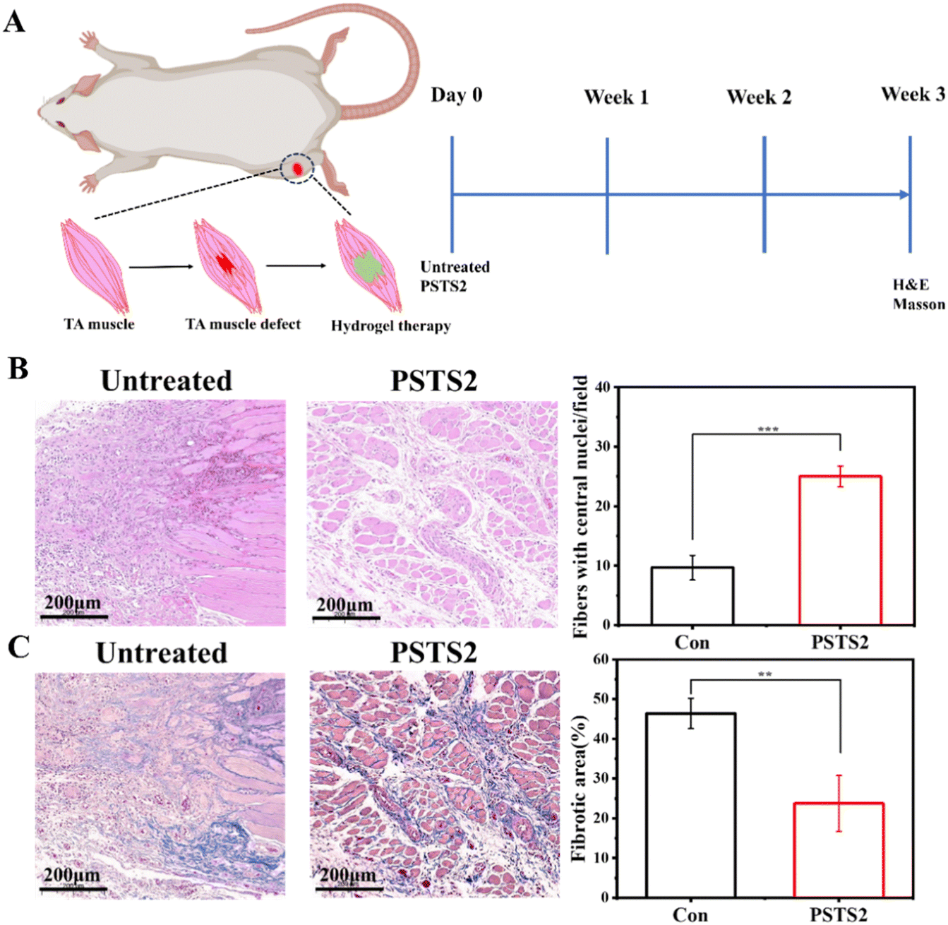 | ||
| Fig. 7 (A) Schematic diagram of the experimental design. (B) Hematoxylin and eosin staining of the injury site three weeks after surgery and quantification of the corresponding central nucleus myofibrils. The stained section was processed and analyzed from the same batch of three mice. (C) Masson trichrome staining of the injury site three weeks postoperatively and the corresponding quantification of the fibrotic area in the neoplastic region. The stained section was processed and analyzed from the same batch of three mice. (**p < 0.01; ***p < 0.001.) | ||
The corrected Fig. 7 is shown here. Meanwhile, corresponding original images of all processed sections are provided in an additional supplementary information file.
The Royal Society of Chemistry apologises for these errors and any consequent inconvenience to authors and readers.
| This journal is © The Royal Society of Chemistry 2025 |