
 Open Access Article
 This Open Access Article is licensed under a Creative Commons Attribution-Non Commercial 3.0 Unported Licence
SIM imaging of bacterial membrane dynamics and lipid peroxidation during photodynamic inactivation with a dual-functional activatable probe†
Yi
Tao
ab,
Qinglong
Qiao
*a,
Yiyan
Ruan
ab,
Xiangning
Fang
ab,
Xiang
Wang
a,
Yinchan
Zhang
ab,
Pengjun
Bao
ab,
Yalin
Huang
ab and
Zhaochao
Xu
 *ab
*ab
aDalian Institute of Chemical Physics, Chinese Academy of Sciences, 457 Zhongshan Road, Dalian 116023, China. E-mail: qqlqiao@dicp.ac.cn; zcxu@dicp.ac.cn
bUniversity of Chinese Academy of Sciences, Beijing 100049, China
First published on 31st March 2025
Abstract
Photodynamic inactivation (PDI) has emerged as a promising antibacterial strategy that mitigates the risk of bacterial resistance. However, the precise morphological dynamics and mechanisms underlying bacterial cell death during PDI remain insufficiently understood. In this study, we developed a dual-functional activatable probe, RDP, which integrated rhodamine B as a fluorophore with moderate brightness and fatty chains for selective bacterial membrane localization. The probe employed an aggregation-disaggregation mechanism to achieve both fluorescence activation and PDI functionality. Using super-resolution fluorescence imaging, we unveiled the selective rupture of bacterial membranes at specific sites during PDI, followed by membrane contraction and internalization, ultimately leading to the formation of lipid-enriched droplets within the bacteria. Further mechanistic investigations confirmed that this membrane rupture was driven by lipid peroxidation. Statistical analysis of bacterial morphological changes during PDI revealed that membrane rupture predominantly occurred at the septum during cell division, whereas in other growth phases, rupture sites were mainly localized at the poles. These findings provide critical insights into the role of selective membrane rupture in bacterial growth and viability, paving the way for the rational design of targeted and highly efficient antibacterial agents.
Introduction
The rise of antibiotic-resistant bacteria poses a critical global health challenge, necessitating the development of innovative antibacterial strategies.1–4 Photodynamic inactivation (PDI) has emerged as a promising approach for eradicating pathogenic microorganisms, with applications in medicine and food safety.5–11 PDI relies on light-activated photosensitizers (PSs) to generate reactive oxygen species (ROS), which oxidize bacterial cellular components, leading to cell death. This strategy exhibits broad-spectrum antibacterial activity, particularly against antibiotic-resistant bacteria.12–15 Unlike conventional antibiotics, PDI induces bacterial membrane disruption through a non-specific, multi-targeted, and irreversible oxidative mechanism, simultaneously damaging other cellular structures. This significantly reduces the likelihood of resistance development, making PDI a promising alternative for antimicrobial therapy.16 However, the bactericidal mechanism of PDI is highly complex, involving multiple biochemical pathways and cellular responses.17 A comprehensive understanding of these mechanisms remains incomplete, which hinders the rational design of PDI agents and their widespread application across various fields.During PDI, PSs primarily target the bacterial plasma membrane or DNA, with the membrane being the primary site of damage and a key determinant of bacterial cell death.18–20 The lipid components of the bacterial membrane play essential roles in nutrient exchange, intercellular communication, and structural integrity.21–24 When ROS oxidize membrane lipids, the resulting structural disruption leads to loss of function and ultimately cell death.25 Therefore, understanding the dynamic changes in the bacterial membrane during PDI is crucial for elucidating its bactericidal mechanism. While previous studies have largely focused on the overall antimicrobial effects of PDI, the detailed morphological and molecular-level changes in bacterial membranes remain poorly understood.26–30 A thorough investigation of bacterial membrane dynamics during PDI would provide critical insights into its mechanism of action, facilitating the design of more effective PDI agents. However, the bacterial membrane is highly dynamic, primarily composed of phospholipids such as phosphatidylethanolamine (PE) and phosphatidylglycerol (PG), which assemble into nanoscale microdomains (2–200 nm) with distinct structural and functional properties.31 Capturing these dynamic changes requires advanced imaging techniques capable of resolving nanoscale membrane structures in real time. Recent advances in super-resolution fluorescence microscopy have provided powerful tools for visualizing bacterial membranes, enabling the analysis of their structural and functional dynamics at nanometer resolution.32–35 Therefore, developing probes that can image bacterial membranes with super-resolution during PDI is essential for elucidating the underlying mechanisms of bacterial cell death.
Despite this need, there remains a significant gap in the development of probes that integrate bacterial membrane imaging with PDI activity, enabling real-time, in situ visualization of membrane dynamics. Current PSs, while effective in bacterial eradication, often lack intrinsic fluorescence, requiring the use of separate fluorescent probes to monitor bacterial membrane changes.29,36,37 This limitation prevents direct visualization of the specific sites of PS action. Although some dual-functional probes with both PDI and fluorescence imaging capabilities have been developed, they often suffer from weak fluorescence intensity or poor photostability, making them unsuitable for long-term, high-resolution imaging.38–40 As a result, they fail to provide real-time insights into bacterial membrane dynamics during PDI-induced cell death. Addressing this limitation requires the development of robust dual-functional probes that simultaneously enable high-resolution imaging of bacterial membranes and efficient PDI, thereby facilitating a deeper understanding of membrane disruptions induced by photosensitizers during bacterial inactivation.
To address these challenges, we developed a rhodamine-derived dual-functional probe, RDP, which enables both fluorescence imaging of the plasma membrane in Gram-positive bacteria and photodynamic inactivation (PDI)-induced bacterial death (Fig. 1). Rhodamine B was chosen as the fluorophore due to its optimal brightness for effective fluorescence imaging and its ability to undergo intersystem crossing, facilitating the generation of ROS.41 To achieve selective membrane targeting, a long fatty acid chain was conjugated to rhodamine B, promoting the formation of fluorescence-quenched aggregates in aqueous solution. Upon disaggregation, RDP monomers anchor to the bacterial plasma membrane via hydrophobic interactions with membrane lipids and electrostatic interactions with membrane phospholipids, leading to fluorescence activation and localized ROS production. This ROS-mediated lipid peroxidation disrupts the membrane structure, ultimately causing bacterial cell death. Using RDP, we achieved real-time SIM imaging of bacterial membrane dynamics during PDI, capturing rupture events that drive lipid peroxidation-induced droplet formation. Super-resolution imaging further enabled us to construct a dynamic model of bacterial membrane behaviours during PDI, revealing that membrane rupture preferentially occurs at the septum during cell division and at the poles in other growth stages. This study advances our understanding of the molecular mechanisms underlying PDI and provides a foundation for the rational design of targeted antibacterial agents.
 | ||
| Fig. 1 Dual-functionality in super-resolution imaging and PDI via aggregation–disaggregation-induced activation of the photosensitizer RDP. | ||
Results and discussion
Aggregation–disaggregation-induced fluorescence activation and ROS generation
To examine the fluorogenicity of RDP towards the bacterial membrane, the spectral properties of RDP in various solvents were first measured (Fig. 2a, b, and S1†). The quantum yield of RDP in PBS was found to be 0.02, accompanied by a broad absorption spectrum, indicating significant fluorescence quenching. However, in the presence of surfactants, the fluorescence intensity was dramatically enhanced, increasing by 156-fold and 139-fold in SDS and TritonX-100, respectively (Fig. 2b). Moreover, both the molar extinction coefficient (ε) and fluorescence quantum yield (Φ) showed significant improvements (Fig. 2c). | ||
| Fig. 2 The photophysical properties of RDP. (a) UV-Vis absorption spectra of RDP (5 μM) in PBS, PBS + SDS, PBS + TritonX-100, and DMSO. (b) Fluorescence emission spectra of RDP (5 μM) in PBS, PBS + SDS, and PBS + TritonX-100. λex/em = 520/590 nm. (c) Maximum absorption wavelength (λabs), maximum emission wavelength (λem), molar extinction coefficient (ε) and fluorescence quantum yield (Φ) of RDP in different solvents. (d) DLS analysis of RDP aggregates (5 μM) in PBS (PDI = 0.307), and fluorescence imaging of RDP aggregates (10 μM) in ultrapure water. (e) Fluorescence spectra of DCFH (2 μM) and RDP (2 μM) monomers in PBS + TritonX-100 solution after irradiation with 561 nm laser (40.6 mW cm−2) for different time intervals. Excitation wavelength: 470 nm. Scale bar: 1 μm. | ||
We hypothesized that the significant increase in fluorescence intensity of RDP in the presence of surfactants could be attributed to two main factors. The first factor is the fluorescence quenching effect due to aggregation. The long fatty acid chains facilitated the formation of RDP aggregates in aqueous solution, leading to fluorescence quenching. This was confirmed by comparing the absorption spectra, which showed a reduced absorbance of RDP in PBS, with an absorption peak at 558 nm corresponding to aggregates. Furthermore, dynamic light scattering (DLS) and fluorescence microscopy confirmed the formation of RDP aggregates in PBS (Fig. S2†). When the concentration of RDP in PBS reached 0.05 μM, aggregate particles began to form, with the particle size increasing gradually as the concentration of RDP was raised (Fig. S2a†). When the concentration of RDP was 5 μM, the DLS results revealed a relatively broad particle size distribution (polydispersity index, PDI = 0.307), indicating the coexistence of larger aggregate particles and smaller particles, with an average particle size of approximately 685.5 nm (Fig. 2d). The second factor is the formation of non-fluorescent rhodamine spironolactone structures within the aggregates, which further contributed to the fluorescence quenching. This was evidenced by the absorption peaks at 275 nm and 321 nm, corresponding to spironolactone, which matched the absorption spectra observed in DMSO and indicated the presence of this non-fluorescent structure within the aggregates (Fig. 2a, green arrow). Additionally, RDP at various concentrations exhibited similar spectral characteristics, with the absorption spectra in PBS showing the characteristic absorption peak of the rhodamine spironolactone structure (Fig. S3†). The fluorescence quenching due to aggregation, along with the formation of non-fluorescent rhodamine spironolactone structures, led to a significant enhancement of fluorescence intensity upon the disaggregation of RDP aggregates into monomers. This suggested that RDP exhibited “fluorescence-on” properties, making it a promising candidate for bacterial wash-free fluorescence imaging applications.
Notably, the quantum yield of RDP in PBS with surfactants was approximately 0.5, suggesting the potential for intersystem crossing (ISC) to the triplet state (T1), which could facilitate energy transfer to molecular oxygen, thereby promoting the generation of ROS. To evaluate ROS generation, we used dichlorofluorescein (DCFH), a fluorescent ROS probe, where ROS presence enhanced fluorescence at 525 nm.42,43 Under 561 nm laser irradiation, DCFH fluorescence gradually increased with RDP, but not with DCFH alone (Fig. 2e and S4†), confirming RDP's ROS-generating capability. To identify the specific ROS generated by RDP, we first used ABDA to detect singlet oxygen (1O2). No change in ABDA absorbance at 380 nm indicated that RDP did not produce 1O2 (Fig. S5†). We then used DCFH with quenchers VC (for ·O2−) and IPA (for ·OH) to detect the generation of ·O2− and ·OH. A significant decrease in DCFH fluorescence with VC, but only a slight decrease with IPA, suggested that RDP primarily generated ·O2− (Fig. S6†). Electron paramagnetic resonance (EPR) tests further confirmed ·O2− generation (Fig. S7,† marked by blue dots), while other peaks, attributed to DMPO oxidation, did not interfere with ·O2− detection. Therefore, the primary ROS generated by RDP was ·O2−, suggesting that RDP absorbed light energy, transitioned from the ground state (S0) to the singlet state (S1), and then underwent ISC to T1. During the electron transfer process, the triplet state of RDP donated an electron to molecular oxygen, reducing it to ·O2− (Type I reaction). These results also demonstrated that RDP could function as a dual-functional PS, capable of both imaging and PDI.
Fluorescence imaging of the plasma membrane of Gram-positive bacteria by RDP
Given the aggregation–disaggregation-induced fluorescence enhancement property of RDP, we investigated its potential as a fluorescent probe for wash-free imaging of living bacteria. We incubated five types of Gram-positive bacteria (B. cereus, B. subtilis, S. aureus, S. epidermidis, and E. faecalis) and three types of Gram-negative bacteria (E. coli, C. freundii, and S. enteritidis) with RDP and DAPI (a commercial nucleic acid dye) for 30 minutes, followed by wash-free confocal imaging. Gram-positive bacteria exhibited strong fluorescence on their membranes with a high signal-to-noise ratio (SNR, 7-fold), while Gram-negative bacteria showed negligible fluorescence (Fig. 3). This discrepancy can be attributed to the structural differences in the bacterial envelopes of Gram-positive and Gram-negative bacteria. In Gram-positive bacteria, the plasma membrane is surrounded by a thick peptidoglycan layer, allowing RDP to easily penetrate and reach the plasma membrane, where it anchored through electrostatic and hydrophobic interactions. In this environment, RDP initially formed non-fluorescent aggregates in bacterial solutions. However, upon targeting the membrane, the aggregates disaggregated into fluorescent rhodamine monomers, activating fluorescence and illuminating the plasma membrane of Gram-positive bacteria. In contrast, Gram-negative bacteria possess not only a thinner peptidoglycan layer but also an outer membrane rich in lipopolysaccharides, which hindered the entry of molecules and prevents RDP from reaching the plasma membrane. As a result, RDP effectively stained the plasma membrane of Gram-positive bacteria but not Gram-negative bacteria.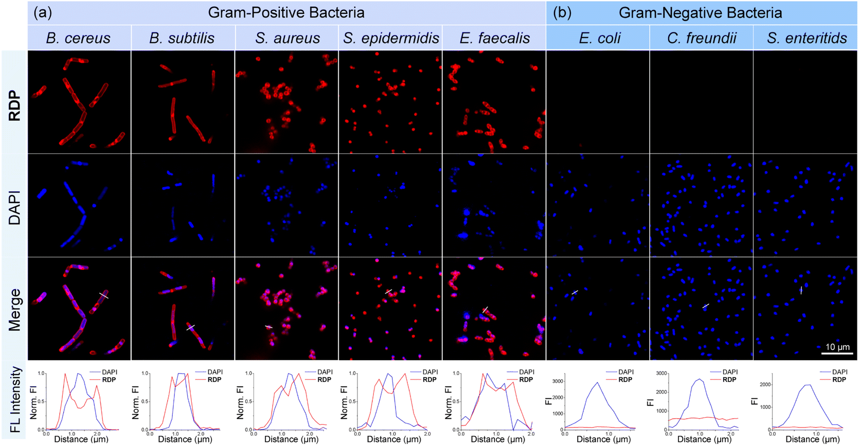 | ||
| Fig. 3 Confocal fluorescence wash-free imaging of bacterial plasma membrane with RDP. (a) Five kinds of Gram-positive bacteria (B. cereus, B. subtilis, S. aureus, S. epidermidis, and E. faecalis) and (b) three kinds of Gram-negative bacteria (E. coli, C. freundii, and S. enteritids) (OD = 1) were pretreated with 2 μM RDP and 2 μM DAPI for 30 minutes, respectively, followed by fluorescence imaging (λex = 405 nm/543 nm). And fluorescence intensity graphs normalized fluorescence profiles along the lines shown in merge images. | ||
Photodynamic inactivation of Gram-positive bacteria using RDP
Meanwhile, we explored the potential of RDP as a photosensitizer for bacterial killing. We selected three common Gram-positive bacteria (B. cereus, S. aureus, and E. faecalis), one multidrug-resistant Gram-positive bacteria (MDR S. aureus), and one Gram-negative bacteria (C. freundii) to assess the bactericidal performance of RDP. The colony-forming unit (CFU) counting method was used to evaluate the photodynamic bactericidal effect. Bacteria labeled with different concentrations of RDP were exposed to 561 nm laser irradiation for 30 minutes, followed by incubation on solid culture plates at 37 °C. After 24 hours, colonies formed on the plates were counted and analyzed (Fig. 4). The results showed that for common Gram-positive bacteria (B. cereus, S. aureus, and E. faecalis), 90% of the bacteria were eradicated at an RDP concentration of 0.625 μM (0.45 μg mL−1) (Fig. 4a–c and f). For drug-resistant Gram-positive bacteria (MDR S. aureus), a higher RDP concentration of 1.25 μM (0.90 μg mL−1) achieved a 99% kill rate (Fig. 4d and f). In contrast, RDP exhibited negligible PDI activity against Gram-negative bacteria (Fig. 4e and f). These findings demonstrated the excellent PDI efficacy of RDP against Gram-positive bacteria. We speculated that once RDP anchored to the plasma membrane of Gram-positive bacteria, laser irradiation triggered ROS production, which disrupted the surrounding membrane structure and ultimately led to bacterial death. In contrast, RDP failed to kill Gram-negative bacteria due to its inability to penetrate and access their plasma membrane.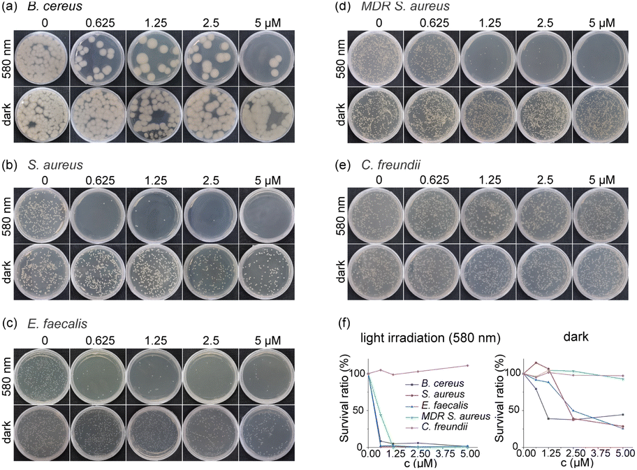 | ||
| Fig. 4 Photodynamic antibacterial activity test results of RDP. (a) B. cereus, (b) S. aureus, (c) E. faecalis, (d) MDR S. aureus and (e) C. freundii were treated with different concentrations of RDP in dark/580 nm irradiation (5 mW cm−2) for 30 min. Pictures of colony counting plates cultured 24 h in a 37 °C incubator. (f) Bacteria survival rates of different kinds of bacteria exposed to RDP (0–5 μM) in dark/580 nm irradiation (5 mW cm−2). | ||
Leveraging the dual functionality of RDP to label the plasma membrane of Gram-positive bacteria and perform PDI, we utilized this probe to investigate the effects of PDI on bacterial plasma membrane using SIM super-resolution microscopy. B. cereus was stained with RDP and irradiated with a 561 nm laser for 15 minutes. A comparison of the imaging results before and after irradiation revealed a reduction in bacterial volume and the formation of spherical fluorescent bright spots inside the bacteria (Fig. 5a). In contrast, B. cereus stained with RDP in the dark or with FM4-64 (a commercial membrane dye) under 561 nm laser irradiation showed no observable changes (Fig. 5a and S8). Based on these imaging results, we hypothesized that the reduction in bacterial volume and shrinkage of RDP-labeled bacteria under laser irradiation resulted from PDI, ultimately leading to bacterial death.
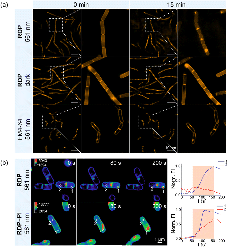 | ||
| Fig. 5 SIM imaging and analysis of RDP photodynamic inactivation of B. cereus. (a) 0 and 15 min images of SIM imaging of B. cereus plasma membrane with RDP (2 μM, 30 min)/FM4-64 (5 μM, 30 min) in dark/561 nm irradiation. (b) Fluorescence intensity changes at different positions of long-term dynamic imaging of B. cereus treated with RDP (2 μM, incubation for 30 min)/RDP + PI (2 μM, incubation for 30 min). The orange area is an intensity mutation area. | ||
To further confirm bacterial death, we simultaneously labeled the bacteria with propidium iodide (PI), a commercial dye specific for marking dead bacteria. As a DNA dye, PI could not penetrate the intact cell membrane of live bacteria but readily entered through damaged membrane in dead bacteria to stain DNA, thereby distinguishing live and dead cells. B. cereus was stained with RDP alone and with a combination of RDP and PI, followed by 561 nm laser irradiation and imaging to monitor the formation of fluorescent bright spots within the bacteria. We extracted and analyzed the fluorescence spectra from regions where the bright spots appeared and other areas within the bacteria during this process (Fig. 5b). In the control group stained only with RDP, the fluorescence intensity of the bright spots increased, while no significant fluorescence enhancement was observed in other regions of the bacteria (Fig. 5b and Movie S1†). In contrast, in the experimental group stained with both RDP and PI, the fluorescence intensity of other regions within the bacteria also showed a significant increase coinciding with the formation of the bright spots (Fig. 5b and Movie S2†). These results indicated that the generation of fluorescent bright spots corresponded to PDI-induced bacterial death, allowing PI to penetrate the damaged bacterial membrane and stain the DNA.
Imaging of plasma membrane dynamics during photodynamic inactivation of Gram-positive bacteria
To better understand the behavior of the bacterial plasma membrane during PDI, we used RDP to monitor the real-time dynamics of the plasma membrane in B. cereus (Fig. 6). During the ROS-induced bacterial death process, two distinct phenomena were observed on the plasma membrane: first, thin filaments appeared, which gradually thickened and condensed into bright spherical fluorescent spots, or second, small spherical fluorescent spots formed on the membrane and progressively grew into larger bright spots (Fig. 6a, Movie S3 and 4†). Three-dimensional structured illumination microscopy (3D-SIM) imaging confirmed that the spherical fluorescent spots were directly associated with the plasma membrane, forming on its surface (Fig. 6b). Simultaneously, bacterial shrinkage and a reduction in plasma membrane size were observed, coinciding with the formation of these fluorescent droplets. Based on these observations, we hypothesized that the fluorescent droplets were a result of membrane rupture, where damaged membrane lipids combined with fluorescent molecules. These damaged lipids likely consisted of lipid peroxides formed through ROS-induced lipid peroxidation driven by the PS.44 To validate this hypothesis, membrane lipids from B. cereus were extracted before and after PDI and analyzed via high-resolution mass spectrometry, confirming damage and peroxidation, resulting in lipid peroxides (Fig. 6c and S9).45 | ||
| Fig. 6 Long-term SIM imaging of the plasma membrane during photodynamic inactivation of B. cereus. (a) B. cereus was pretreated with 2 μM RDP for 30 min, followed by SIM imaging and imaged for 15 min. (b) 3D images of dead B. cereus from different views. (c) Structure and HRMS analysis of PE and peroxidized PE on the plasma membrane of B. cereus before and after PDI. (d) The ratio of the distance between the fluorescent droplet and the septum or poles to the length of the bacterial cell. (e) Model of the dynamic death process of B. cereus. (f) S. aureus was pretreated with 2 μM RDP for 30 min, followed by SIM imaging and imaged for 15 min. | ||
Moreover, extensive super-resolution imaging allowed for detailed analysis of the locations where lipid-condensed droplets formed. For non-contracted, dividing bacteria, most droplets were located at the septum, whereas for contracted, non-dividing bacteria, droplet formation appeared more random, with a concentration near the poles (Fig. 6d). This suggested that the sites where lipid-condensed droplets formed corresponded to regions of membrane rupture. Statistical analysis indicated that ROS were more likely to target the bacterial plasma membrane near the septum and poles, areas potentially more vulnerable on the bacterial surface. Additionally, scanning electron microscopy (SEM) further demonstrated that bacterial membrane subjected to PDI exhibited rupture, particularly at the poles and septa, with leakage of intracellular contents from these sites (Fig. S10†). Based on these observations, we summarized the dynamic changes in the plasma membrane of B. cereus during PDI and proposed a model to describe this process (Fig. 6e). We concluded that two types of changes might occur on the plasma membrane: the first involves the appearance of filaments that transform into spherical fluorescent lipid droplets, and the second involves the growth of small spherical fluorescent droplets into larger ones. Both processes are more likely to occur near the septum and poles and are accompanied by gradual contraction and reduction of the plasma membrane's size. Similar phenomena were also observed in S. aureus, where fluorescent lipid-condensed droplets predominantly formed at the septum of dividing bacteria (Fig. 6f and Movie S5†).
Conclusions
In conclusion, we have designed and synthesized an aggregation–disaggregation-regulated fluorescent probe, RDP, which selectively targeted the plasma membrane of Gram-positive bacteria through electrostatic and hydrophobic interactions, enabling effective fluorescence imaging. Additionally, RDP functioned as a photosensitizer (PS) for photodynamic inactivation (PDI), effectively inducing bacterial death through lipid peroxidation. By leveraging this dual-functionality, we developed a probe that both labels bacterial membranes for imaging and facilitates bacterial killing via PDI. Furthermore, we visualized the morphological changes in the plasma membrane of Gram-positive bacteria during cell death induced by lipid peroxidation. Our analysis highlighted that the regions most vulnerable to ROS attack were located near the septum and poles of the bacterial plasma membrane, which might represent key sites for bacterial inactivation. We believe this work provides valuable insights into the PDI bactericidal mechanism and holds promise for the development of new strategies for combating bacterial infections.Data availability
All the data supporting this article have been included in the main text and the ESI.†Author contributions
Y. Tao, Q. L. Qiao and Z. C. Xu conceived the methodology and supervised the project. Y. Tao, Y. Y. Ruan and X. Wang performed the biological experiments and statistical analyses. X. N. Fang, Y. C. Zhang, P. J. Bao and Y. L. Huang carried out the synthetic experimental work.Conflicts of interest
There are no conflicts to declare.Acknowledgements
This work is supported by the National Natural Science Foundation of China (22225806, 22078314, 22278394, 22378385) and Dalian Institute of Chemical Physics (DICPI202142, DICPI202436).Notes and references
- J. L. Martínez, F. Baquero and D. I. Andersson, Nat. Rev. Microbiol., 2007, 5, 958–965 CrossRef.
- E. M. Darby, E. Trampari, P. Siasat, M. S. Gaya, I. Alav, M. A. Webber and J. M. A. Blair, Nat. Rev. Microbiol., 2022, 21, 280–295 CrossRef PubMed.
- D. Meziane-Cherif and P. Courvalin, Nature, 2014, 510, 477–478 CrossRef CAS.
- E. D. Brown and G. D. Wright, Nature, 2016, 529, 336–343 CAS.
- S. M. Lashkari, H. Kariminezhad, H. Amani, P. Mataji and M. Rahimnejad, Photodiagn. Photodyn. Ther., 2019, 25, 336–343 CrossRef CAS.
- A. Späth, C. Leibl, F. Cieplik, K. Lehner, J. Regensburger, K.-A. Hiller, W. Bäumler, G. Schmalz and T. Maisch, J. Med. Chem., 2014, 57, 5157–5168 CrossRef PubMed.
- M. Asnaashari, H. Homayuni and P. Paymanpour, J. Lasers Med. Sci., 2016, 7, 238–242 CrossRef.
- V. S. Ghate, W. Zhou and H. G. Yuk, Compr. Rev. Food Sci. Food Saf., 2019, 18, 402–424 CrossRef.
- Y. Liu, S. Wang, J. Wu, G. Qi, G. Chen, H. Li and H. Wang, Food Bioprocess Technol., 2023, 17, 1294–1308 CrossRef.
- W. Dai, J. Hu, B. K. Tan and S. Lin, Food Chem., 2025, 464, 141925 CrossRef CAS.
- L. Chen, Y. Zhao, W. Wu, Q. Zeng and J. J. Wang, Compr. Rev. Food Sci. Food Saf., 2023, 22, 3814–3846 CAS.
- J. F. Lovell, T. W. B. Liu, J. Chen and G. Zheng, Chem. Rev., 2010, 5, 2839–2857 CrossRef PubMed.
- J. Sun, X. Cai, C. Wang, K. Du, W. Chen, F. Feng and S. Wang, J. Am. Chem. Soc., 2021, 143, 868–878 CAS.
- W. Zhu, Y. Li, S. Guo, W.-J. Guo, T. Peng, H. Li, B. Liu, H.-Q. Peng and B. Z. Tang, Nat. Commun., 2022, 13, 7026 Search PubMed.
- X. Li, S. Lee and J. Yoon, Chem. Soc. Rev., 2018, 47, 1174–1188 CAS.
- M. R. Hamblin and T. Hasan, Photochem. Photobiol. Sci., 2004, 3, 436–450 CrossRef CAS PubMed.
- T. Dai, Y.-Y. Huang and M. R. Hamblin, Photodiagn. Photodyn. Ther., 2009, 6, 170–188 CrossRef CAS.
- T. Maisch, R.-M. Szeimies, G. Jori and C. Abels, Photochem. Photobiol. Sci., 2004, 3, 907–917 CrossRef CAS.
- H. Ma, Y. Ma, L. Lei, W. Yin, Y. Yang, T. Wang, P. Yin, Z. Lei, M. Yang, Y. Qin and S. Zhang, ACS Sustain. Chem. Eng., 2018, 6, 15064–15071 Search PubMed.
- D. P. Valenzeno, Photochem. Photobiol., 1987, 46, 147–160 CrossRef CAS.
- H. Strahl and J. Errington, Annu. Rev. Microbiol., 2017, 71, 519–538 CrossRef CAS PubMed.
- L. M. Y. Mitchison-Field and B. J. Belin, Curr. Opin. Microbiol., 2023, 74, 102315 Search PubMed.
- I. Barák and K. Muchová, Int. J. Mol. Sci., 2013, 14, 4050–4065 Search PubMed.
- T.-Y. Lin and D. B. Weibel, Appl. Microbiol. Biotechnol., 2016, 100, 4255–4267 Search PubMed.
- E. Alves, T. Melo, C. Simões, M. A. F. Faustino, J. P. C. Tomé, M. G. P. M. S. Neves, J. A. S. Cavaleiro, Â. Cunha, N. C. M. Gomes, P. Domingues, M. R. M. Domingues and A. Almeida, Rapid Commun. Mass Spectrom., 2013, 27, 1607–1618 Search PubMed.
- X. Pang, D. Li, J. Zhu, J. Cheng and G. Liu, Nano-Micro Lett., 2020, 12, 144 CrossRef CAS.
- K. Li, Y.-Y. Zhang, G.-Y. Jiang, Y.-J. Hou, B.-W. Zhang, Q.-X. Zhou and X.-S. Wang, Chem. Commun., 2015, 51, 7923–7926 RSC.
- J. Y. Hyun, C.-H. Lee, H. Lee, W.-D. Jang and I. Shin, ACS Macro Lett., 2020, 9, 1429–1432 CrossRef CAS PubMed.
- A. Tavares, C. M. B. Carvalho, M. A. Faustino, M. G. P. M. S. Neves, J. P. C. Tomé, A. C. Tomé, J. A. S. Cavaleiro, Â. Cunha, N. C. M. Gomes, E. Alves and A. Almeida, Mar. Drugs, 2010, 8, 91–105 CrossRef CAS.
- J. Tang, B. Chu, J. Wang, B. Song, Y. Su, H. Wang and Y. He, Nat. Commun., 2019, 10, 4057 CrossRef.
- G. Meer, D. R. Voelker and G. W. Feigenson, Nat. Rev. Mol. Cell Biol., 2008, 9, 112–124 CrossRef PubMed.
- A. Carsten, M. Wolters and M. Aepfelbacher, Mol. Microbiol., 2023, 121, 646–658 Search PubMed.
- R. K. G. Tank, V. A. Lund, S. Kumar, R. D. Turner, L. Lafage, L. Pasquina Lemonche, P. A. Bullough, A. Cadby, S. J. Foster and J. K. Hobbs, ACS Nano, 2021, 15, 16011–16018 CAS.
- D. Jeong, M. J. Kim, Y. Park, J. Chung, H.-S. Kweon, N.-G. Kang, S. J. Hwang, S. H. Youn, B. K. Hwang and D. Kim, BMC Biol., 2022, 20, 270 CAS.
- N. Pende, A. Sogues, D. Megrian, A. Sartori-Rupp, P. England, H. Palabikyan, S. K. M. R. Rittmann, M. Graña, A. M. Wehenkel, P. M. Alzari and S. Gribaldo, Nat. Commun., 2021, 12, 3214 CAS.
- B. Mai, X. Wang, Q. Liu, A. W. Leung, X. Wang, C. Xu and P. Wang, Lasers Surg. Med., 2016, 48, 400–408 Search PubMed.
- L. Huang, M. Krayer, J. G. S. Roubil, Y.-Y. Huang, D. Holten, J. S. Lindsey and M. R. Hamblin, J. Photochem. Photobiol., B, 2014, 141, 119–127 CAS.
- H.-R. Jia, Y.-X. Zhu, Z. Chen and F.-G. Wu, ACS Appl. Mater. Interfaces, 2017, 9, 15943–15951 CAS.
- K. L. Smitten, H. M. Southam, J. B. de la Serna, M. R. Gill, P. J. Jarman, C. G. W. Smythe, R. K. Poole and J. A. Thomas, ACS Nano, 2019, 13, 5133–5146 CAS.
- E. Lee, X. Li, J. Oh, N. Kwon, G. Kim, D. Kim and J. Yoon, Chem. Sci., 2020, 11, 5735–5739 CAS.
- L. Li, Y. Chen, W. Chen, Y. Tan, H. Chen and J. Yin, Chin. Chem. Lett., 2019, 30, 1689–1703 CrossRef CAS.
- M. D. J. F. Curtin and T. G. Cotter, J. Immunol. Methods, 2002, 265, 49–72 CrossRef PubMed.
- J. Yu, C.-H. Hsu, C.-C. Huang and P.-Y. Chang, ACS Appl. Mater. Interfaces, 2014, 7, 432–441 CrossRef.
- P. S. H. Wilker Caetano, R. Itri, D. Severino, V. C. Vieira, M. S. Baptista, A. ́ P. Schröder and C. M. Marques, Langmuir, 2007, 23, 1307–1314 Search PubMed.
- M. L. Y. N. A. Busch and M. Toner, Biophys. J., 1998, 75, 2956–2970 Search PubMed.
Footnote |
| † Electronic supplementary information (ESI) available. See DOI: https://doi.org/10.1039/d5sc00858a |
| This journal is © The Royal Society of Chemistry 2025 |