
 Open Access Article
 This Open Access Article is licensed under a
 Creative Commons Attribution 3.0 Unported Licence
Metal-based immunogenic cell death inducers for cancer immunotherapy
Jiao Xia
Zou†
a,
Meng Rui
Chang†
a,
Nikita A.
Kuznetsov†
b,
Jia Xuan
Kee
 a,
Maria V.
Babak
*b and
Wee Han
Ang
*ac
a,
Maria V.
Babak
*b and
Wee Han
Ang
*ac
aDepartment of Chemistry, National University of Singapore, 4 Science Drive 2, Singapore 117544, Singapore. E-mail: ang.weehan@nus.edu.sg
bDrug Discovery Lab, Department of Chemistry, City University of Hong Kong, 83 Tat Chee Avenue, Hong Kong SAR 999077, People's Republic of China. E-mail: mbabak@cityu.edu.hk
cNUS Graduate School – Integrative Science and Engineering Programme (ISEP), National University of Singapore, 21 Lower Kent Ridge Rd, Singapore 119077, Singapore
First published on 25th February 2025
Abstract
Immunogenic cell death (ICD) has attracted enormous attention over the past decade due to its unique characteristics in cancer cell death and its role in activating innate and adaptive immune responses against tumours. Many efforts have been dedicated to screening, identifying and discovering ICD inducers, resulting in the validation of several based on metal complexes. In this review, we provide a comprehensive summary of current metal-based ICD inducers, their molecular mechanisms for triggering ICD initiation and subsequent protective antitumour immune responses, along with considerations for validating ICD both in vitro and in vivo. We also aim to offer insights into the future development of metal complexes with enhanced ICD-inducing properties and their applications in potentiating antitumour immunity.
 Jiao Xia Zou | Jiao Xia Zou received her MSc in Pharmacy from Lanzhou University (2020) and is currently a final year PhD candidate in Prof. Wee Han Ang's lab at the National University of Singapore. She is working on the development of chemical probes for studying enzyme biology and functions, and for targeting identification and mechanism elucidation of ICD. |
 Meng Rui Chang | Dr Meng Rui Chang received her PhD in Chemistry from National University of Singapore (2025) under the supervision of Prof. Wee Han Ang at National University of Singapore. Her research mainly focused on developing cyclometalated gold complexes as ICD inducers to treat cold tumors. |
 Nikita A. Kuznetsov | Nikita Kuznetsov received his BSc from D. I. Mendeleev University of Chemical Technology of Russia (2023). He is a research assistant in Prof Maria V. Babak's laboratory at City University of Hong Kong, working on the development of synthetic analogues of naturally occurring compounds, as well as the design of Au and Ru anticancer compounds. |
 Jia Xuan Kee | Jia Xuan Kee graduated with a BSc (Hons) in Chemistry from the National University of Singapore with the Singapore National Institute of Chemistry Prize (2020). He is currently a PhD candidate in Prof. Wee Han Ang's lab at the National University of Singapore under the President's Graduate Fellowship, focusing on the development of novel Pt(IV) prodrugs and the elucidation of their mechanisms of actions. |
 Maria V. Babak | Prof. Maria (Masha) V. Babak received her BSc/MSc with Honors from Higher Chemical College of Russian Academy of Sciences (2010) and obtained her PhD from the University of Vienna (2014) under the supervision of Prof. Bernhard K. Keppler and Prof. Christian G. Hartinger. After working as a postdoctoral research fellow under the supervision of Prof. Wee Han Ang at the National University of Singapore and completing High Impact Cancer Research program at Harvard Medical School, she assumed the position of principal investigator at City University of Hong Kong (2020). Her current research interests focus on experimental oncology, with a specific emphasis on in vivo mechanisms of anticancer drug candidates and AI-assisted drug discovery. |
 Wee Han Ang | Prof. Wee Han ANG received his BSc (Hons) from Imperial College of London (1995) and obtained his PhD from École Polytechnique Fédérale de Lausanne (2007) under the supervision of Prof. Paul Dyson. After a postdoctoral stint with Prof. Stephan Lippard at Massachusetts Institute of Technology, he joined the National University of Singapore (2009) as a principal investigator and has been there since. His current research focusses on the investigation and development of therapeutic metal complexes, particularly those based on platinum group metals, as well as the development of analytical techniques to study them. |
1. Introduction
The landscape of clinical cancer treatments has undergone a significant transformation with the advent of immunotherapy, driven by the rise of revolutionary technologies, such as immune checkpoint blockade therapy,1 adoptive T-cell therapy,2–4 and cancer vaccines.5 The concept of harnessing the body's immune system to combat cancerous cells dates back to the 1800s, when physicians Fehleisen and Busch observed tumour regression in cancer patients infected with Streptococcus pyogenes-induced erysipelas.6,7 In 1891, William Bradley Coley, acknowledged as the Father of Immunotherapy, first injected inactivated bacteria (“Coley's toxins”) to activate the immune system for treating bone cancer, thereby pioneering the field of cancer therapy.8Immunotherapy offers a distinct advantage over conventional cancer treatment modalities, such as surgery, chemotherapy, and radiotherapy, due to its systemic tumour-targeting capability and its potential to confer sustained, long-term immunity against tumours.9 However, the success of immunotherapies is heavily linked to the state of an individual's immune system and the immunogenicity of the tumour.10 The complexity of the immune response, negative feedback loops, immune evasion checkpoints, cancer heterogeneity, and other factors further complicate the efficacy of immunotherapies.11–18 In light of the challenges and limitations of cancer immunotherapy, tremendous efforts have been made to identify key determinants of anticancer immune responses to improve immunotherapy outcomes.19 Within the cancer-immunity cycle, a crucial factor in initiating an immune response against cancer involves the recognition of cancer antigens by the immune system.20 However, tumours can reduce their immunogenicity through multiple mechanisms, such as upregulating PD-L1, secreting immunosuppressive factors, and establishing an immunosuppressive tumour microenvironment (TME) that is inaccessible to immune cells.15,21–24
Enhancing tumour immunogenicity has emerged as a promising strategy to combat tumour-induced immunosuppression. Immunogenic cell death (ICD), defined by the Nomenclature Committee on Cell Death as a form of regulated cell death (RCD) that is sufficient to activate an adaptive immune response in immunocompetent syngeneic hosts,25,26 has been at the forefront of this approach. ICD was first recognized in 2005 by Kroemer and coworkers, who found that doxorubicin (DOX)-induced apoptotic tumour cell death is immunogenic. They demonstrated that DOX-treated tumour cells can serve as cancer vaccines to elicit antitumour immune responses mediated by dendritic cells (DCs) and cytotoxic CD8+ T-cells in immunocompetent mice.27 This study first linked apoptosis inflicted by certain chemotherapeutic agents to ICD. Apoptosis, traditionally considered a physiological form of cell death, was believed to be immunogenically silent or even immunosuppressive for many years.28–30 Following the discovery of anthracycline-induced ICD phenomenon, other chemotherapeutic agents, such as mitoxantrone (MTX)31,32 and oxaliplatin (OXP),33 cardiac glycosides,34 as well as some physical anticancer therapies, including γ-irradiation35 and photodynamic therapy,36,37 were also reported to induce ICD, resulting in antitumour immune response in vivo. These studies on ICD open the possibility of fully using ICD as an effective strategy to modulate the innate and adaptive immune systems, with the aim of preventing tumour recurrence and metastasis.
Over the last decade, numerous studies have been conducted to discover ICD inducers for therapeutic applications,38–50 owing to their ability to directly eradicate cancer cells and concomitantly stimulate adaptive immune responses for tumour eradication. Mechanistically, ICD involves the release of damage-associated molecular patterns (DAMPs) from dying cancer cells.38,39,50,51 These DAMPs, including the cell surface translocation of calreticulin (CRT), the extracellular release of high mobility group box 1 (HMGB1), and the extracellular secretion of adenosine triphosphate (ATP), augment the immunogenicity of cancer cells and initiate the cancer-immunity cycle. These processes lead to the recruitment of mature, activated immune cells to the tumour site, ensuring effective antigen capture and presentation (Fig. 1).38,41,49,50,52
 | ||
| Fig. 1 Activation of antitumour immune response following ICD induction. Upon treatment with ICD inducers, tumour cells undergo ICD and emit DAMPs. DAMPs recruit immune cells to the ICD site and interact with their corresponding receptors (i.e. CRT-LRP1, HMGB1-TLR2/4 and ATP-P2X2/P2Y7) on APCs, facilitating antigen uptake and processing. Mature APCs then present tumour antigens to T-cells while concurrently secreting cytokines, such as IL-1β, consequently stimulating T-cell activation and proliferation. Ultimately, cytotoxic T-cells are generated, capable of producing INFγ to eradicate tumour cells. In the meantime, memory T-cells are formed, indicative of the establishment of immunological memory. | ||
This review focuses on the unique characteristics of metal complexes and provides an in-depth examination of current metal-based ICD inducers, with a particular emphasis on their molecular targets, mode of mechanism, and roles in enhancing antitumour immune responses. Additionally, we outline the benchmark methods and models employed to validate ICD both in vitro and in vivo. Unresolved yet significant issues and challenges encountered in the development of metal-based ICD inducers are also discussed. Finally, we conclude this review by highlighting the potential applications of ICD inducers for cancer immunotherapy that could pave the way for future explorations.
2. ICD mechanism
ICD is a unique event where cancer cells undergo programmed death while becoming immunogenic and activating adaptive immune response.26,49 Consequently, dying cancer cells treated with ICD inducers can be used as a vaccine to prevent tumour proliferation and activate a cancer-specific immune response.53 Various cell stressors, including certain traditional chemotherapeutic agents,27,32,34,54,55 infective pathogens,56–59 and some physical therapeutic modalities, such as photodynamic therapy,60,61 extracorporeal photochemotherapy,62 electrochemotherapy,63 photothermal therapy,64,65 radiotherapy,35,66–68 high hydrostatic pressure,69 and many more,70–74 can provoke ICD.ICD inducers are a type of chemotherapeutic agent that can cause cancer cells to undergo ICD and can be largely divided into two main types: Type I and Type II ICD inducers.51,73 The classification of ICD inducers mainly depends on whether they act directly on the endoplasmic reticulum (ER). Type I ICD inducers primarily act on intracellular components other than ER and generate ER stress as secondary or collateral effects. In contrast, Type II ICD inducers target the ER directly, which results in ER stress, thereby initiating ICD.
The ER stress in question typically arises from perturbation in proteostasis and is characterized by an accumulation of misfolded proteins within the ER, which can occur under the influence of ICD inducers.75–77 Beyond a tolerable ER stress threshold, an unfolded protein response (UPR) is provoked to restore protein folding capacity. UPR is mediated by three ER stress sensors: protein kinase R-like ER kinase (PERK), inositol-requiring enzyme 1 alpha (IRE1α), and activating transcription factor 6 (ATF6).78,79 The PERK pathway, in particular, is crucial for the initiation of ICD.80,81 Multiple studies have underscored the importance of ER stress and UPR for ICD induction.75,77,80 For example, ER stress was shown to restore the immunogenicity of cisplatin (CDDP)-induced cancer cell death.82 Most ICD inducers trigger ER stress for the initiation of the ICD process, with schweinfurthin alkaloids being the notable exception as they can induce ICD without eliciting ER stress.83 Moreover, Type II ICD inducers are typically considered more effective than their Type I counterparts, and the ER-targeting strategy has been shown to be an effective approach to reinforce ICD effects.65,84–86
In the process of ICD, DAMPs may be surface exposed, released or secreted.38,50,51 Most DAMPs are immunologically silent until they are released into the extracellular environment and serve as either adjuvant or danger signals to the immune system. These emitted DAMPs can be recognized by pattern-recognition receptors (PRRs) on immune cells, such as toll-like and nucleotide oligomerization domain (NOD)-like receptors.87 The interactions between DAMPs and PRRs facilitate the uptake, processing and presenting of cancer antigens by antigen-presenting cells (APCs), leading to the activation of APCs and T-cells. These activated T-cells then infiltrate into the tumour sites and eradicate the cancer cells (Fig. 1).
2.1 Hallmarks of ICD
The induction of ICD is characterized by the emission of DAMPs, namely the translocation of CRT to the outer cell membrane and extracellular secretion of HMGB1 and ATP.26,88–90 The concurrent manifestation of these events serves as an indicator of ICD induction in vitro. Beyond these classical ICD hallmarks, ICD is also associated with other biological activities, such as cell surface exposure to heat-shock proteins (HSP70 and HSP90)36,91,92 and enhanced expression of Type I interferons (IFNs)93 and interleukin-1 (IL-1) family cytokines,94 which are observed in certain instances.Extracellular ATP acts as a “find me” signal for myeloid cells by binding to purinergic receptor P2Y2 and ionotropic receptor P2X7, thereby recruiting them to the site of dying tumour cells.109,111,118 This promotes their local differentiation and the subsequent effective uptake of tumour antigens in situ.108,110,119 ATP binding to P2X7 leads to an efflux of K+ and Ca2+, which then activate the nucleotide-binding domain, leucine-rich – containing family, and pyrin domain – containing-3 (NLRP3) inflammasomes. This activation drives the secretion of IL-1β, which is essential for the stimulation of IFN-γ producing CD8+ T-cells and the tumour-specific adaptive immune system.118,120,121 ATP is critical for exerting an effective ICD effect and subsequent immune response activation because depleting extracellular ATP by overexpressing ATPase on the cell surface abolished the immunogenicity of dying tumour cells.122 Despite the indispensability of extracellular ATP in ensuring robust anticancer immune response, it is important to note that ATP secretion alone is insufficient for inducing effective ICD, as it may also be present during non-immunogenic cell death.123,124
In the context of ICD, HMGB1 is passively released at a late stage by dying tumour cells as a “danger” signal.51,90 Extracellular HMGB1 can interact with several receptors, including toll-like receptor 2 or 4 (TLR2/4) on DCs, and the receptor for advanced glycation end products (RAGE) to mediate inflammatory responses.136–138 The binding between HMGB1 and TLR4 is particularly important for the effective processing and presentation of tumour antigens by DCs, which is essential for the cross-priming of tumour-specific T-cells.136,137 The significance of HMGB1-TLR4 interaction has been presented in many studies, showing that the immune responses are compromised through HMGB1 deletion or blockade in chemotherapy-treated dying tumour cells with TLR4 gene disruption.136,137 However, it is important to note that the presence of increased extracellular HMGB1 level alone is not indicative of ICD initiation, as it may also be observed when the plasma membrane loses its integrity owing to cell damage.139
2.2 Identification and validation of ICD inducers
 | ||
| Fig. 2 Current screening procedures to identify ICD inducers. Potential ICD candidates are usually screened by determining ICD characteristic biomarkers in vitro using the depicted methods and techniques, followed by in vitro immune response activation and, ultimately, in vivo vaccination studies. | ||
In contrast to measuring the residual pool of intracellular HMGB1 indirectly, extracellular HMGB1 can be directly quantified via enzyme-linked immunosorbent assay (ELISA).32,69,147 Likewise, extracellular ATP can be directly quantified by bioluminescent ATP detection assay, in which ATP is consumed by luciferase enzyme to catalyse the light-emitting oxidation of luciferin.145,148 However, this direct measuring method can be confounded by expression of ATP degrading enzymes, such as CD39, in some cell lines.122
Despite the reliability of these assays, they are generally time-consuming and tedious. Therefore, to accelerate the discovery of new ICD inducers, several platforms for screening ICD biomarkers have been established.34,82,141,142,149–151 Particularly, researchers have engineered human osteosarcoma U2OS cells with diverse visualizable or detectable indicators, such as CRT-GFP chimera,34 CRT-HaloTag fusion protein,82,152 HMGB1-GFP chimera,34,142 HMGB1-SBP-GFP (SBP, streptavidin-binding peptide),141 and implemented ATP-specific fluorescence resonance energy transfer (FRET)-based reporters,34,145,151 for high throughput screening (879 candidates included in the NCI Mechanistic Diversity Set) at different concentrations.149
The engulfment of dying cancer cells and their corpses can be assessed by phagocytosis assay.54,154–158 In this assay, mononuclear phagocytes (e.g. macrophages, monocytes) and cancer cells are labelled separately using non-toxic fluorescent dyes or expression of different reporter fluorescent proteins, and co-cultured after the cancer cells are treated with ICD candidates. Phagocytes with engulfed treated tumour cells exhibit dual fluorescence signals, which can be subsequently quantified by flow cytometry or fluorescence microscopy. The percentage of phagocytes with dual fluorescence emissions represents the degree of phagocyte activation. Subsequently, the co-culture experiment could be repeated in the presence of a CRT-specific antibody or CRT-binding peptide to block the interaction of the phagocytes with the treated tumour cells.99 A statistically significant reduction in phagocyte activation would implicate CRT in the phagocytic response, as expected in ICD induction, and rule out other non-specific causes. Markers of DC maturation, such as CD80, CD83, and CD86, can be detected using immunostaining techniques.32,69,159 The migratory ability of these cells is often assessed using a trans-well migration assay.160
The proliferation and activation of T-cells can be assessed by isolating T-cells that have been co-incubated with cancer cells and subsequently analyzed via flow cytometry.26,161 Meanwhile, the profiling of cytokines in the supernatant, such as IL-1β, IL-6, IL-12, and IL-23 produced by APCs, or IFN-γ by T-cells, can be conducted post-coculture to appraise the activation status of the immune cells.32,69 Cytokine levels are then measured using specific ELISA kits or flow cytometry. These comprehensive in vitro approaches allow for a detailed understanding of the immune response elicited by potential ICD inducers.
Another approach that is also being explored as a viable cancer vaccine strategy is to use DCs that have been exposed to ICD-succumbing cancer cells instead.164–166 Complementary assessment and comparison of tumour growth and immune response in immunocompetent and immunodeficient mice can be conducted to verify the role of the immune system in tumour prevention. Finally, the abscopal response model is used as an alternate model to validate ICD inducers.167–170 For the abscopal response model, two lesions (i.e. primary tumour and secondary tumour) are generated at two different sites in mice. The primary tumour is subjected to localized treatment, while the secondary or distant tumour is monitored for any signs of tumour growth and metastasis, which would indicate the induction of ICD.
3. Metal complexes as ICD inducers
Inorganic metal complexes exhibit unique characteristics that stem from the varied interactions between metallic and non-metallic elements (Fig. 3).171–173 First, metal-containing molecules are usually positively charged, but the overall complexes can be cationic, anionic or neutral depending on their associated ligands and counterions. The overall charge of a metal complex can significantly influence its biological activities and therapeutic outcomes. Second, metal ion centers can chelate with diverse ligands and display distinct coordination geometry. Structural modification can be achieved simply by replacing ligands, giving rise to a wide variety of inorganic complexes with markedly distinct activities. Third, metal ions possess different oxidation states that can readily interconvert through redox processes.174–176 Hence, under physiological conditions, metal complexes can disrupt intracellular redox balance through several possible mechanisms: (1) directly initiating ROS generation via Fenton reaction (Mn+ + H2O2 → M(n+z)+ + OH− + ˙OH) as catalysts, such as Fe, Cu, Co, Mn, Ag, and Ru;177 (2) producing H2O2 by catalyzing hydride transfer from NADH to oxygen;178 (3) interacting with intracellular antioxidants, such as glutathione (GSH), thioredoxin reductases (TrxR), and glutathione peroxidases (GPx) owing to their nucleophilicity and high electron affinity.174–176,179–181 These modes of action confer potent cytotoxicity to metal complexes in combating neoplastic cells.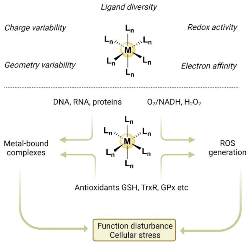 | ||
| Fig. 3 The unique properties of metal complexes and potential modes of action for ICD action. | ||
Ideally, an ICD inducer should activate multiple ICD-associated pathways to ensure robust and effective ICD effects. Metal-based complexes represent a class of compounds that have the potential for this capability owing to their diverse modes of action. These include not only their binding affinity to DNA but also to a multitude of proteins, which can effectively cause cellular stress and physiological disturbance. The capacity of metal-based agents to disturb intracellular redox balance stands out in the search quest for ICD inducers. This is because cellular stress induced by reactive oxygen species (ROS), especially ROS-mediated ER stress, is highly associated with ICD induction.75–77,82,182 Furthermore, beyond their direct cytotoxic effects, accumulating evidence supports the importance of certain metal-based agents in promoting ICD-driven antitumour immunity.183–186
To date, a wide variety of metal-based ICD inducers have been discovered with different metals, including platinum (Pt), iridium (Ir), gold (Au), ruthenium (Ru), copper (Cu), rhenium (Re), and manganese (Mn). Most of them can be classified as Type II ICD inducers. For ease of reference, we categorize these metal-based ICD inducers based on their metal centers, followed by their coordination chemistry. We further examine their efficacies and activities using reported in vitro and in vivo results and study their design strategies. We discuss their molecular targets and mechanisms of action, where applicable, and consider their potential application for cancer therapy.
3.1 Pt-based ICD inducers
 | ||
| Fig. 4 Molecular structures of reported Pt-based ICD inducers. Counter anions are omitted for clarity. | ||
| Trial title | Treatment | Indication | Phase | Status | Reference |
|---|---|---|---|---|---|
| OXP-based clinical trials | |||||
| Study of S 95005 in combination with oxaliplatin in metastatic colorectal cancer | OXP + trifluridine + bevacizumab + nivolumab | Metastatic colorectal cancer | I | Completed | NCT02848443 |
| Dendritic cell vaccine and chemotherapy for patients with pancreatic cancer (PancVax) | OXP + FA + irinotecan + 5-FU + PTX + gemcitabine + a DC-based vaccine | Pancreatic cancer | I | Terminated | NCT02548169 |
| Nivolumab (anti-PD1 antibody) and ipilimumab (anti-CTLA4 antibody) in combination with immunogenic chemotherapy for patients with advanced non-small cell lung cancer | OXP + nivolumab + ipilimumab | Advanced NSCLC | II | Active | NCT04043195 |
| Chemotherapy and immunotherapy as treatment for MSS metastatic colorectal cancer with high immune infiltrate (POCHI) | OXP + capecitabine + bevacizumab + pembrolizumab | Metastatic colorectal cancer | II | Recruiting | NCT04262687 |
| METIMMOX: colorectal cancer metastasis – shaping anti-tumour immunity by oxaliplatin (METIMMOX) | OXP + 5-FU + leucovorin + nivolumab | Metastatic colorectal cancer | II | Active | NCT03388190 |
| Safety and efficacy of pembrolizumab (MK-3475) in combination with TS-1 + CDDP or TS-1 + oxaliplatin as first line chemotherapy in gastric cancer (MK-3475-659/KEYNOTE-659) | OXP + pembrolizumab or CDDP + TS-1 | Gastric cancer | II | Completed | NCT03382600 |
| Rectal artery infusion chemotherapy of oxaliplatin plus capecitabine combined with anti-PD1 antibody after induction chemotherapy for microsatellite stable locally advanced rectal cancer: a prospective single-arm phase II study | OXP + rectectomy + capecitabine + anti-PD-1 monoclonal antibody | Advanced rectal cancer | II | Recruiting | NCT05307198 |
| Neoadjuvant arterial embolization chemotherapy combined PD-1 inhibitor for locally advanced rectal cancer (NECI) | OXP + capecitabine + tislelizumab | Rectal cancer | II | Recruiting | NCT05420584 |
| METIMMOX-2: metastatic pMMR/MSS colorectal cancer – shaping anti-tumour immunity by oxaliplatin (METIMMOX-2) | OXP + nivolumab | Metastatic colorectal cancer | II | Recruiting | NCT05504252 |
| Trial comparing two strategies of chemotherapy for metastatic colorectal cancer | OXP + 5-FU + irinotecan + leucovorin | Colorectal cancer | III | Completed | NCT00126256 |
![[thin space (1/6-em)]](https://www.rsc.org/images/entities/char_2009.gif) |
|||||
| PT-112-based clinical trials | |||||
| A phase 1/2a dose-finding study of PT-112 in patients with relapsed or refractory multiple myeloma | PT-112 | Multiple myeloma | I | Completed | NCT03288480 |
| PT-112 in subjects with thymoma and thymic carcinoma | PT-112 | Thymoma and thymic carcinoma | II | Recruiting | NCT05104736 |
| A study evaluating the safety, pharmacokinetics, and clinical effects of intravenously administered | PT-112 | Thymoma and thymic carcinoma; metastatic castrate-resistant prostate cancer | II | Active | NCT02266745 |
| PT-112 injection in subjects with advanced solid tumours and subsequent dose expansion cohorts | |||||
| An open-label phase I/II clinical trial of PT-112 injection for advanced solid tumours and advanced hepatocellular carcinoma | PT-112 | Hepatocellular carcinoma (HCC) | II | Unknown | NCT03439761 |
| An open-label phase I/II clinical study of PT-112 in combination with docetaxel in subjects with advanced solid tumour in a phase I dose escalation study and in subjects with non-small cell lung cancer (NSCLC) in a phase II dose confirmation study | PT-112 + docetaxel | Advanced solid tumours and NSCLC | II | Unknown | NCT02884479 |
| PT-112 (phosplatin's platinum) combined with gemcitabine injection for advanced solid tumours | PT-112 + gemcitabine | Biliary tract cancer | II | Unknown | NCT05357196 |
| A dose escalation and confirmation study of PT-112 in advanced solid tumours in combination with avelumab (PAVE-1) | PT-112 + anti-PD-L1 antibody (avelumab) | NSCLC | II | Completed | NCT03409458 |
The discovery of OXP as a potent ICD inducer fuels further research into optimizing OXP-based cancer chemo-immunotherapy with enhanced antitumour efficacy, developing novel Pt-based ICD inducers and understanding the underlying associated molecular mechanisms. For example, several OXP-based macromolecules combined with immune checkpoint inhibitors (ICIs) or other immuno-modulatory agents have been constructed with the potential to reverse immunosuppressive TME, minimizing undesirable adverse effects and boosting immunotherapeutic efficacy.191–199 Compared to OXP alone, these nanostructures generally exhibited higher cytotoxicity, lower side effects and superior antitumour immunity. One such example is the nano-FOLFOX delivery system that released [Pt(DACH)(H2O)2]2+, the active form of OXP, and folinic acid (FnA) to form OXP-FnA adducts that could inflict ICD and exhibit anticancer activity.200
Given the role of the 1R,2R-diaminocyclohexane (DACH) ligand in the activity of OXP, a few studies have explored the relationship between ligand structures and ICD-inducing activity. A study in 2012 suggested that adding methyl groups to DACH ligands (KP1537 and KP1691) could influence its side effects and ICD-inducing capacity.201 Interestingly, in contrast to OXP, which elicited ICD-driven antitumour immune response only in immunocompetent but not immunocompromised mice, KP1537 and KP1691 exhibited anticancer activity in both types of mice. Another study investigated the abilities of distinct diaminocycloalkanes chelating OXP analogues to induce ICD-associated DAMPs emission.202 Efforts to achieve the optimal structural modifications of the DACH ligand motif to yield new OXP analogs may be valuable for the development of new ICD inducers.
PT-112 is the subject of several ongoing clinical investigations in patients with solid tumours and hematologic malignancies, either as monotherapy (NCT02266745) or in combination therapy with PD-L1 inhibitor avelumab (NCT03409458). PT-112 was developed as a novel ICD inducer by replacing the oxalate ligand in OXP with a pyrophosphate group.203–205 Although PT-112 shares structural similarity with OXP as it retained the Pt-(DACH) pharmacophore (Fig. 4), the pyrophosphate moiety not only enhanced its pharmacokinetic and pharmacodynamic properties but also reduced its toxicity. PT-112 exerts its cytotoxicity on cancer cells through different mechanisms of action from traditional DNA-damaging Pt agents. In a murine breast carcinoma TSA cell model, PT-112 was compared with a well-established organic small molecule ICD inducer MTX and showed superior ability to stimulate immunostimulatory DAMP-accompanied ICD induction and the establishment of long-term immunologic anticancer memory in vivo.203 Impressively, in a vaccination model, PT-112-treated breast carcinoma TSA cells conferred 100% immunological protection against the subsequent injection of living TSA cells. Anti-tumour protection exhibited good durability when the mice were rechallenged after 60 days. PT-112 combined with ICBs achieved tumour elimination, enhanced cytotoxic T lymphocytes (CTLs) infiltration and reduced immunosuppressive CD25+FOXP3+ regulatory T-cells (Tregs) and tumour-associated macrophages (TAMs) in the TME. These results demonstrated that PT-112 exhibited remarkable therapeutic efficacy in eradicating tumour and establishing long-term antitumour immunity. Several Phase I/II trials for PT-112 as a monotherapy (NCT05104736, NCT02266745, NCT03288480, and NCT03439761) and in combination with the PD-L1 inhibitor avelumab (NCT03409458) in treating immunologically “cold” advanced metastatic castration-resistant prostate cancer, or with other chemotherapeutic agents, including docetaxel (NCT02884479) and gemcitabine (NCT05357196), have yielded promising results. Ongoing trials investigating PT-112-induced ICD for cancer treatment are shown in Table 1.
In addition to the cyclometallated Pt scaffold examples, other Pt(II)-carbene complexes have been explored for their capacities for evoking ICD in HCC. Liu group designed and evaluated a series of Pt(II)-carbene complexes derived from 4,5-diarylimidazole. One of 19 compounds, Pt1, triggered DAMPs emission and anti-HCC immune response.212 In a follow-up study, replacing iodide ligands with other halogen atoms did not affect the ICD-inducing capacity of the 4,5-diarylimidazole-based Pt(II)-NHC scaffold, indicating the importance of carbene ligands.213 Notably, the reported Pt-carbene complexes were Type II ICD inducers and triggered ROS-associated ER stress-driven ICD, suggesting the high relevance of carbene ligands in the development of novel Pt-based Type II ICD inducers.
As previously discussed, toll-like receptors (TLRs) are essential pattern recognition receptors on DCs and macrophages for recognizing ICD-associated DAMPs and initiating an immune response.136–138 Given the important role of TLRs, Wang group fabricated OXP-based Pt(IV) prodrug Pt5 conjugated with a TLR7 agonist.219 As expected, in addition to instigating CRT translocation and ATP secretion, Pt5 promotes DC activation in vitro characterized by enhanced secretion of proinflammatory cytokines IFN-γ, TNF-α, IL-6, and IL-12 and clearly increased percentages of intratumourally infiltrated CD8+ T-cells in vivo compared to OXP or the TLR7 agonist itself.
In contrast to activation by reductants in the aforementioned studies, a photoactivatable Pt(IV) prodrug complex Pt6 bearing coumarin axial ligands induced ICD upon photoirradiation.220Pt6 exhibited superior phototoxicity towards multiple cell lines, including two CDDP-resistant ones. Upon photoactivation of Pt6, distinct ICD biomarkers were observed in the treated A549cisR cells. In contrast, no detectable ICD effects were observed upon Pt6 treatment in the dark. Pt6-treated A549cisR cells largely promoted T-cell proliferation in mixed leukocyte reactions. Another photoactivatable Pt(IV)-azido prodrug Pt7 was capable of inducing ROS and reactive nitrogen species (RNS) production and, simultaneously, releasing cytotoxic Pt(II) species.221 Under blue light irradiation, Pt7 induces autophagic cell death accompanying 3 characteristic ICD signatures and promotes phagocytosis of Pt-treated CT26 carcinoma cells by J774.A1 macrophages as well.
3.2 Ir-based ICD inducers
 | ||
| Fig. 5 Molecular structures of reported Ir-based ICD inducers. Counter anions are omitted for clarity. | ||
Recently, Liang et al. reported cyclometalated Ir(III) complex Ir4 based on isoquinoline alkaloid-induced autophagy-dependent ferroptosis and ICD response in triple-negative breast cancer (TNBC) cells, thereby triggering the emission of DAMPs.225Ir4 exerted its cytotoxic effect via the generation of ROS that induced ferroptosis and downregulation of indoleamine 2,3-dioxygenase (IDO), an immunosuppressive enzyme. It also activated CD8+ T-cells and reduced regulatory T-cells (Tregs). Ir4 showed superior efficacy compared to traditional chemotherapy agents, such as OXP. Similar to Ir2, the combination of Ir4 with anti-PD1 therapy significantly improved tumour inhibition.
A recent study showcased two Ir(III) complexes, Ir5a and Ir5b, as effective inducers when they were delivered to the ER using a liposome-based encapsulation strategy.226 Liposomal encapsulation greatly enhanced the cellular uptake of Ir5a and Ir5b, which preferentially accumulated in the ER and triggered oxidative stress and apoptotic ICD. Without facilitated delivery, Ir5a and Ir5b alone exhibited weak cytotoxicity and CRT exposure owing to low cellular uptake efficiencies. Despite the further structural optimization required, this study demonstrated the versatility of carrier-aided strategies to enhance ICD effects.
Another study by Zou group reported an ER stress-inducing cyclometalated Ir(III)-bis NHC complex (Ir6) that can elicit ICD hallmarks both in vitro and in vivo using the vaccination model.227 The innovative use of a specially designed clickable photoaffinity probe showed that Ir6 could directly bind with and subsequently inhibit BiP, a key regulator of the UPR pathway that functions as a protein chaperone, aiding in protein proper folding and assembly.228 This work was significant as it was the first time that the molecular target of an ICD inducer was systematically uncovered using chemical biology approaches.
Ir-pbt-Bpa Ir8 was developed for two-photon excitation photodynamic immunotherapy by replacing the ancillary ligand 2-phenylpyridine with 2-phenylbenzo[d]-thiazole.232 This modification enhanced two-photon absorption, increased ROS production, and shifted the primary subcellular target from the ER to the mitochondria, leading to cell death in melanoma cells via ferroptosis. This stress response was enhanced by Ca2+ release from the ER, resulting in significant detection of ICD biomarkers and a significant reduction of both primary and distant melanoma tumours, even though only the primary tumour was directly treated. Histological examinations showed enhanced DC maturation and inhibition of tumour immunosuppression, as indicated by a favorable CD8+/Foxp3+ ratio. This study highlighted the significance of ligand modification in influencing their subcellular localization and biological activity, and the importance of targeting other organelles such as mitochondria, in addition to ER, for inducing ICD.
The incorporation of the carbonic anhydrase IX (CAIX)-targeting group into phenylpyridine-, difluorophenylpyridine-, and phenylquinoline-based Ir(III) complexes was investigated as an approach for the treatment of HT29 colon cancer cells via PDT.233 Upon irradiation with light (λex at 425 nm), phenylquinoline-based Ir9 induced pyroptosis under hypoxic conditions. Ir9 targeted CAIX, an enzyme highly expressed in hypoxic tumours, leading to its degradation. This, in turn, downregulated the expression of hypoxia-inducible factor 1α (HIF-1α) levels and vascular endothelial growth factor (VEGF) expression, improving the cancer immune microenvironment.
Another Ir(III) photosensitizer Ir10 with a phenanthroline ligand modified with a hydrophobic long-chain ER targeting N-phenethylsuccinamide moiety generated ROS upon irradiation in oral squamous cell carcinoma (OSCC), which elicited ER stress, leading to ICD and an upregulation of PD-L1 expression.234 The combination with PD-L1 inhibitor was particularly effective in converting “cold” tumours (with low immune activity) into “hot” tumours (with high immune activity) in vivo. The combination significantly upregulated the level of mature DCs (MHC II+ and CD80+CD86+ DCs), T-cell infiltration (CD4+ and CD8+) and cytokines (TNF-α and IFN-γ) while downregulating immunosuppressive inflammatory cytokine IL-6, signifying transformation into “hot tumour”.
A cyclopentadienyl Ir(III) complex with natural product betulin, Ir13, activated the ferroptosis cascade through ferritinophagy and iron homeostasis regulation.237Ir13 activated PERK/eIF2α pathway and CRT exposure, and HMGB1 and ATP release were detected in vitro in A549 cancer cells. RNA sequence analysis indicated that ferroptosis and nuclear factor kappa light chain enhancer of activated B cells (NF-κB) activation further amplified the antitumour effect. The in vivo vaccination model showed that Ir13 inhibited tumour growth and upregulated the expression of proinflammatory cytokines and cytotoxic T-cells to stimulate a robust immune response.
3.3 Au-based ICD inducers
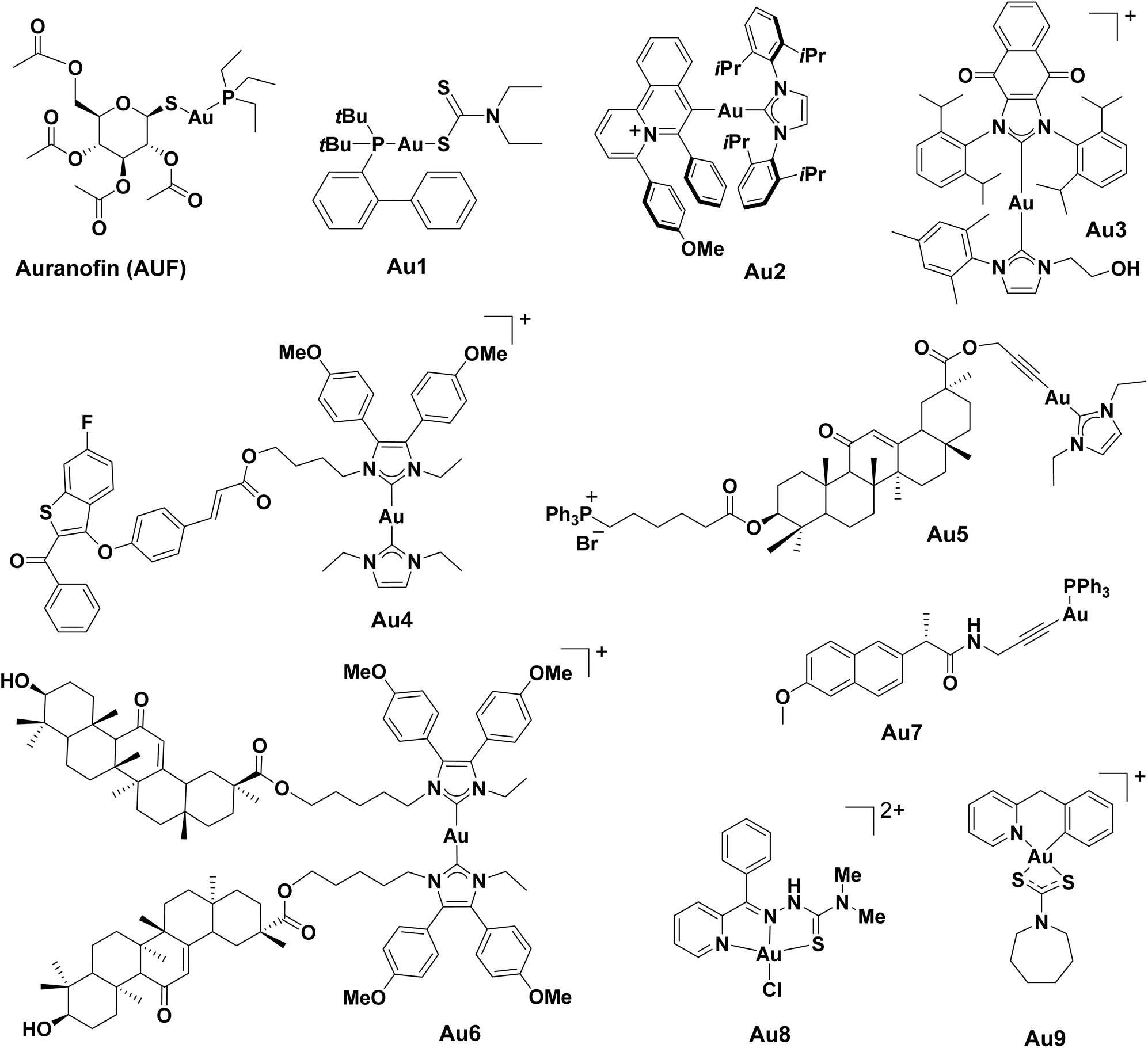 | ||
| Fig. 6 Molecular structures of reported Au-based ICD inducers. Counter anions are omitted for clarity. | ||
Boullosa et al. reported that the FDA-approved auranofin (AUF) triggered ICD by inducing both apoptosis and ferroptosis in mutant p53 NSCLC cells in vitro.240 The treatment of AUF significantly induced DAMPs emission, and the co-culture of AUF-treated cancer cells with immature DCs led to their maturation. AUF further improved the innate immune response, as evidenced by the enhanced killing of cancer cells when they were co-cultured with natural killer cells. The group also reported that the combination of AUF and cold atmospheric plasma-treated PBS resulted in a synergistic ICD response in the glioblastoma cell culture.
A rationally designed redox-active Au(I)-NHC complex Au3 exhibited potent ICD induction efficacy in vitro and in vivo.242 The authors postulated that dual targeting of the cancer antioxidant network through TrxR inhibition by the redox-active Au(I)-NHC motif and redox cycling via the embedded naphthoquinone moiety could increase ROS generation and ER stress to promote ICD induction. In a vaccination model using mice inoculated with treated CT26 cells, low dose Au3 (10 μM) demonstrated a significantly higher percentage of tumour-free mice compared to high dose OXP (150 μM), even after extended periods of recovery post-challenge (42 days).
Moreover, several Au(I) complexes targeting the TrxR-ROS-ERS-ICD axis were reported by Liu and co-workers. For example, the in vitro ICD effects of Au(I)-NHC complex incorporating selective estrogen receptor degrader (SERD) moiety (G1T48) Au4 were studied in human MCF7 cells.243 The Au(I)-NHC moiety in Au4 was designed to inhibit TrxR activity, which consequently triggered ROS generation and ICD-associated DMAP emission, including CRT exposure, HMGB1 release, and ATP secretion. In addition, an NHC-Au(I) complex with liver-targeting scaffold 18β-glycyrrhetinic acid and mitochondria-directing triphenyl-phosphonium group (TPP+) Au5 was discovered to simultaneously induce both ICD and cGAS-STING pathways to trigger an immune response.244In vivo vaccination studies with Au5 produced a stable population of 50% tumour-free mice after 30 days. Another Au(I)-NHC with the same 18β-glycyrrhetinic acid ligand Au6 displayed a significant emission of DAMPs in Hepa1-6 cells after treatment.245 In particular, the Au6-treated cells saw no tumour growth for 30 days in an in vivo vaccination model. The treatment also increased the number of CD8+ T-cells and CD4+ T-cells by 3.9-fold and 5.6-fold, respectively.
Alkynyl ligands are widely used to stabilize Au(I) complexes owing to their strong electron donating abilities. Besides Au6, Liu and co-workers also designed a series of Au(I)-alkynyl complexes conjugated to nonsteroidal anti-inflammatory drugs (NSAID) with the aim of inhibiting TrxR activity and disrupting redox balance.246 Amongst these 7 Au(I)-NSAID complexes, naproxen-containing Au7 triggered oxidative stress and ICD-associated DAMPs emission in human A2780 cells and elicited a more effective immune response, inducing the downregulation of cyclooxygenase-2 (COX-2) and PD-L1, DC maturation and increased infiltration of CTLs.
Babak, Berger and Ang et al. utilized novel Au(III)-thiocarbamate scaffolds to develop ICD inducers with superior efficacy and can reverse immunosuppressive TME. By applying a combinatorial coordination chemistry approach, a library of 35 cyclometalated Au(III)-thiocarbamate complexes was constructed, and their ability to inflict ICD effects was assessed in a malignant pleural mesothelioma (MPM) cell model.248 A systematic structure–activity relationship study revealed that the cyclometalated scaffold and the overall lipophilicity of the complexes are crucial for the phagocytosis of immunologically “cold” MPM cells upon treatment. A bona fide Au(III)-based inducer Au9 was successfully identified from the library, as evidenced by a robust antitumour immune response against MPM in immunocompetent mice. Protective antitumour immunity was observed for more than 6 months in these mice, demonstrating the viability of this Au(III) scaffold as a discovery platform for ICD inducers.
3.4 Ru-based ICD inducers
 | ||
| Fig. 7 Molecular structures of reported Ru-based ICD inducers. Counter anions are omitted for clarity. | ||
Another study described the ability of Ru(II)-polypyridyl complex Ru2 in increasing ecto-CRT in mice although other ICD hallmarks were not measured.262 Intriguingly, the combination of Ru2 and natural killer (NK) cells led to the surprising finding that this combination treatment could foster NK cell infiltration, potentiate NK cell immunotherapy, and improve therapeutic efficacy against breast tumour in vivo. This study highlighted the immunoregulatory effects of Ru2 as a potential ICD inducer and provided an innovative angle for applying ICD inducers to augment immunotherapy.
Chao and co-workers designed 3 cyclometalated Ru(II) complexes and found that Ru3 targeted mitochondria and nucleus, leading to oncosis accompanied by ICD induction.153 Its mechanism of action involved DNA damage causing activation of polyADP-ribose polymerase 1 (PARP1), associated ATP depletion and porimin activation, as well as concurrent mitochondria damage and ER stress. More importantly, macrophage M1 polarization was observed, indicating the activation of innate immune response on top of adaptive T-cell response. However, the limited solubility and bioavailability of Ru3 necessitated an encapsulation approach in vivo.
3.5 Other metal-based ICD inducers
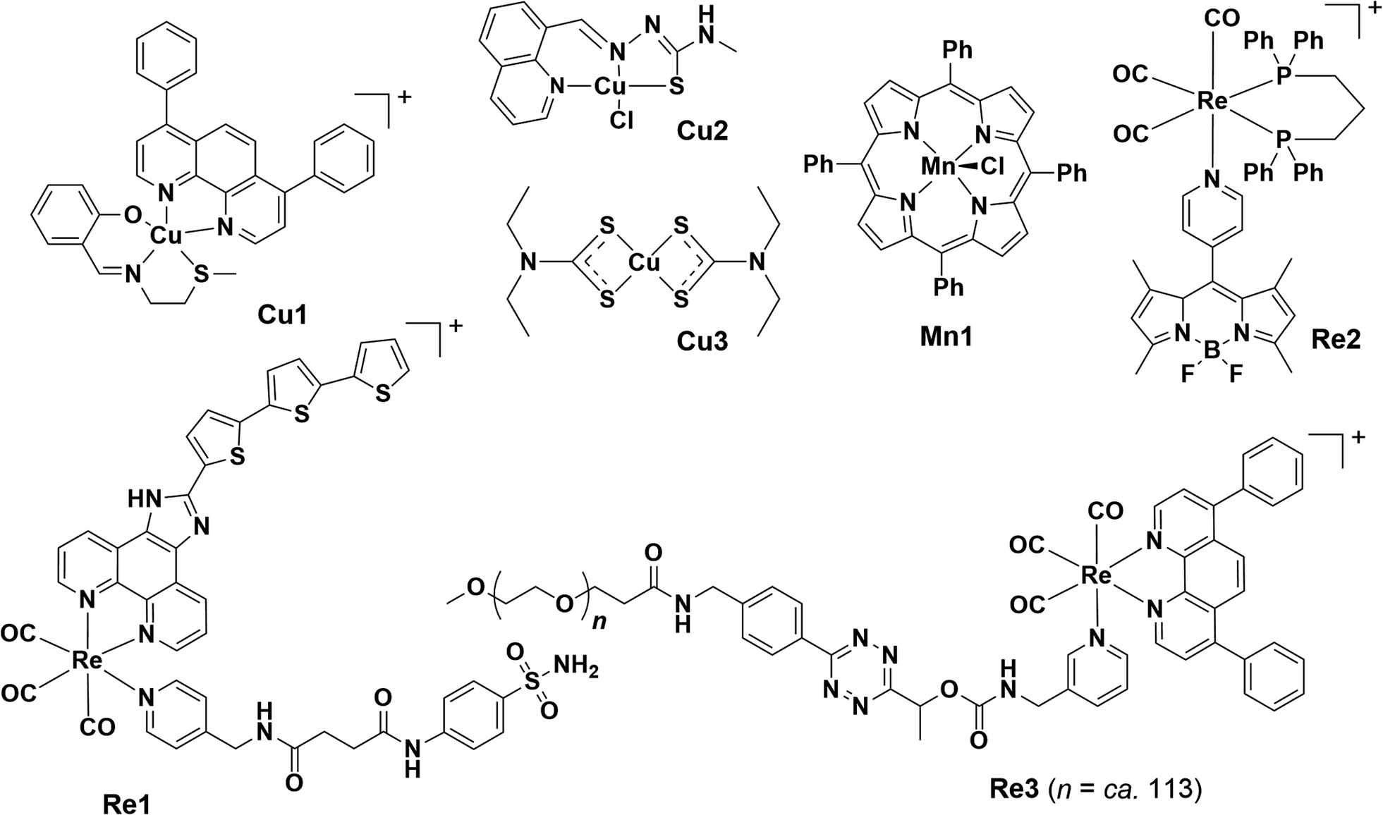 | ||
| Fig. 8 Molecular structures of reported Re, Cu and Mn-based ICD inducers. Counter anions are omitted for clarity. | ||
Tan et al. presented a theranostic Re(I) complex Re2 appended with 4,4-difluoroboradiazaindacene (BODIPY) moiety, which was used for viscosity measuring and imaging.269Re2 was preferentially localized in the ER, causing ER stress and, eventually, necrosis, and could be simultaneously used to monitor ER viscosity. Re2 was described as a Type II ICD inducer based on its ability to induce the 3 classical ICD biomarkers.
A Re(I) aminomethylpyridine complex Re3, modified with a cleavable tetrazine moiety which could be triggered by trans-cyclooct-4-enol (TCO-OH), underwent a click-to-release reaction, giving rise to a more cytotoxic Re(I) ICD inducer.270 In the presence of TCO-OH and light, the released Re(I) compound led to substantial ROS generation, lysosome rupture, autophagy inhibition, necrosis, and ICD induction with accompanying biomarkers. Notably, ATP secretion was observed even when autophagosome formation was blocked in the process, in contrast to a previous claim that ATP secretion in ICD relied on the autophagic process. Remarkable increases in the proportion of mature DCs (CD80+CD86+) and cytotoxic T-cells (CD3+CD8+), and the expression level of TNF-α were observed in a co-incubation of treated-MDA-MB-231 with hPBMCs, further proving its ability of immune activation.
A Re(I) photosensitizer Re4, constructed by coordinating [Re(CO)3]+ to g-C3N4 nanosheets (Re(I)–g-C3N4), was demonstrated as a Type II ICD inducer with ER-specific accumulation.271 Upon two-photon excitation, Re4 triggered robust ROS (˙O2− and ˙OH)-driven ER stress, with different cell death modes, including apoptosis, ferroptosis, and pyroptosis, and most importantly, ICD-related DAMPs emission. Activation of antitumour immune responses together with inhibited growth of primary and secondary distant tumour in mice was observed.
Another recent study also showcased the superior ICD inducing capacity of Cu(II) complexes which can be attributed to their redox activities.274Cu2 depleted GSH forming monovalent Cu+ species that catalyzed ˙OH production via Fenton-like reaction. Replacing the center metal with Co, Pt or Pd resulted in the loss of cytotoxicity. Cu2-induced ICD was ferroptosis-dependent and enabled significant tumour growth prevention and effective antitumour immune response (increased CD8+ T-cell infiltration and decreased Foxp3+ T-cells) in vaccinated c57BL/6 mice challenged with colorectal cancer. Importantly, Cu2 exhibited cytotoxic specificity towards cancer cells only.
Another form of Cu-based ICD inducer is the combination of CuCl2 with an anti-alcoholism drug disulfiram (DSF/Cu).169,275–279 DSF was repurposed as an anticancer agent that readily formed active metabolite Cu-diethyldithiocarbamate complex Cu3, significantly enhancing the anti-tumour effects of DSF.279–281 DSF/Cu treatment was found to induce potent ICD in multiple cancers and ICD-based immune response against primary and rechallenged tumours.169,275,277–279 The ICD evoked by DSF/Cu was associated with cuproptosis, a newly characterized cell death characterized by the accumulation of Cu in mitochondria.282,283 Multiple lines of evidence showed that cuproptosis elicited ICD and enhanced the immunogenicity of dying tumour cells.284–286 In addition, DSF/Cu treatment drove the reprogramming and reversal of the immunosuppressive TME in humanized mice.169,281 It could be used in combination with αPD-L1 to enhance cancer immunotherapy276 and trigger radiation therapy-induced ICD when combined with radiation and chemotherapeutic agents.169,277,278 Some DSF/Cu combination therapies were investigated in clinical trials, such as NCT02671890 (Phase I, solid tumours and pancreatic cancer), NCT02715609 (Phase I/II, glioblastoma), and NCT02678975 (Phase II/III, glioblastoma).
4. Current challenges and limitations
4.1 Understanding the molecular targets
Genome-wide CRISPR screening, RNA interference (RNAi), and multi-omic techniques (e.g. genomics, transcriptomics, proteomics and metabolomics) have been frequently used to investigate targets of metallodrugs.227,264,288–294 However, there are only few studies on target identification and validation for ICD complexes. A noteworthy example reported by Zou and co-workers disclosed binding immunoglobulin protein (i.e. Bip/GRP78), an abundant ER chaperone regulating protein homeostasis in the ER, as a potential therapeutic target in ICD induction by photoaffinity-based target profiling.227KP1339, OXP and a cyclometalated Ir(III)-bisNHC complex Ir6 were found to interact with BiP, as evidenced by shifted Tm values in cellular thermal shift assay (CETSA). Apart from Bip, the functional proteins in the ER that could be promising targets for ICD inducers remains elusive.Compounds with different metals will likely have different molecular targets and MOA, while ROS generation and ER stress are believed to be strongly associated with ICD induction.75–77,82,182 Multiple targets might be implicated in the continuous generation of ROS and ER stress provoked by metal complexes. The mystery of the molecular targets of respective metal-based ICD inducers and the relationship between their targets and ROS-driven ER stress-based ICD remains an intriguing topic in this field.
Meanwhile, because of the lack of understanding of the respective molecular targets in the induction pathway of ICD, the rational design of potent metallic ICD inducers based on structural optimization is challenging. Thus, the target profiling of respective metallo molecules is highly demanding and of great significance in accelerating the understanding of mechanistic mysteries in ICD. Notably, ICD inducers that simultaneously target multiple pathways to provoke ER stress can potentiate ICD effects. This suggests that the design of novel ICD inducers with multiple targets could be reasonable. Overall, unraveling the molecular targets of metal-based ICD inducers to aid in the rational design of more effective and potent ICD inducers is needed to bridge this research gap.
4.2 Comprehensive structure–activity relationship (SAR) studies
Most of the development approaches for metallo-ICD inducers rely on screening and only few studies have attempted a systematic investigation of the effects of structural changes on their ICD-inducing capacity (e.g.Pt-NHC and Au-NHC).207,209,241 The scarcity of SAR studies occurs for several reasons. First, identification of a potential ICD inducer in vitro requires successful detection of multiple DAMPs as well as effective activation of immune cells, but related experimental procedures are highly laborious and resource-intensive. Second, to ensure robust ICD induction, it is necessary to monitor DAMPs at different time points and several drug concentrations.26,34,89 Despite the establishment of transgenic screening platforms, screening for large compound libraries at different concentrations with different treatment durations is challenging in practice.149 Third, building up a reliable screening platform by genetic manipulation is not easy. Finally, owing to the complexity of immune regulatory pathways,87 overall immunogenicity derived from ICD and the degree of activated antitumour immunity in vivo varies depending on the immunostimulatory and immunoinhibitory DAMPs balance, which complicates analyses. For example, gemcitabine triggers immunostimulatory DAMPs emission but also concurrently promotes the release of prostaglandin E2 as an immunoinhibitory signal, thus failing to provoke an effective antitumour immune response in vivo.295 An integrated screening platform that enables high throughput and systematic evaluation of ICD candidates would accelerate the drug discovery process and facilitate more robust mechanistic investigations, shedding light on their unique SAR.4.3 Better cell and animal test models for ICD
In the design and screening of ICD inducers, the type of cell line models used is also crucial. Certain cell lines overexpressing ATP-hydrolyzing enzymes (i.e. CD39 and CD73) are likely to exhibit compromised immunostimulatory activity of DAMPs.122,296 This is because CD39 and CD73 reduce extracellular ATP by converting ATP to adenosine monophosphate (AMP) and adenosine, respectively.296,297 The accumulation of immunosuppressive extracellular adenosine, together with the decrease in the level of chemotactic and immunostimulatory ATP, weakens antitumour immunity.296 As ATP release is a pivotal marker of ICD, the activity of these enzymes might compromise the outcomes of ICD induction. Depending on whether the focus is on enhancing immune activation or studying immune evasion, it is key to consider the type of cell line carefully when designing the model. Next, human and mouse cell lines may exhibit differing responses to ICD inducers. A comprehensive ICD study should include both human and murine cell lines to compare the immunogenic response or ICD markers and ensure translational relevance because the ultimate goal of ICD research is to develop effective treatments for human patients in clinical settings.Beyond in vitro assays, ICD should be validated in vivo to assess the induction of an immune response.162,163,295 However, validation can only be conducted in animal models with murine cell lines, as human cancer cells are intrinsically incompatible with in vivo immunological studies. Although attempts are being made to allow proper evaluation of ICD in the human system, the current state-of-the-art approach to identifying bona fide ICD inducers is through vaccination assays with immunocompetent syngeneic mice. Numerous studies despite showing promising in vitro results often validate through subcutaneous tumour models,219,223,234,243,244,246,247,262,298 where the primary focus is on analyzing the tumour growth curve to infer immunogenicity. Although informative, this approach may not fully capture the complexity of the immune response induced by ICD inducers. We therefore encourage researchers to standardize their validation processes by utilizing vaccination assays in syngeneic models because this will improve the reliability of ICD studies and better guide the development of novel ICD inducers.
5. Conclusion and future perspectives
This review highlights the significant advancements that have been made in the investigation, development and understanding of metal complexes for ICD in the past 2 decades since the ICD phenomenon was originally discovered. Given the strong interest in this field of research in recent years, this trend is expected to continue. Moving forward, we anticipate that the focus of effort will be channeled towards rationalizing the design of ICD complexes and developing specific clinical applications.One strategy to rationalize the design of ICD complexes is to consider the indispensable role of continuous ER stress in ICD initiation and, hence, to reinforce ER stress precisely via an ER targeting manner.65,84–86,226,299–302 In response to stressors, cancer cells initiate UPR to relieve stress and maintain ER homeostasis. Stressors can target and remain in the ER, invoke persistent ER stress and counteract stress relief. Multiple lines of evidence have shown the effectiveness of this method by modifying the ligand environment of metal compounds to ensure their accumulation in the ER86,222,234,299,303 or constructing ER targeting delivery systems to direct them into the ER.65,85,226,301,302
The effectiveness of ICD in potentiating antitumour immunity depends on tumour immunogenicity and the host immune system. Another strategy is to develop complexes that act on immune cells, such as DCs and T-cells, and facilitate their detection, recognition and interaction of cancer antigens, which can boost ICD-primed immune response. Such functional molecules, aptly called “ICD enhancers” by Kroemer and co-workers, include hexokinase-2 inhibitors (immunometabolic modifiers) and ligands of pattern recognition receptor 3 (TLR3).304 Overall, the augmentation of ICD effects can be achieved by amplifying ICD in cancer cells or enhancing the perception of ICD by immune cells, suggesting promising ways to augment ICD and improve ICD-based therapies.
ICD-primed antitumour immune responses were also observed in the context of non-apoptotic RCD, such as ferroptosis,305–307 necroptosis,308,309 cuproptosis,285,286 and pyroptosis.303,310,311 These new RCDs could provide the basis and inspiration for the design of new ICD complexes. For example, cancer cells undergoing glutathione peroxidase 4 inhibition-induced ferroptosis were found to be immunogenic and can elicit robust antitumour immunity in vivo.305,306 Necroptotic cancer cells generated by genetic manipulation emit DAMPs, promote DC maturation and cross-priming of CTLs, and trigger an antigen-specific antitumour immune response.308 Pyroptosis is viewed as an ICD modality characterized by DAMPs emission that can enhance antitumour immune responses. Despite the poor direct killing of cancer cells, the pyroptotic cell death activator (i.e. GSDMD agonist) is an effective booster when synergized with other cancer immunotherapy.310 Cuproptosis, a newly described cell death modality, occurs owing to the overload of Cu in mitochondria and has been linked to trigger ICD effects.285,286 Altogether, these studies highlight the significance of non-apoptotic RCD in ICD initiation and suggest a promising way to discover ICD inducers that trigger immunogenic non-apoptotic cell death. Although agents that lead to non-apoptotic RCD may not guarantee the discovery of bona fide ICD inducers, they are more likely to yield a successful ICD induction and effective antitumour immunity.303,312–314
Regarding clinical applications, a promising avenue for ICD inducers may lie in complementing existing T-cell-based immunotherapy specifically targeting cold tumours.45,189,190,196,248,315–318 Cold tumours, also known as immune-excluded tumours, are characterized generally by a low expression of PD-L1 and lack of tumour-infiltrating lymphocytes, and are thus poorly responsive to T-cell-based therapies, such as immune checkpoint inhibitors.319 This represents a significant gap in cancer immunotherapy. ICD is one of the therapeutic strategies that can promote T-cell priming in “cold” tumours. ICD inducers can change TME from a “cold” to “hot” immune status by enhancing the immunogenicity of the dead cells through the production of neoantigens, DAMPs and cytokines to recruit and activate APCs and effector T-cells (CD4+ and CD8+).45,196,203,204,315,318,319 The efficient release of antigen, along with antigen processing and presentation, can improve T-cell priming. Thus, this enables T-cells to be available in the tumour for future cancer cell elimination.
One example of ICD inducers described previously, PT-112, is effective against “cold” tumour.203–205 Its combination with ICIs such as CTLA4, PD1 and PD-L1 blockers has shown a synergistic effect and can induce a more potent immune response compared to either therapy alone in vivo. The study also shows that PT-112 favored the establishment of an immunostimulatory tumour microenvironment. Collectively, the combination of PT-112 with immune checkpoint inhibitors suggests a promising immunotherapy with improved clinical safety and efficacy for overcoming “cold” tumour. Besides PT-112, the cyclometalated Au(III) complex Au9 has demonstrated the ability to boost the immune response against MPM cells, which can be classified as immunologically “cold tumours” owing to poor responses to the immune checkpoint blockade combination treatment. In a preclinical study involving vaccinated mice, Au9 extended the tumour-free survival period to 5–7 months. Although lacking clinical investigation, this example highlights the potential of ICD inducers in targeting cold tumours that are less receptive to immunotherapies and hence are limited to chemotherapy. Apart from PT-112 and Au9, a few metallic ICD inducers have been shown to upregulate the level of PD-L1 in cancer cell lines that are classified as “cold” tumour in recent years.198,199,223,225,234,246 The combination of these complexes and PD-1 displayed a synergistic effect in vivo, with the combination significantly upregulating the level of mature DCs, T-cell infiltration and cytokines while downregulating immunosuppressive inflammatory cytokines, signifying the transformation into “hot” tumour.
These studies underscore the potential of ICD inducers as promising agents for the treatment of cancers that display limited sensitivity to ICIs. Thus, enhancing the immunogenicity with ICD inducers while simultaneously reducing immunosuppression through ICIs offers a promising approach to convert the TME from an immunosuppressive “cold” to an immunostimulatory “hot” environment that can generate a robust immune response.
Data availability
No primary research results, software or code have been included and no new data were generated or analysed as part of this review.Author contributions
Jiao Xia Zou – original draft, figures and table, data procuring, editing, citing, formatting; Meng Rui Chang – original draft, data procuring: Nikita A. Kuznetsov – writing; Jia Xuan Kee – writing, review & editing. Prof. Wee Han Ang and Prof. Maria V. Babak: writing, formatting, review & editing.Conflicts of interest
There are no conflicts to declare.Acknowledgements
W. H. A. acknowledges financial support from the Singapore Ministry of Education (A-8002492-00-00). M. V. B. acknowledges financial support from the Pneumoconiosis Compensation Fund Board of Hong Kong (Project No. 9211315). The authors thank Clemen Yu Jie Ong (National University of Singapore) and Ho-Jung Choe (City University of Hong Kong) for their assistance in preparing the figures and graphics.References
- P. Sharma, S. Goswami, D. Raychaudhuri, B. A. Siddiqui, P. Singh, A. Nagarajan, J. Liu, S. K. Subudhi, C. Poon, K. L. Gant, S. M. Herbrich, S. Anandhan, S. Islam, M. Amit, G. Anandappa and J. P. Allison, Cell, 2023, 186, 1652 CrossRef CAS.
- M. Morotti, A. Albukhari, A. Alsaadi, M. Artibani, J. D. Brenton, S. M. Curbishley, T. Dong, M. L. Dustin, Z. Hu, N. McGranahan, M. L. Miller, L. Santana-Gonzalez, L. W. Seymour, T. Shi, P. Van Loo, C. Yau, H. White, N. Wietek, D. N. Church, D. C. Wedge and A. A. Ahmed, Br. J. Cancer, 2021, 124, 1759 CrossRef.
- C. M. Southam, A. Brunschwig, A. G. Levin and Q. S. Dizon, Cancer, 1966, 19, 1743 CrossRef CAS PubMed.
- A. D. Waldman, J. M. Fritz and M. J. Lenardo, Nat. Rev. Immunol., 2020, 20, 651 Search PubMed.
- C. Guo, M. H. Manjili, J. R. Subjeck, D. Sarkar, P. B. Fisher and X. Y. Wang, Adv. Cancer Res., 2013, 119, 421 CrossRef CAS PubMed.
- P. Dobosz and T. Dzieciatkowski, Front. Immunol., 2019, 10, 2965 CrossRef CAS PubMed.
- S. J. Oiseth and M. S. Aziz, J. Cancer Metastasis Treat., 2017, 3, 250 CrossRef CAS.
- E. F. McCarthy, Iowa Orthop. J., 2006, 26, 154 Search PubMed.
- J. Couzin-Frankel, Science, 2013, 342, 1432 CrossRef CAS PubMed.
- A. Haslam and V. Prasad, JAMA Netw. Open, 2019, 2, e192535 CrossRef.
- L. B. Alexandrov, S. Nik-Zainal, D. C. Wedge, S. A. Aparicio, S. Behjati, A. V. Biankin, G. R. Bignell, N. Bolli, A. Borg, A. L. Borresen-Dale, S. Boyault, B. Burkhardt, A. P. Butler, C. Caldas, H. R. Davies, C. Desmedt, R. Eils, J. E. Eyfjord, J. A. Foekens, M. Greaves, F. Hosoda, B. Hutter, T. Ilicic, S. Imbeaud, M. Imielinski, N. Jager, D. T. Jones, D. Jones, S. Knappskog, M. Kool, S. R. Lakhani, C. Lopez-Otin, S. Martin, N. C. Munshi, H. Nakamura, P. A. Northcott, M. Pajic, E. Papaemmanuil, A. Paradiso, J. V. Pearson, X. S. Puente, K. Raine, M. Ramakrishna, A. L. Richardson, J. Richter, P. Rosenstiel, M. Schlesner, T. N. Schumacher, P. N. Span, J. W. Teague, Y. Totoki, A. N. Tutt, R. Valdes-Mas, M. M. van Buuren, L. van't Veer, A. Vincent-Salomon, N. Waddell, L. R. Yates, I. Australian Pancreatic Cancer Genome, I. B. C. Consortium, I. M.-S. Consortium, I. PedBrain, J. Zucman-Rossi, P. A. Futreal, U. McDermott, P. Lichter, M. Meyerson, S. M. Grimmond, R. Siebert, E. Campo, T. Shibata, S. M. Pfister, P. J. Campbell and M. R. Stratton, Nature, 2013, 500, 415 CrossRef CAS PubMed.
- S. Bagchi, R. Yuan and E. G. Engleman, Annu. Rev. Pathol., 2021, 16, 223 CrossRef CAS PubMed.
- C. Kandoth, M. D. McLellan, F. Vandin, K. Ye, B. Niu, C. Lu, M. Xie, Q. Zhang, J. F. McMichael, M. A. Wyczalkowski, M. D. M. Leiserson, C. A. Miller, J. S. Welch, M. J. Walter, M. C. Wendl, T. J. Ley, R. K. Wilson, B. J. Raphael and L. Ding, Nature, 2013, 502, 333 CrossRef CAS PubMed.
- H. Nishikawa and S. Sakaguchi, Curr. Opin. Immunol., 2014, 27, 1 CrossRef CAS PubMed.
- M. J. Smyth, G. P. Dunn and R. D. Schreiber, Adv. Immunol., 2006, 90, 1 CAS.
- S. Spranger and T. F. Gajewski, Annu. Rev. Cancer Biol., 2018, 2, 213 CrossRef.
- S. Tang, Q. Ning, L. Yang, Z. Mo and S. Tang, Int. Immunopharmacol., 2020, 86, 106700 CrossRef CAS PubMed.
- M. D. Vesely, M. H. Kershaw, R. D. Schreiber and M. J. Smyth, Annu. Rev. Immunol., 2011, 29, 235 CrossRef CAS PubMed.
- P. S. Hegde and D. S. Chen, Immunity, 2020, 52, 17 CrossRef CAS PubMed.
- D. S. Chen and I. Mellman, Immunity, 2013, 39, 1 CrossRef CAS PubMed.
- J. Galon and D. Bruni, Nat. Rev. Drug Discovery, 2019, 18, 197 CrossRef CAS PubMed.
- M. Pickup, S. Novitskiy and H. L. Moses, Nat. Rev. Cancer, 2013, 13, 788 CrossRef CAS PubMed.
- G. Willimsky, M. Czeh, C. Loddenkemper, J. Gellermann, K. Schmidt, P. Wust, H. Stein and T. Blankenstein, J. Exp. Med., 2008, 205, 1687 CrossRef CAS PubMed.
- S. T. Workenhe, J. Pol and G. Kroemer, Oncoimmunology, 2021, 10, 1893466 CrossRef PubMed.
- L. Galluzzi, I. Vitale, S. A. Aaronson, J. M. Abrams, D. Adam, P. Agostinis, E. S. Alnemri, L. Altucci, I. Amelio, D. W. Andrews, M. Annicchiarico-Petruzzelli, A. V. Antonov, E. Arama, E. H. Baehrecke, N. A. Barlev, N. G. Bazan, F. Bernassola, M. J. M. Bertrand, K. Bianchi, M. V. Blagosklonny, K. Blomgren, C. Borner, P. Boya, C. Brenner, M. Campanella, E. Candi, D. Carmona-Gutierrez, F. Cecconi, F. K. Chan, N. S. Chandel, E. H. Cheng, J. E. Chipuk, J. A. Cidlowski, A. Ciechanover, G. M. Cohen, M. Conrad, J. R. Cubillos-Ruiz, P. E. Czabotar, V. D'Angiolella, T. M. Dawson, V. L. Dawson, V. De Laurenzi, R. De Maria, K. M. Debatin, R. J. DeBerardinis, M. Deshmukh, N. Di Daniele, F. Di Virgilio, V. M. Dixit, S. J. Dixon, C. S. Duckett, B. D. Dynlacht, W. S. El-Deiry, J. W. Elrod, G. M. Fimia, S. Fulda, A. J. Garcia-Saez, A. D. Garg, C. Garrido, E. Gavathiotis, P. Golstein, E. Gottlieb, D. R. Green, L. A. Greene, H. Gronemeyer, A. Gross, G. Hajnoczky, J. M. Hardwick, I. S. Harris, M. O. Hengartner, C. Hetz, H. Ichijo, M. Jaattela, B. Joseph, P. J. Jost, P. P. Juin, W. J. Kaiser, M. Karin, T. Kaufmann, O. Kepp, A. Kimchi, R. N. Kitsis, D. J. Klionsky, R. A. Knight, S. Kumar, S. W. Lee, J. J. Lemasters, B. Levine, A. Linkermann, S. A. Lipton, R. A. Lockshin, C. Lopez-Otin, S. W. Lowe, T. Luedde, E. Lugli, M. MacFarlane, F. Madeo, M. Malewicz, W. Malorni, G. Manic, J. C. Marine, S. J. Martin, J. C. Martinou, J. P. Medema, P. Mehlen, P. Meier, S. Melino, E. A. Miao, J. D. Molkentin, U. M. Moll, C. Munoz-Pinedo, S. Nagata, G. Nunez, A. Oberst, M. Oren, M. Overholtzer, M. Pagano, T. Panaretakis, M. Pasparakis, J. M. Penninger, D. M. Pereira, S. Pervaiz, M. E. Peter, M. Piacentini, P. Pinton, J. H. M. Prehn, H. Puthalakath, G. A. Rabinovich, M. Rehm, R. Rizzuto, C. M. P. Rodrigues, D. C. Rubinsztein, T. Rudel, K. M. Ryan, E. Sayan, L. Scorrano, F. Shao, Y. Shi, J. Silke, H. U. Simon, A. Sistigu, B. R. Stockwell, A. Strasser, G. Szabadkai, S. W. G. Tait, D. Tang, N. Tavernarakis, A. Thorburn, Y. Tsujimoto, B. Turk, T. Vanden Berghe, P. Vandenabeele, M. G. Vander Heiden, A. Villunger, H. W. Virgin, K. H. Vousden, D. Vucic, E. F. Wagner, H. Walczak, D. Wallach, Y. Wang, J. A. Wells, W. Wood, J. Yuan, Z. Zakeri, B. Zhivotovsky, L. Zitvogel, G. Melino and G. Kroemer, Cell Death Differ., 2018, 25, 486 CrossRef PubMed.
- L. Galluzzi, I. Vitale, S. Warren, S. Adjemian, P. Agostinis, A. B. Martinez, T. A. Chan, G. Coukos, S. Demaria, E. Deutsch, D. Draganov, R. L. Edelson, S. C. Formenti, J. Fucikova, L. Gabriele, U. S. Gaipl, S. R. Gameiro, A. D. Garg, E. Golden, J. Han, K. J. Harrington, A. Hemminki, J. W. Hodge, D. M. S. Hossain, T. Illidge, M. Karin, H. L. Kaufman, O. Kepp, G. Kroemer, J. J. Lasarte, S. Loi, M. T. Lotze, G. Manic, T. Merghoub, A. A. Melcher, K. L. Mossman, F. Prosper, O. Rekdal, M. Rescigno, C. Riganti, A. Sistigu, M. J. Smyth, R. Spisek, J. Stagg, B. E. Strauss, D. Tang, K. Tatsuno, S. W. van Gool, P. Vandenabeele, T. Yamazaki, D. Zamarin, L. Zitvogel, A. Cesano and F. M. Marincola, J. Immunother. Cancer, 2020, 8, e000337 CrossRef.
- N. Casares, M. O. Pequignot, A. Tesniere, F. Ghiringhelli, S. Roux, N. Chaput, E. Schmitt, A. Hamai, S. Hervas-Stubbs, M. Obeid, F. Coutant, D. Metivier, E. Pichard, P. Aucouturier, G. Pierron, C. Garrido, L. Zitvogel and G. Kroemer, J. Exp. Med., 2005, 202, 1691 CrossRef CAS PubMed.
- R. S. Wong, J. Exp. Clin. Cancer Res., 2011, 30, 87 CrossRef CAS PubMed.
- R. E. Cocco and D. S. Ucker, Mol. Biol. Cell, 2001, 12, 919 CrossRef CAS PubMed.
- V. A. Fadok, D. L. Bratton, A. Konowal, P. W. Freed, J. Y. Westcott and P. M. Henson, J. Clin. Invest., 1998, 101, 890 CrossRef CAS PubMed.
- T. Panaretakis, N. Joza, N. Modjtahedi, A. Tesniere, I. Vitale, M. Durchschlag, G. M. Fimia, O. Kepp, M. Piacentini, K. U. Froehlich, P. van Endert, L. Zitvogel, F. Madeo and G. Kroemer, Cell Death Differ., 2008, 15, 1499 CrossRef CAS PubMed.
- J. Fucikova, P. Kralikova, A. Fialova, T. Brtnicky, L. Rob, J. Bartunkova and R. Spisek, Cancer Res., 2011, 71, 4821 CrossRef CAS PubMed.
- A. Tesniere, F. Schlemmer, V. Boige, O. Kepp, I. Martins, F. Ghiringhelli, L. Aymeric, M. Michaud, L. Apetoh, L. Barault, J. Mendiboure, J. P. Pignon, V. Jooste, P. van Endert, M. Ducreux, L. Zitvogel, F. Piard and G. Kroemer, Oncogene, 2010, 29, 482 CrossRef CAS PubMed.
- L. Menger, E. Vacchelli, S. Adjemian, I. Martins, Y. Ma, S. Shen, T. Yamazaki, A. Q. Sukkurwala, M. Michaud, G. Mignot, F. Schlemmer, E. Sulpice, C. Locher, X. Gidrol, F. Ghiringhelli, N. Modjtahedi, L. Galluzzi, F. André, L. Zitvogel, O. Kepp and G. Kroemer, Sci. Transl. Med., 2012, 4, 143ra99 Search PubMed.
- C. A. Perez, A. Fu, H. Onishko, D. E. Hallahan and L. Geng, Int. J. Radiat. Biol., 2009, 85, 1126 CrossRef CAS PubMed.
- A. D. Garg, D. V. Krysko, P. Vandenabeele and P. Agostinis, Cancer Immunol., Immunother., 2012, 61, 215 CrossRef CAS PubMed.
- M. Korbelik, W. Zhang and S. Merchant, Cancer Immunol., Immunother., 2011, 60, 1431 CrossRef CAS PubMed.
- A. Ahmed and S. W. G. Tait, Mol. Oncol., 2020, 14, 2994 CrossRef CAS PubMed.
- M. Choi, J. Shin, C. E. Lee, J. Y. Chung, M. Kim, X. Yan, W. H. Yang and J. H. Cha, BMB Rep., 2023, 56, 275 CrossRef CAS PubMed.
- K. P. Fabian, B. Wolfson and J. W. Hodge, Front. Oncol., 2021, 11, 728018 Search PubMed.
- L. Galluzzi, E. Guilbaud, D. Schmidt, G. Kroemer and F. M. Marincola, Nat. Rev. Drug Discovery, 2024, 23, 445 CrossRef CAS PubMed.
- A. D. Garg, S. More, N. Rufo, O. Mece, M. L. Sassano, P. Agostinis, L. Zitvogel, G. Kroemer and L. Galluzzi, Oncoimmunology, 2017, 6, e1386829 CrossRef PubMed.
- Y. Han, X. Tian, J. Zhai and Z. Zhang, Front. Cell Dev. Biol., 2024, 12, 1363121 CrossRef PubMed.
- Y. Li, X. Liu, X. Zhang, W. Pan, N. Li and B. Tang, Chem. Commun., 2021, 57, 12087 Search PubMed.
- Z. Li, X. Lai, S. Fu, L. Ren, H. Cai, H. Zhang, Z. Gu, X. Ma and K. Luo, Adv. Sci., 2022, 9, e2201734 CrossRef PubMed.
- I. Vanmeerbeek, J. Sprooten, D. De Ruysscher, S. Tejpar, P. Vandenberghe, J. Fucikova, R. Spisek, L. Zitvogel, G. Kroemer, L. Galluzzi and A. D. Garg, Oncoimmunology, 2020, 9, 1703449 CrossRef PubMed.
- J. Zhai, X. Gu, Y. Liu, Y. Hu, Y. Jiang and Z. Zhang, Front. Pharmacol, 2023, 14, 1152934 CrossRef CAS PubMed.
- L. Galluzzi, O. Kepp, E. Hett, G. Kroemer and F. M. Marincola, J. Transl. Med., 2023, 21, 162 CrossRef CAS PubMed.
- G. Kroemer, C. Galassi, L. Zitvogel and L. Galluzzi, Nat. Immunol., 2022, 23, 487 Search PubMed.
- L. Galluzzi, A. Buque, O. Kepp, L. Zitvogel and G. Kroemer, Nat. Rev. Immunol., 2017, 17, 97 CrossRef CAS PubMed.
- D. V. Krysko, A. D. Garg, A. Kaczmarek, O. Krysko, P. Agostinis and P. Vandenabeele, Nat. Rev. Cancer, 2012, 12, 860 CrossRef CAS.
- S. Janssens, S. Rennen and P. Agostinis, Immunol. Rev., 2024, 321, 350 CrossRef CAS PubMed.
- M. Z. Jin and X. P. Wang, Front. Immunol., 2021, 12, 697964 CrossRef PubMed.
- C. Pozzi, A. Cuomo, I. Spadoni, E. Magni, A. Silvola, A. Conte, S. Sigismund, P. S. Ravenda, T. Bonaldi, M. G. Zampino, C. Cancelliere, P. P. Di Fiore, A. Bardelli, G. Penna and M. Rescigno, Nat. Med., 2016, 22, 624 CrossRef CAS PubMed.
- Z. S. Guo, P. Kalinski, H. Chen and Z. Zhu, Clin. Transl. Discovery, 2022, 2, e69 CrossRef.
- T. Huang, S. Li, G. Li, Y. Tian, H. Wang, L. Shi, G. Perez-Cordon, L. Mao, X. Wang, J. Wang and H. Feng, PLoS One, 2014, 9, e110826 Search PubMed.
- C. Sun, H. Wang, S. Mao, J. Liu, S. Li and J. Wang, Immunol. Lett., 2015, 164, 65 CrossRef CAS PubMed.
- A. Melacarne, V. Ferrari, L. Tiraboschi, M. Mishto, J. Liepe, M. Aralla, L. Marconato, M. Lizier, C. Pozzi, O. Zeira, G. Penna and M. Rescigno, Cell Rep., 2021, 36, 109312 CrossRef CAS PubMed.
- J. Huang, F. Duan, C. Xie, J. Xu, Y. Zhang, Y. Wang, Y. P. Tang and E. L. Leung, Immunol. Rev., 2024, 321, 128 CrossRef CAS PubMed.
- R. Alzeibak, T. A. Mishchenko, N. Y. Shilyagina, I. V. Balalaeva, M. V. Vedunova and D. V. Krysko, J. Immunother. Cancer, 2021, 9, e001926 Search PubMed.
- A. D. Garg, D. V. Krysko, P. Vandenabeele and P. Agostinis, Oncoimmunology, 2012, 1, 786 Search PubMed.
- K. Tatsuno, T. Yamazaki, D. Hanlon, P. Han, E. Robinson, O. Sobolev, A. Yurter, F. Rivera-Molina, N. Arshad, R. L. Edelson and L. Galluzzi, Cell Death Dis., 2019, 10, 578 Search PubMed.
- C. Y. Calvet, D. Famin, F. M. Andre and L. M. Mir, Oncoimmunology, 2014, 3, e28131 Search PubMed.
- E. E. Sweeney, J. Cano-Mejia and R. Fernandes, Small, 2018, 14, e1800678 CrossRef PubMed.
- W. Li, J. Yang, L. Luo, M. Jiang, B. Qin, H. Yin, C. Zhu, X. Yuan, J. Zhang, Z. Luo, Y. Du, Q. Li, Y. Lou, Y. Qiu and J. You, Nat. Commun., 2019, 10, 3349 Search PubMed.
- E. B. Golden, D. Frances, I. Pellicciotta, S. Demaria, M. Helen Barcellos-Hoff and S. C. Formenti, Oncoimmunology, 2014, 3, e28518 CrossRef PubMed.
- P. Schildkopf, B. Frey, O. J. Ott, Y. Rubner, G. Multhoff, R. Sauer, R. Fietkau and U. S. Gaipl, Radiother. Oncol., 2011, 101, 109 CrossRef CAS PubMed.
- E. B. Golden and L. Apetoh, Semin. Radiat. Oncol., 2015, 25, 11 CrossRef PubMed.
- J. Fucikova, I. Moserova, I. Truxova, I. Hermanova, I. Vancurova, S. Partlova, A. Fialova, L. Sojka, P. F. Cartron, M. Houska, L. Rob, J. Bartunkova and R. Spisek, Int. J. Cancer, 2014, 135, 1165 CrossRef CAS PubMed.
- E. Freund, K. R. Liedtke, J. van der Linde, H. R. Metelmann, C. D. Heidecke, L. I. Partecke and S. Bekeschus, Sci. Rep., 2019, 9, 634 CrossRef PubMed.
- D. Xie, Q. Wang and G. Wu, Front. Immunol., 2022, 13, 1017400 CrossRef CAS PubMed.
- I. Adkins, L. Sadilkova, N. Hradilova, J. Tomala, M. Kovar and R. Spisek, Oncoimmunology, 2017, 6, e1311433 CrossRef PubMed.
- A. M. Dudek, A. D. Garg, D. V. Krysko, D. De Ruysscher and P. Agostinis, Cytokine Growth Factor Rev., 2013, 24, 319 CrossRef CAS PubMed.
- J. Zhou, G. Wang, Y. Chen, H. Wang, Y. Hua and Z. Cai, J. Cell. Mol. Med., 2019, 23, 4854 CrossRef PubMed.
- O. Kepp, L. Menger, E. Vacchelli, C. Locher, S. Adjemian, T. Yamazaki, I. Martins, A. Q. Sukkurwala, M. Michaud, L. Senovilla, L. Galluzzi, G. Kroemer and L. Zitvogel, Cytokine Growth Factor Rev., 2013, 24, 311 CrossRef CAS PubMed.
- G. Di Conza, P. C. Ho, J. R. Cubillos-Ruiz and S. C. Huang, Nat. Rev. Immunol., 2023, 23, 546 CrossRef CAS PubMed.
- N. Rufo, A. D. Garg and P. Agostinis, Trends Cancer, 2017, 3, 643 CrossRef CAS PubMed.
- C. Hetz, Nat. Rev. Mol. Cell Biol., 2012, 13, 89 CrossRef CAS PubMed.
- C. Hetz, K. Zhang and R. J. Kaufman, Nat. Rev. Mol. Cell Biol., 2020, 21, 421 CrossRef CAS PubMed.
- L. Zitvogel, O. Kepp, L. Senovilla, L. Menger, N. Chaput and G. Kroemer, Clin. Cancer Res., 2010, 16, 3100 CrossRef CAS PubMed.
- T. Panaretakis, O. Kepp, U. Brockmeier, A. Tesniere, A. C. Bjorklund, D. C. Chapman, M. Durchschlag, N. Joza, G. Pierron, P. van Endert, J. Yuan, L. Zitvogel, F. Madeo, D. B. Williams and G. Kroemer, EMBO J., 2009, 28, 578 CrossRef CAS PubMed.
- I. Martins, O. Kepp, F. Schlemmer, S. Adjemian, M. Tailler, S. Shen, M. Michaud, L. Menger, A. Gdoura, N. Tajeddine, A. Tesniere, L. Zitvogel and G. Kroemer, Oncogene, 2011, 30, 1147 CrossRef CAS PubMed.
- R. Zhang, J. D. Neighbors, T. D. Schell and R. J. Hohl, Oncoimmunology, 2022, 11, 2104551 Search PubMed.
- J. R. Cubillos-Ruiz, S. E. Bettigole and L. H. Glimcher, Cell, 2017, 168, 692 CrossRef CAS PubMed.
- H. Deng, Z. Zhou, W. Yang, L. S. Lin, S. Wang, G. Niu, J. Song and X. Chen, Nano Lett., 2020, 20, 1928 Search PubMed.
- Y. Liu, H.-R. Jia, X. Han and F.-G. Wu, Smart Mater. Med., 2021, 2, 334 Search PubMed.
- N. Yatim, S. Cullen and M. L. Albert, Nat. Rev. Immunol., 2017, 17, 262 CrossRef CAS PubMed.
- J. Fucikova, O. Kepp, L. Kasikova, G. Petroni, T. Yamazaki, P. Liu, L. Zhao, R. Spisek, G. Kroemer and L. Galluzzi, Cell Death Dis., 2020, 11, 1013 CrossRef CAS PubMed.
- O. Kepp, L. Senovilla, I. Vitale, E. Vacchelli, S. Adjemian, P. Agostinis, L. Apetoh, F. Aranda, V. Barnaba, N. Bloy, L. Bracci, K. Breckpot, D. Brough, A. Buque, M. G. Castro, M. Cirone, M. I. Colombo, I. Cremer, S. Demaria, L. Dini, A. G. Eliopoulos, A. Faggioni, S. C. Formenti, J. Fucikova, L. Gabriele, U. S. Gaipl, J. Galon, A. Garg, F. Ghiringhelli, N. A. Giese, Z. S. Guo, A. Hemminki, M. Herrmann, J. W. Hodge, S. Holdenrieder, J. Honeychurch, H. M. Hu, X. Huang, T. M. Illidge, K. Kono, M. Korbelik, D. V. Krysko, S. Loi, P. R. Lowenstein, E. Lugli, Y. Ma, F. Madeo, A. A. Manfredi, I. Martins, D. Mavilio, L. Menger, N. Merendino, M. Michaud, G. Mignot, K. L. Mossman, G. Multhoff, R. Oehler, F. Palombo, T. Panaretakis, J. Pol, E. Proietti, J. E. Ricci, C. Riganti, P. Rovere-Querini, A. Rubartelli, A. Sistigu, M. J. Smyth, J. Sonnemann, R. Spisek, J. Stagg, A. Q. Sukkurwala, E. Tartour, A. Thorburn, S. H. Thorne, P. Vandenabeele, F. Velotti, S. T. Workenhe, H. Yang, W. X. Zong, L. Zitvogel, G. Kroemer and L. Galluzzi, Oncoimmunology, 2014, 3, e955691 Search PubMed.
- A. D. Garg, L. Galluzzi, L. Apetoh, T. Baert, R. B. Birge, J. M. Bravo-San Pedro, K. Breckpot, D. Brough, R. Chaurio, M. Cirone, A. Coosemans, P. G. Coulie, D. De Ruysscher, L. Dini, P. de Witte, A. M. Dudek-Peric, A. Faggioni, J. Fucikova, U. S. Gaipl, J. Golab, M. L. Gougeon, M. R. Hamblin, A. Hemminki, M. Herrmann, J. W. Hodge, O. Kepp, G. Kroemer, D. V. Krysko, W. G. Land, F. Madeo, A. A. Manfredi, S. R. Mattarollo, C. Maueroder, N. Merendino, G. Multhoff, T. Pabst, J. E. Ricci, C. Riganti, E. Romano, N. Rufo, M. J. Smyth, J. Sonnemann, R. Spisek, J. Stagg, E. Vacchelli, P. Vandenabeele, L. Vandenberk, B. J. Van den Eynde, S. Van Gool, F. Velotti, L. Zitvogel and P. Agostinis, Front. Immunol., 2015, 6, 588 Search PubMed.
- S. T. A Melcher, N. Hardwick, M. Ford, M. Jacobson and R. G. Vile, Nat. Med., 1998, 4, 581 CrossRef PubMed.
- B. Zunino, C. Rubio-Patino, E. Villa, O. Meynet, E. Proics, A. Cornille, S. Pommier, L. Mondragon, J. Chiche, J. M. Bereder, M. Carles and J. E. Ricci, Oncogene, 2016, 35, 261 CrossRef CAS PubMed.
- A. Sistigu, T. Yamazaki, E. Vacchelli, K. Chaba, D. P. Enot, J. Adam, I. Vitale, A. Goubar, E. E. Baracco, C. Remedios, L. Fend, D. Hannani, L. Aymeric, Y. Ma, M. Niso-Santano, O. Kepp, J. L. Schultze, T. Tuting, F. Belardelli, L. Bracci, V. La Sorsa, G. Ziccheddu, P. Sestili, F. Urbani, M. Delorenzi, M. Lacroix-Triki, V. Quidville, R. Conforti, J. P. Spano, L. Pusztai, V. Poirier-Colame, S. Delaloge, F. Penault-Llorca, S. Ladoire, L. Arnould, J. Cyrta, M. C. Dessoliers, A. Eggermont, M. E. Bianchi, M. Pittet, C. Engblom, C. Pfirschke, X. Preville, G. Uze, R. D. Schreiber, M. T. Chow, M. J. Smyth, E. Proietti, F. Andre, G. Kroemer and L. Zitvogel, Nat. Med., 2014, 20, 1301 CrossRef CAS PubMed.
- S. J. Martin, FEBS J., 2016, 283, 2599 CrossRef CAS PubMed.
- A. Schcolnik-Cabrera, B. Oldak, M. Juarez, M. Cruz-Rivera, A. Flisser and F. Mendlovic, Apoptosis, 2019, 24, 245 CrossRef CAS PubMed.
- C. Hong, X. Qiu, Y. Li, Q. Huang, Z. Zhong, Y. Zhang, X. Liu, L. Sun, P. Lv and X. M. Gao, J. Immunol., 2010, 185, 4561 CrossRef CAS PubMed.
- M. Obeid, T. Panaretakis, N. Joza, R. Tufi, A. Tesniere, P. van Endert, L. Zitvogel and G. Kroemer, Cell Death Differ., 2007, 14, 1848 CrossRef CAS PubMed.
- A. D. Garg, D. V. Krysko, T. Verfaillie, A. Kaczmarek, G. B. Ferreira, T. Marysael, N. Rubio, M. Firczuk, C. Mathieu, A. J. Roebroek, W. Annaert, J. Golab, P. de Witte, P. Vandenabeele and P. Agostinis, EMBO J., 2012, 31, 1062 CrossRef PubMed.
- M. Obeid, A. Tesniere, F. Ghiringhelli, G. M. Fimia, L. Apetoh, J. L. Perfettini, M. Castedo, G. Mignot, T. Panaretakis, N. Casares, D. Metivier, N. Larochette, P. van Endert, F. Ciccosanti, M. Piacentini, L. Zitvogel and G. Kroemer, Nat. Med., 2007, 13, 54 CrossRef CAS PubMed.
- P. Giglio, M. Gagliardi, N. Tumino, F. Antunes, S. Smaili, D. Cotella, C. Santoro, R. Bernardini, M. Mattei, M. Piacentini and M. Corazzari, Oncoimmunology, 2018, 7, e1466765 CrossRef PubMed.
- L. Bezu, A. Sauvat, J. Humeau, L. C. Gomes-da-Silva, K. Iribarren, S. Forveille, P. Garcia, L. Zhao, P. Liu, L. Zitvogel, L. Senovilla, O. Kepp and G. Kroemer, Cell Death Differ., 2018, 25, 1375 CrossRef CAS PubMed.
- L. Bezu, A. Sauvat, J. Humeau, M. Leduc, O. Kepp and G. Kroemer, Oncoimmunology, 2018, 7, e1431089 CrossRef PubMed.
- O. Kepp, M. Semeraro, J. M. Bravo-San Pedro, N. Bloy, A. Buque, X. Huang, H. Zhou, L. Senovilla, G. Kroemer and L. Galluzzi, Semin. Cancer Biol., 2015, 33, 86 CrossRef CAS PubMed.
- S. J. Gardai, K. A. McPhillips, S. C. Frasch, W. J. Janssen, A. Starefeldt, J. E. Murphy-Ullrich, D. L. Bratton, P. A. Oldenborg, M. Michalak and P. M. Henson, Cell, 2005, 123, 321 CrossRef CAS PubMed.
- J. Fucikova, I. Truxova, M. Hensler, E. Becht, L. Kasikova, I. Moserova, S. Vosahlikova, J. Klouckova, S. E. Church, I. Cremer, O. Kepp, G. Kroemer, L. Galluzzi, C. Salek and R. Spisek, Blood, 2016, 128, 3113 CrossRef CAS PubMed.
- I. Truxova, L. Kasikova, C. Salek, M. Hensler, D. Lysak, P. Holicek, P. Bilkova, M. Holubova, X. Chen, R. Mikyskova, M. Reinis, M. Kovar, B. Tomalova, J. P. Kline, L. Galluzzi, R. Spisek and J. Fucikova, Haematologica, 2020, 105, 1868 CrossRef CAS PubMed.
- L. Kasikova, M. Hensler, I. Truxova, P. Skapa, J. Laco, L. Belicova, I. Praznovec, S. Vosahlikova, M. J. Halaska, T. Brtnicky, L. Rob, J. Presl, J. Kostun, I. Cremer, A. Ryska, G. Kroemer, L. Galluzzi, R. Spisek and J. Fucikova, J. Immunother. Cancer, 2019, 7, 312 CrossRef PubMed.
- J. Stagg and M. J. Smyth, Oncogene, 2010, 29, 5346 CrossRef CAS PubMed.
- M. R. Elliott, F. B. Chekeni, P. C. Trampont, E. R. Lazarowski, A. Kadl, S. F. Walk, D. Park, R. I. Woodson, M. Ostankovich, P. Sharma, J. J. Lysiak, T. K. Harden, N. Leitinger and K. S. Ravichandran, Nature, 2009, 461, 282 CAS.
- K. S. Ravichandran, Immunity, 2011, 35, 445 Search PubMed.
- I. Martins, A. Tesniere, O. Kepp, M. Michaud, F. Schlemmer, L. Senovilla, C. Seror, D. Metivier, J. L. Perfettini, L. Zitvogel and G. Kroemer, Cell Cycle, 2009, 8, 3723 CrossRef CAS PubMed.
- I. M. Mickaël Michaud, A. Q. Sukkurwala, S. Adjemian, Y. Ma, P. Pellegatti, S. Shen, O. Kepp, M. Scoazec, G. Mignot, S. Rello-Varona, M. Tailler, L. Menger, E. Vacchelli, L. Galluzzi, F. Ghiringhelli, F. di Virgilio, L. Zitvogel and G. Kroemer, Science, 2011, 334, 1573 CrossRef PubMed.
- I. Martins, M. Michaud, A. Q. Sukkurwala, S. Adjemian, Y. Ma, S. Shen, O. Kepp, L. Menger, E. Vacchelli, L. Galluzzi, L. Zitvogel and G. Kroemer, Autophagy, 2012, 8, 413 CrossRef CAS PubMed.
- Y. Wang, I. Martins, Y. Ma, O. Kepp, L. Galluzzi and G. Kroemer, Autophagy, 2013, 9, 1624 CrossRef CAS PubMed.
- I. Martins, Y. Wang, M. Michaud, Y. Ma, A. Q. Sukkurwala, S. Shen, O. Kepp, D. Metivier, L. Galluzzi, J. L. Perfettini, L. Zitvogel and G. Kroemer, Cell Death Differ., 2014, 21, 79 CrossRef CAS PubMed.
- F. B. Chekeni, M. R. Elliott, J. K. Sandilos, S. F. Walk, J. M. Kinchen, E. R. Lazarowski, A. J. Armstrong, S. Penuela, D. W. Laird, G. S. Salvesen, B. E. Isakson, D. A. Bayliss and K. S. Ravichandran, Nature, 2010, 467, 863 CrossRef CAS PubMed.
- S. Wang, G. Wang, W. Wu, Z. Xu, J. Yang, M. Cao, Q. Wang, J. Wang, C. Yang and W. Zhang, Front. Immunol., 2022, 13, 968686 CrossRef CAS PubMed.
- S. Mariathasan, D. S. Weiss, K. Newton, J. McBride, K. O'Rourke, M. Roose-Girma, W. P. Lee, Y. Weinrauch, D. M. Monack and V. M. Dixit, Nature, 2006, 440, 228 CrossRef CAS PubMed.
- Y. Ma, S. Adjemian, S. R. Mattarollo, T. Yamazaki, L. Aymeric, H. Yang, J. P. Portela Catani, D. Hannani, H. Duret, K. Steegh, I. Martins, F. Schlemmer, M. Michaud, O. Kepp, A. Q. Sukkurwala, L. Menger, E. Vacchelli, N. Droin, L. Galluzzi, R. Krzysiek, S. Gordon, P. R. Taylor, P. Van Endert, E. Solary, M. J. Smyth, L. Zitvogel and G. Kroemer, Immunity, 2013, 38, 729 CrossRef CAS PubMed.
- K. V. Swanson, M. Deng and J. P. Ting, Nat. Rev. Immunol., 2019, 19, 477 CrossRef CAS PubMed.
- F. Ghiringhelli, L. Apetoh, A. Tesniere, L. Aymeric, Y. Ma, C. Ortiz, K. Vermaelen, T. Panaretakis, G. Mignot, E. Ullrich, J. L. Perfettini, F. Schlemmer, E. Tasdemir, M. Uhl, P. Genin, A. Civas, B. Ryffel, J. Kanellopoulos, J. Tschopp, F. Andre, R. Lidereau, N. M. McLaughlin, N. M. Haynes, M. J. Smyth, G. Kroemer and L. Zitvogel, Nat. Med., 2009, 15, 1170 CrossRef CAS PubMed.
- M. Michaud, A. Q. Sukkurwala, I. Martins, S. Shen, L. Zitvogel and G. Kroemer, Oncoimmunology, 2012, 1, 393 CrossRef PubMed.
- M. V. Zamaraeva, R. Z. Sabirov, E. Maeno, Y. Ando-Akatsuka, S. V. Bessonova and Y. Okada, Cell Death Differ., 2005, 12, 1390 CrossRef CAS PubMed.
- H. L. Zhang, D. Sandai, Z. W. Zhang, Z. J. Song, D. Babu, Y. Tabana, S. S. Dahham, M. Adam Ahmed Adam, Y. Wang, W. Wang, H. L. Zhang, R. Zhao, K. Barakat, M. S. R. Harun, S. N. M. Shapudin and B. Lok, World J. Clin. Oncol., 2023, 14, 549 CrossRef PubMed.
- S. Muller, L. Ronfani and M. E. Bianchi, J. Intern. Med., 2004, 255, 332 CrossRef CAS PubMed.
- H. Yang, H. Wang, S. S. Chavan and U. Andersson, Mol. Med., 2015, 21(1), S6 CAS.
- M. F. W. Guoqian Chen, A. E. Sama and H. Wang, J. Interferon Cytokine Res., 2004, 24, 329 CrossRef PubMed.
- H. W. Ulf Andersson, K. Palmblad, A.-C. Aveberger, O. Bloom, H. Erlandsson-Harris, A. Janson, R. Kokkola, M. Zhang, H. Yang and K. J. Tracey, J. Exp. Med., 2000, 192, 565 CrossRef PubMed.
- T. M. Paola Scaffidi and M. E. Bianchi, Nature, 2002, 418, 191 CrossRef PubMed.
- S. Jube, Z. S. Rivera, M. E. Bianchi, A. Powers, E. Wang, I. Pagano, H. I. Pass, G. Gaudino, M. Carbone and H. Yang, Cancer Res., 2012, 72, 3290 CrossRef CAS PubMed.
- P. Rovere-Querini, A. Capobianco, P. Scaffidi, B. Valentinis, F. Catalanotti, M. Giazzon, I. E. Dumitriu, S. Muller, M. Iannacone, C. Traversari, M. E. Bianchi and A. A. Manfredi, EMBO Rep., 2004, 5, 825 CrossRef CAS PubMed.
- H. Kazama, J. E. Ricci, J. M. Herndon, G. Hoppe, D. R. Green and T. A. Ferguson, Immunity, 2008, 29, 21 CrossRef CAS PubMed.
- R. Palumbo, M. Sampaolesi, F. De Marchis, R. Tonlorenzi, S. Colombetti, A. Mondino, G. Cossu and M. E. Bianchi, J. Cell Biol., 2004, 164, 441 CrossRef CAS PubMed.
- D. Tang, R. Kang, K. M. Livesey, C. W. Cheh, A. Farkas, P. Loughran, G. Hoppe, M. E. Bianchi, K. J. Tracey, H. J. Zeh 3rd and M. T. Lotze, J. Cell Biol., 2010, 190, 881 CrossRef CAS PubMed.
- E. Venereau, M. Casalgrandi, M. Schiraldi, D. J. Antoine, A. Cattaneo, F. De Marchis, J. Liu, A. Antonelli, A. Preti, L. Raeli, S. S. Shams, H. Yang, L. Varani, U. Andersson, K. J. Tracey, A. Bachi, M. Uguccioni and M. E. Bianchi, J. Exp. Med., 2012, 209, 1519 CrossRef CAS PubMed.
- L. Apetoh, F. Ghiringhelli, A. Tesniere, M. Obeid, C. Ortiz, A. Criollo, G. Mignot, M. C. Maiuri, E. Ullrich, P. Saulnier, H. Yang, S. Amigorena, B. Ryffel, F. J. Barrat, P. Saftig, F. Levi, R. Lidereau, C. Nogues, J. P. Mira, A. Chompret, V. Joulin, F. Clavel-Chapelon, J. Bourhis, F. Andre, S. Delaloge, T. Tursz, G. Kroemer and L. Zitvogel, Nat. Med., 2007, 13, 1050 CrossRef CAS PubMed.
- L. Apetoh, F. Ghiringhelli, A. Tesniere, A. Criollo, C. Ortiz, R. Lidereau, C. Mariette, N. Chaput, J. P. Mira, S. Delaloge, F. Andre, T. Tursz, G. Kroemer and L. Zitvogel, Immunol. Rev., 2007, 220, 47 CrossRef CAS PubMed.
- J. S. Park, F. Gamboni-Robertson, Q. He, D. Svetkauskaite, J. Y. Kim, D. Strassheim, J. W. Sohn, S. Yamada, I. Maruyama, A. Banerjee, A. Ishizaka and E. Abraham, Am. J. Physiol. Cell Physiol., 2006, 290, C917 CrossRef CAS PubMed.
- R. Chen, R. Kang and D. Tang, Exp. Mol. Med., 2022, 54, 91 CrossRef CAS PubMed.
- P. Liu, L. Zhao, O. Kepp and G. Kroemer, Methods Enzymol., 2020, 632, 1 CAS.
- L. Zhao, P. Liu, O. Kepp and G. Kroemer, Methods Enzymol., 2019, 629, 177 CAS.
- O. K. Isabelle Martins, L. Menger, M. Michaud, S. Adjemian, A. Qader Sukkurwala, E. Vacchelli, L. Galluzzi and G. Kroemer, Methods Mol. Biol., 2013, 1004, 43 CrossRef PubMed.
- Y. Zhang, R. Thangam, S. H. You, R. D. Sultonova, A. Venu, J. J. Min and Y. Hong, Cancers, 2021, 13, 2801 CrossRef CAS PubMed.
- J. H. Sabrina Forveille, A. Sauvat, L. Bezu, G. Kroemer and O. Kepp, Methods Enzymol., 2019, 629, 103 Search PubMed.
- T. H. K. A. Vessey, A. I. Jobling, A. Y. Wang and E. L. Fletcher, Methods Mol. Biol., 2020, 2041, 209 CrossRef PubMed.
- S. Forveille, J. Humeau, A. Sauvat, L. Bezu, G. Kroemer and O. Kepp, Methods Enzymol., 2019, 629, 103 CAS.
- S. Barnay-Verdier, C. Gaillard, M. Messmer, C. Borde, S. Gibot and V. Marechal, Cytokine, 2011, 55, 4 CrossRef CAS PubMed.
- B. L. Strehler and J. R. Totter, Arch. Biochem. Biophys., 1952, 40, 28 CrossRef CAS PubMed.
- A. Q. Sukkurwala, S. Adjemian, L. Senovilla, M. Michaud, S. Spaggiari, E. Vacchelli, E. E. Baracco, L. Galluzzi, L. Zitvogel, O. Kepp and G. Kroemer, Oncoimmunology, 2014, 3, e28473 CrossRef PubMed.
- P. Liu, L. Zhao, F. Loos, K. Iribarren, S. Lachkar, H. Zhou, L. C. Gomes-da-Silva, G. Chen, L. Bezu, G. Boncompain, F. Perez, L. Zitvogel, O. Kepp and G. Kroemer, Sci. Rep., 2017, 7, 14915 CrossRef PubMed.
- H. Imamura, K. H. Huynh Nhat, H. Togawa, K. Saito, R. Iino, Y. Kato-Yamada, T. Nagai and H. Noji, Proc. Natl. Acad. Sci. U. S. A., 2009, 106, 15651 CrossRef CAS PubMed.
- C. Groer, T. Zhang, R. Lu, S. Cai, D. Mull, A. Huang, M. Forrest, C. Berkland, D. Aires and M. L. Forrest, Mol. Pharm., 2020, 17, 4334 CrossRef CAS PubMed.
- T. Feng, Z. Tang, J. Shu, X. Wu, H. Jiang, Z. Chen, Y. Chen, L. Ji and H. Chao, Angew. Chem., Int. Ed., 2024, 63, e202405679 CrossRef CAS PubMed.
- C. R. Nascimento, N. A. Rodrigues Fernandes, L. A. Gonzalez Maldonado and C. Rossa Junior, Biochem. Biophys. Rep., 2022, 32, 101383 CAS.
- G. H. Nam, Y. Hong, Y. Choi, G. B. Kim, Y. K. Kim, Y. Yang and I. S. Kim, J. Immunol. Methods, 2019, 470, 27 CrossRef CAS PubMed.
- C. W. Chou, C. N. Hung, C. H. Chiu, X. Tan, M. Chen, C. C. Chen, M. Saeed, C. W. Hsu, M. A. Liss, C. M. Wang, Z. Lai, N. Alvarez, P. A. Osmulski, M. E. Gaczynska, L. L. Lin, V. Ortega, N. B. Kirma, K. Xu, Z. Liu, A. P. Kumar, J. A. Taverna, G. V. N. Velagaleti, C. L. Chen, Z. Zhang and T. H. Huang, Nat. Commun., 2023, 14, 6569 CrossRef CAS PubMed.
- C. Xu, H. Wu, Y. Liu, F. Li, R. K. Manne and H. K. Lin, STAR Protoc., 2023, 4, 101940 CrossRef CAS PubMed.
- M. Feng, J. Y. Chen, R. Weissman-Tsukamoto, J. P. Volkmer, P. Y. Ho, K. M. McKenna, S. Cheshier, M. Zhang, N. Guo, P. Gip, S. S. Mitra and I. L. Weissman, Proc. Natl. Acad. Sci. U. S. A., 2015, 112, 2145 CrossRef CAS PubMed.
- L. Kulzer, Y. Rubner, L. Deloch, A. Allgauer, B. Frey, R. Fietkau, J. Dorrie, N. Schaft and U. S. Gaipl, J. Immunotoxicol., 2014, 11, 328 CrossRef CAS PubMed.
- C. R. Justus, M. A. Marie, E. J. Sanderlin and L. V. Yang, Methods Mol. Biol., 2023, 2644, 349 CrossRef CAS PubMed.
- Y. Ma, L. Aymeric, C. Locher, S. R. Mattarollo, N. F. Delahaye, P. Pereira, L. Boucontet, L. Apetoh, F. Ghiringhelli, N. Casares, J. J. Lasarte, G. Matsuzaki, K. Ikuta, B. Ryffel, K. Benlagha, A. Tesniere, N. Ibrahim, J. Dechanet-Merville, N. Chaput, M. J. Smyth, G. Kroemer and L. Zitvogel, J. Exp. Med., 2011, 208, 491 CrossRef CAS PubMed.
- K. Tatsuno, P. Han, R. Edelson and D. Hanlon, Methods Mol. Biol., 2021, 2255, 171 CrossRef CAS PubMed.
- S. L. Juliette Humeau, G. Kroemer and J. G. Pol, Methods Mol. Biol., 2019, 1884, 297 CrossRef PubMed.
- Y. Ma, E. Vacchelli, E. E. Baracco, A. Sistigu, D. P. Enot, F. Pietrocola, H. Yang, S. Adjemian, K. Chaba, M. Semeraro, M. Signore, A. De Ninno, V. Lucarini, F. Peschiaroli, L. Businaro, A. Gerardino, G. Manic, T. Ulas, P. Günther, J. L. Schultze, O. Kepp, G. Stoll, C. Lefebvre, C. Mulot, F. Castoldi, S. Rusakiewicz, S. Ladoire, L. Apetoh, J. M. Bravo-San Pedro, M. Lucattelli, C. Delarasse, V. Boige, M. Ducreux, S. Delaloge, C. Borg, F. André, G. Schiavoni, I. Vitale, P. Laurent-Puig, F. Mattei, L. Zitvogel and G. Kroemer, Science, 2015, 350, 972 Search PubMed.
- C. Bauer, F. Bauernfeind, A. Sterzik, M. Orban, M. Schnurr, H. A. Lehr, S. Endres, A. Eigler and M. Dauer, Gut, 2007, 56, 1275 Search PubMed.
- S. K. Wculek, J. Amores-Iniesta, R. Conde-Garrosa, S. C. Khouili, I. Melero and D. Sancho, J. Immunother. Cancer, 2019, 7, 100 CrossRef PubMed.
- M. E. Rodriguez-Ruiz, I. Rodriguez, S. Garasa, B. Barbes, J. L. Solorzano, J. L. Perez-Gracia, S. Labiano, M. F. Sanmamed, A. Azpilikueta, E. Bolanos, A. R. Sanchez-Paulete, M. A. Aznar, A. Rouzaut, K. A. Schalper, M. Jure-Kunkel and I. Melero, Cancer Res., 2016, 76, 5994 CrossRef CAS PubMed.
- C. Vanpouille-Box, J. M. Diamond, K. A. Pilones, J. Zavadil, J. S. Babb, S. C. Formenti, M. H. Barcellos-Hoff and S. Demaria, Cancer Res., 2015, 75, 2232 CrossRef CAS PubMed.
- W. Guo, L. Jia, L. Xie, J. G. Kiang, Y. Wang, F. Sun, Z. Lin, E. Wang, Y. Zhang, P. Huang, T. Sun, X. Zhang, Z. Bian, T. Tang, J. Guo, S. Ferrone and X. Wang, Cell Death Dis., 2024, 15, 298 CrossRef CAS PubMed.
- M. E. Rodriguez-Ruiz, I. Rodriguez, L. Mayorga, T. Labiano, B. Barbes, I. Etxeberria, M. Ponz-Sarvise, A. Azpilikueta, E. Bolanos, M. F. Sanmamed, P. Berraondo, F. A. Calvo, M. H. Barcelos-Hoff, J. L. Perez-Gracia and I. Melero, Mol. Cancer Ther., 2019, 18, 621 CrossRef CAS PubMed.
- E. J. Anthony, E. M. Bolitho, H. E. Bridgewater, O. W. L. Carter, J. M. Donnelly, C. Imberti, E. C. Lant, F. Lermyte, R. J. Needham, M. Palau, P. J. Sadler, H. Shi, F. X. Wang, W. Y. Zhang and Z. Zhang, Chem. Sci., 2020, 11, 12888 RSC.
- P. J. Sadler, Adv. Inorg. Chem., 1991, 36, 1 CrossRef CAS.
- R. K. Sodhi, Cancer Ther. Oncol. Int. J., 2019, 14, 555883 Search PubMed.
- I. Romero-Canelon and P. J. Sadler, Inorg. Chem., 2013, 52, 12276 CrossRef CAS PubMed.
- P. Zhang and P. J. Sadler, Eur. J. Inorg. Chem., 2017, 2017, 1541 Search PubMed.
- U. Jungwirth, C. R. Kowol, B. K. Keppler, C. G. Hartinger, W. Berger and P. Heffeter, Antioxid. Redox Signaling, 2011, 15, 1085 CrossRef CAS PubMed.
- Y. Liu and J. Wang, Chem. Eng. J., 2023, 466, 143147 CrossRef CAS.
- M. Clemente, I. H. Polat, J. Albert, R. Bosque, M. Crespo, J. Granell, C. López, M. Martínez, J. Quirante, R. Messeguer, C. Calvis, J. Badía, L. Baldomà, M. Font-Bardia and M. Cascante, Organometallics, 2018, 37, 3502 CrossRef CAS.
- Z. Liu, I. Romero-Canelon, B. Qamar, J. M. Hearn, A. Habtemariam, N. P. Barry, A. M. Pizarro, G. J. Clarkson and P. J. Sadler, Angew. Chem., Int. Ed., 2014, 53, 3941 CrossRef CAS PubMed.
- R. Franco, M. I. Panayiotidis and J. A. Cidlowski, J. Biol. Chem., 2007, 282, 30452 CrossRef CAS PubMed.
- S. Abdolmaleki, S. Khaksar, A. Aliabadi, A. Panjehpour, E. Motieiyan, D. Marabello, M. H. Faraji and M. Beihaghi, Toxicology, 2023, 492, 153516 CrossRef CAS PubMed.
- Y. Zhang, L. Liu, L. Jin, X. Yi, E. Dang, Y. Yang, C. Li and T. Gao, J. Invest. Dermatol., 2014, 134, 183 CrossRef CAS PubMed.
- L. Zhang, N. Montesdeoca, J. Karges and H. Xiao, Angew. Chem., Int. Ed., 2023, 62, e202300662 CrossRef CAS PubMed.
- S. Sen, M. Won, M. S. Levine, Y. Noh, A. C. Sedgwick, J. S. Kim, J. L. Sessler and J. F. Arambula, Chem. Soc. Rev., 2022, 51, 1212 Search PubMed.
- S. Sen, K. Karoscik, E. Maier and J. F. Arambula, Curr. Opin. Chem. Biol., 2023, 73, 102277 Search PubMed.
- A. Terenzi, C. Pirker, B. K. Keppler and W. Berger, J. Inorg. Biochem., 2016, 165, 71 CrossRef CAS PubMed.
- P. M. Bruno, Y. Liu, G. Y. Park, J. Murai, C. E. Koch, T. J. Eisen, J. R. Pritchard, Y. Pommier, S. J. Lippard and M. T. Hemann, Nat. Med., 2017, 23, 461 CrossRef CAS PubMed.
- J. Wang, H. Zhang, X. Yin and Y. Bian, J. Oncol., 2022, 2022, 3760766 Search PubMed.
- F. Sun, L. Cui, T. Li, S. Chen, J. Song and D. Li, J. Recept. Signal Transduction Res., 2019, 39, 208 Search PubMed.
- H. Zhu, Y. Shan, K. Ge, J. Lu, W. Kong and C. Jia, Cell. Oncol., 2020, 43, 1203 CrossRef CAS PubMed.
- Z. Huang, Y. Chen, J. Zhang, W. Li, M. Shi, M. Qiao, X. Zhao, H. Hu and D. Chen, ACS Appl. Mater. Interfaces, 2021, 13, 39934 CrossRef CAS PubMed.
- S. W. Zhu, M. Ye, X. Ma, Z. Z. Wu, S. C. Wan, S. C. Yang, H. Li, Z. Xu and Z. J. Sun, Acta Biomater., 2022, 154, 497 CrossRef CAS PubMed.
- J. U. Choi, R. Maharjan, R. Pangeni, S. K. Jha, N. K. Lee, S. Kweon, H. K. Lee, K. Y. Chang, Y. K. Choi, J. W. Park and Y. Byun, J. Controlled Release, 2020, 322, 13 Search PubMed.
- J. Zheng, J. Sun, J. Chen, S. Zhu, S. Chen, Y. Liu, L. Hao, Z. Wang and S. Chang, J. Controlled Release, 2021, 332, 448 CrossRef CAS PubMed.
- B. Feng, B. Hou, Z. Xu, M. Saeed, H. Yu and Y. Li, Adv. Mater., 2019, 31, e1902960 CrossRef PubMed.
- F. Zhou, B. Feng, H. Yu, D. Wang, T. Wang, Y. Ma, S. Wang and Y. Li, Adv. Mater., 2019, 31, e1805888 CrossRef PubMed.
- F. Shen, L. Feng, Y. Zhu, D. Tao, J. Xu, R. Peng and Z. Liu, Biomaterials, 2020, 255, 120190 CrossRef CAS PubMed.
- L. Song, Y. Hao, C. Wang, Y. Han, Y. Zhu, L. Feng, L. Miao and Z. Liu, J. Controlled Release, 2022, 350, 922 CrossRef CAS PubMed.
- W. Du, C. Chen, P. Sun, S. Zhang, J. Zhang, X. Zhang, Y. Liu, R. Zhang, C. Yan, C. Fan, J. Wu and X. Jiang, Nanoscale, 2020, 12, 3317 RSC.
- J. Guo, Z. Yu, M. Das and L. Huang, ACS Nano, 2020, 14, 5075 CrossRef CAS PubMed.
- U. Jungwirth, D. N. Xanthos, J. Gojo, A. K. Bytzek, W. Korner, P. Heffeter, S. A. Abramkin, M. A. Jakupec, C. G. Hartinger, U. Windberger, M. Galanski, B. K. Keppler and W. Berger, Mol. Pharmacol., 2012, 81, 719 CrossRef CAS PubMed.
- V. Novohradsky, L. Markova, H. Kostrhunova, J. Kasparkova, J. Hoeschele and V. Brabec, J. Inorg. Biochem., 2022, 226, 111628 CrossRef CAS PubMed.
- T. Yamazaki, A. Buque, T. D. Ames and L. Galluzzi, Oncoimmunology, 2020, 9, 1721810 Search PubMed.
- D. D. Karp, D. R. Camidge, J. R. Infante, T. D. Ames, J. M. Jimeno and A. H. Bryce, Ann. Oncol., 2018, 29, viii143 Search PubMed.
- D. D. Karp, D. R. Camidge, J. R. Infante, T. D. Ames, M. R. Price, J. Jimeno and A. H. Bryce, eClinicalMedicine, 2022, 49, 101430 CrossRef PubMed.
- P. M. Yang, Y. Y. Hsieh, J. L. Du, S. C. Yen and C. F. Hung, Biomolecules, 2020, 10, 643 CrossRef CAS PubMed.
- D. Y. Wong, W. W. Ong and W. H. Ang, Angew. Chem., Int. Ed., 2015, 54, 6483 CrossRef CAS PubMed.
- T. Zou, C. N. Lok, Y. M. Fung and C. M. Che, Chem. Commun., 2013, 49, 5423 RSC.
- M. J. R. Tham, M. V. Babak and W. H. Ang, Angew. Chem., Int. Ed., 2020, 59, 19070 CrossRef CAS PubMed.
- B. Wang, D. Tang, J. Karges, M. Cui and H. Xiao, Adv. Funct. Mater., 2023, 33, 2214824 CrossRef CAS.
- H. Y. Mu, Y. N. N. Ta, M. J. R. Tham, F. F. Hsu, Y. C. Lin, H. C. Huang, Y. C. Sung, C. I. Huang, C. L. Wu, C. H. Chang, S. Yang, T. Y. Lee, D. Wan, J. Wang, D. G. Duda, Y. Boucher, J. H. Huang, W. H. Ang and Y. Chen, Adv. Funct. Mater., 2023, 34, 2303033 CrossRef.
- M. Bian, R. Fan, Z. Yang, Y. Chen, Z. Xu, Y. Lu and W. Liu, J. Med. Chem., 2022, 65, 1848 CrossRef CAS PubMed.
- L. Tang, X. Chang, J. Shi, Z. Wen, C. Bi and W. Liu, Eur. J. Med. Chem., 2025, 282, 117014 CrossRef CAS PubMed.
- K. B. Huang, F. Y. Wang, H. W. Feng, H. Luo, Y. Long, T. Zou, A. S. C. Chan, R. Liu, H. Zou, Z. F. Chen, Y. C. Liu, Y. N. Liu and H. Liang, Chem. Commun., 2019, 55, 13066 RSC.
- C. Li, L. Tu, Y. Xu, M. Li, J. Du, P. J. Stang, Y. Sun and Y. Sun, Angew. Chem., Int. Ed., 2024, 63, e202406392 CrossRef CAS PubMed.
- J. Liang, F. Wei and H. Chao, Smart Mol., 2024, 2, e20240030 CrossRef.
- X. Wang, X. Wang, S. Jin, N. Muhammad and Z. Guo, Chem. Rev., 2019, 119, 1138 CrossRef CAS PubMed.
- Y. Sun, E. Yin, Y. Tan, T. Yang, D. Song, S. Jin, Z. Guo and X. Wang, Dalton Trans., 2021, 50, 3516 RSC.
- L. Tang, D. Cai, M. Qin, S. Lu, M. H. Hu, S. Ruan, G. Jin and Z. Wang, ACS Omega, 2020, 5, 726 CrossRef CAS PubMed.
- Z. Deng, N. Wang, Y. Liu, Z. Xu, Z. Wang, T. C. Lau and G. Zhu, J. Am. Chem. Soc., 2020, 142, 7803 CrossRef CAS PubMed.
- V. Novohradsky, J. Pracharova, J. Kasparkova, C. Imberti, H. E. Bridgewater, P. J. Sadler and V. Brabec, Inorg. Chem. Front., 2020, 7, 4150 RSC.
- L. Wang, R. Guan, L. Xie, X. Liao, K. Xiong, T. W. Rees, Y. Chen, L. Ji and H. Chao, Angew. Chem., Int. Ed., 2021, 60, 4657 CrossRef CAS PubMed.
- Y. Rong, Z. Fan, Z. Yu, L. Wei, H. Shen, H. Huang, X. Hao, Z. Zhao and J. Wang, Inorg. Chem. Front., 2023, 10, 5278 RSC.
- J. Liao, Y. Zhang, M. Huang, Z. Liang, Y. Gong, B. Liu, Y. Li, J. Chen, W. Wu, Z. Huang and J. Sun, Bioorg. Chem., 2023, 140, 106837 CrossRef CAS PubMed.
- Y. Lu, S. S. Wang, M.-Y. Li, R. Liu, M. F. Zhu, L. M. Yang, F. Y. Wang, K. B. Huang and H. Liang, Acta Pharm. Sin. B, 2024, 15, 424 CrossRef PubMed.
- Y. Chen, Y. Gu, H. Hu, H. Liu, W. Li, C. Huang, J. Chen, L. Liang and Y. Liu, J. Inorg. Biochem., 2023, 241, 112134 CrossRef CAS PubMed.
- X. Xiong, K. B. Huang, Y. Wang, B. Cao, Y. Luo, H. Chen, Y. Yang, Y. Long, M. Liu, A. S. C. Chan, H. Liang and T. Zou, J. Am. Chem. Soc., 2022, 144, 10407 CrossRef CAS PubMed.
- J. Dudek, J. Benedix, S. Cappel, M. Greiner, C. Jalal, L. Muller and R. Zimmermann, Cell. Mol. Life Sci., 2009, 66, 1556 CrossRef CAS PubMed.
- L. Zhang and D. Ding, View, 2021, 2, 20200179 CrossRef CAS.
- W. Li, C. Shi, X. Wu, Y. Zhang, H. Liu, X. Wang, C. Huang, L. Liang and Y. Liu, J. Inorg. Biochem., 2022, 236, 111977 CrossRef CAS PubMed.
- G. Vigueras, L. Markova, V. Novohradsky, A. Marco, N. Cutillas, H. Kostrhunova, J. Kasparkova, J. Ruiz and V. Brabec, Inorg. Chem. Front., 2021, 8, 4696 RSC.
- L. Wang, J. Karges, F. Wei, L. Xie, Z. Chen, G. Gasser, L. Ji and H. Chao, Chem. Sci., 2023, 14, 1461 RSC.
- Y. Y. Ling, Y. J. Kong, L. Hao, Z. Y. Pan, Z. W. Mao and C. P. Tan, Inorg. Chem. Front., 2023, 10, 3284 RSC.
- J. Y. Zhou, Q. H. Shen, X. J. Hong, W. Y. Zhang, Q. Su, W. G. Li, B. Cheng, C. P. Tan and T. Wu, Chem. Eng. J., 2023, 474, 145516 CrossRef CAS.
- W. J. Wang, Y. Y. Ling, Y. M. Zhong, Z. Y. Li, C. P. Tan and Z. W. Mao, Angew. Chem., Int. Ed., 2022, 61, e202115247 CrossRef CAS PubMed.
- Y. Y. Ling, W. J. Wang, L. Hao, X. W. Wu, J. H. Liang, H. Zhang, Z. W. Mao and C. P. Tan, Small, 2022, 18, e2203659 CrossRef PubMed.
- M. Lv, Y. Zheng, J. Wu, Z. Shen, B. Guo, G. Hu, Y. Huang, J. Zhao, Y. Qian, Z. Su, C. Wu, X. Xue, H. K. Liu and Z. W. Mao, Angew. Chem., Int. Ed., 2023, 62, e202312897 CrossRef CAS PubMed.
- A. Bindoli, M. P. Rigobello, G. Scutari, C. Gabbiani, A. Casini and L. Messori, Coord. Chem. Rev., 2009, 253, 1692 CrossRef CAS.
- H. V. Le, M. V. Babak, M. A. Ehsan, M. Altaf, L. Reichert, A. L. Gushchin, W. H. Ang and A. A. Isab, Dalton Trans., 2020, 49, 7355 RSC.
- L. Freire Boullosa, J. Van Loenhout, T. Flieswasser, J. De Waele, C. Hermans, H. Lambrechts, B. Cuypers, K. Laukens, E. Bartholomeus, V. Siozopoulou, W. H. De Vos, M. Peeters, E. L. J. Smits and C. Deben, Redox Biol., 2021, 42, 101949 CrossRef CAS PubMed.
- R. D. Mule, A. Kumar, S. P. Sancheti, B. Senthilkumar, H. Kumar and N. T. Patil, Chem. Sci., 2022, 13, 10779 RSC.
- S. Sen, S. Hufnagel, E. Y. Maier, I. Aguilar, J. Selvakumar, J. E. DeVore, V. M. Lynch, K. Arumugam, Z. Cui, J. L. Sessler and J. F. Arambula, J. Am. Chem. Soc., 2020, 142, 20536 CrossRef CAS PubMed.
- Y. Lu, X. Sheng, C. Liu, Z. Liang, X. Wang, L. Liu, Z. Wen, Z. Yang, Q. Du and W. Liu, Pharmacol. Res., 2023, 190, 106731 CrossRef CAS PubMed.
- F. Li, Z. Wen, C. Wu, Z. Yang, Z. Wang, W. Diao, D. Chen, Z. Xu, Y. Lu and W. Liu, J. Med. Chem., 2024, 67, 1982 CrossRef CAS PubMed.
- Z. Yang, M. Bian, L. Lv, X. Chang, Z. Wen, F. Li, Y. Lu and W. Liu, J. Med. Chem., 2023, 66, 3934 CrossRef CAS PubMed.
- Z. Xu, Q. Lu, M. Shan, G. Jiang, Y. Liu, Z. Yang, Y. Lu and W. Liu, J. Med. Chem., 2023, 66, 7813 CrossRef CAS PubMed.
- W. J. Li, S. H. Li, X. Y. Man, G. Xu, Z. L. Zhang, Y. Zhang, H. Liang and F. Yang, Rare Met., 2024, 44, 430 CrossRef.
- M. R. Chang, E. M. Matnurov, C. Wu, J. Arakelyan, H. J. Choe, V. Kushnarev, J. Y. Yap, X. Xuan Soo, M. J. Chow, W. Berger, W. H. Ang and M. V. Babak, J. Am. Chem. Soc., 2025, 147, 7908 Search PubMed.
- D. Wernitznig, K. Kiakos, G. Del Favero, N. Harrer, H. Machat, A. Osswald, M. A. Jakupec, A. Wernitznig, W. Sommergruber and B. K. Keppler, Metallomics, 2019, 11, 1044 CrossRef CAS PubMed.
- C. G. Hartinger, S. Zorbas-Seifried, M. A. Jakupec, B. Kynast, H. Zorbas and B. K. Keppler, J. Inorg. Biochem., 2006, 100, 891 Search PubMed.
- A. Blazevic, A. A. Hummer, P. Heffeter, W. Berger, M. Filipits, G. Cibin, B. K. Keppler and A. Rompel, Sci. Rep., 2017, 7, 40966 CrossRef CAS PubMed.
- R. Trondl, P. Heffeter, C. R. Kowol, M. A. Jakupec, W. Berger and B. K. Keppler, Chem. Sci., 2014, 5, 2925 RSC.
- C. G. Hartinger, M. A. Jakupec, S. Zorbas-Seifried, M. Groessl, A. Egger, W. Berger, H. Zorbas, P. J. Dyson and B. K. Keppler, Chem. Biodivers., 2008, 5, 2140 CrossRef CAS PubMed.
- L. S. Flocke, R. Trondl, M. A. Jakupec and B. K. Keppler, Invest. New Drugs, 2016, 34, 261 CrossRef CAS PubMed.
- B. Neuditschko, A. A. Legin, D. Baier, A. Schintlmeister, S. Reipert, M. Wagner, B. K. Keppler, W. Berger, S. M. Meier-Menches and C. Gerner, Angew. Chem., Int. Ed., 2021, 60, 5063 CrossRef CAS PubMed.
- P. Heffeter, K. Bock, B. Atil, M. A. Reza Hoda, W. Korner, C. Bartel, U. Jungwirth, B. K. Keppler, M. Micksche, W. Berger and G. Koellensperger, J. Biol. Inorg. Chem., 2010, 15, 737 CrossRef CAS PubMed.
- B. Schoenhacker-Alte, T. Mohr, C. Pirker, K. Kryeziu, P. S. Kuhn, A. Buck, T. Hofmann, C. Gerner, G. Hermann, G. Koellensperger, B. K. Keppler, W. Berger and P. Heffeter, Cancer Lett., 2017, 404, 79 CrossRef CAS PubMed.
- F. Zhang, F. Chen, C. Yang, L. Wang, H. Hu, X. Li, X. Zheng, Z. Wang, Z. Chang, T. Li, L. Li, M. Ge, J. Du, W. Sun, W. F. Dong and D. Shao, Small, 2021, 17, e2100006 Search PubMed.
- P. Konda, J. A. Roque III, L. M. Lifshits, A. Alcos, E. Azzam, G. Shi, C. G. Cameron, S. A. McFarland and S. Gujar, Am. J. Cancer Res., 2022, 12, 210 CAS.
- P. Konda, L. M. Lifshits, J. A. Roque 3rd, H. D. Cole, C. G. Cameron, S. A. McFarland and S. Gujar, Oncoimmunology, 2020, 10, 1863626 CrossRef PubMed.
- L. M. Lifshits, J. A. Roque Iii, P. Konda, S. Monro, H. D. Cole, D. von Dohlen, S. Kim, G. Deep, R. P. Thummel, C. G. Cameron, S. Gujar and S. A. McFarland, Chem. Sci., 2020, 11, 11740 RSC.
- Q. Chen, L. He, X. Li, L. Xu and T. Chen, Biomaterials, 2022, 281, 121371 CrossRef CAS PubMed.
- D. Wernitznig, S. M. Meier-Menches, K. Cseh, S. Theiner, D. Wenisch, A. Schweikert, M. A. Jakupec, G. Koellensperger, A. Wernitznig, W. Sommergruber and B. K. Keppler, Metallomics, 2020, 12, 2121 CrossRef CAS PubMed.
- S. M. Meier, D. Kreutz, L. Winter, M. H. M. Klose, K. Cseh, T. Weiss, A. Bileck, B. Alte, J. C. Mader, S. Jana, A. Chatterjee, A. Bhattacharyya, M. Hejl, M. A. Jakupec, P. Heffeter, W. Berger, C. G. Hartinger, B. K. Keppler, G. Wiche and C. Gerner, Angew. Chem., Int. Ed., 2017, 56, 8267 CrossRef CAS PubMed.
- S. M. Meier-Menches, K. Zappe, A. Bileck, D. Kreutz, A. Tahir, M. Cichna-Markl and C. Gerner, Metallomics, 2019, 11, 118 CrossRef CAS PubMed.
- Z. Xu, M. Xu, X. Wu, S. Guo, Z. Tian, D. Zhu, J. Yang, J. Fu, X. Li, G. Song, Z. Liu and X. Song, ChemMedChem, 2023, 18, e202300131 CrossRef CAS PubMed.
- L. Tu, C. Li, Q. Ding, A. Sharma, M. Li, J. Li, J. S. Kim and Y. Sun, J. Am. Chem. Soc., 2024, 146, 8991 CrossRef CAS PubMed.
- X. Su, W. J. Wang, Q. Cao, H. Zhang, B. Liu, Y. Ling, X. Zhou and Z. W. Mao, Angew. Chem., Int. Ed., 2022, 61, e202115800 CrossRef CAS PubMed.
- X. X. Chen, X. Y. Rao, Q. X. Guan, P. Wang and C. P. Tan, Chem. Biomed. Imaging, 2024, 2, 64 CrossRef CAS PubMed.
- G. X. Xu, L. C. Lee, P. K. Leung, E. C. Mak, J. Shum, K. Y. Zhang, Q. Zhao and K. K. Lo, Chem. Sci., 2023, 14, 13508 RSC.
- F. Wei, J. Liang, Z. Tan, S. Tang, H. Xu, H. Liang, X. C. Shen and H. Chao, Chem. Eng. J., 2024, 485, 150154 CrossRef CAS.
- P. Kaur, A. Johnson, J. Northcote-Smith, C. Lu and K. Suntharalingam, ChemBioChem, 2020, 21, 3618 CrossRef CAS PubMed.
- G. Passeri, J. Northcote-Smith and K. Suntharalingam, RSC Adv., 2022, 12, 5290 RSC.
- K. B. Huang, F. Y. Wang, Y. Lu, L. M. Yang, N. Long, S. S. Wang, Z. Xie, M. Levine, T. Zou, J. L. Sessler and H. Liang, Proc. Natl. Acad. Sci. U. S. A., 2024, 121, e2404668121 CrossRef CAS PubMed.
- X. Gao, H. Huang, C. Pan, Z. Mei, S. Yin, L. Zhou and S. Zheng, Cancers, 2022, 14, 4715 CrossRef CAS PubMed.
- B. Guo, F. Yang, L. Zhang, Q. Zhao, W. Wang, L. Yin, D. Chen, M. Wang, S. Han, H. Xiao and N. Xing, Adv. Mater., 2023, 35, e2212267 CrossRef PubMed.
- J. Guo, Y. Ma, T. Tang, Z. Bian, Q. Li, L. Tang, Z. Li, M. Li, L. Wang, A. Zeng, S. Huang and W. Guo, J. Cancer, 2024, 15, 1523 CrossRef CAS PubMed.
- T. Sun, W. Yang, S. M. Toprani, W. Guo, L. He, A. B. DeLeo, S. Ferrone, G. Zhang, E. Wang, Z. Lin, P. Hu and X. Wang, Cell Commun. Signaling, 2020, 18, 36 CrossRef CAS PubMed.
- S. Y. You, W. Rui, S. T. Chen, H. C. Chen, X. W. Liu, J. Huang and H. Y. Chen, Biochem. Biophys. Res. Commun., 2019, 513, 891 CrossRef CAS PubMed.
- Z. Skrott, M. Mistrik, K. K. Andersen, S. Friis, D. Majera, J. Gursky, T. Ozdian, J. Bartkova, Z. Turi, P. Moudry, M. Kraus, M. Michalova, J. Vaclavkova, P. Dzubak, I. Vrobel, P. Pouckova, J. Sedlacek, A. Miklovicova, A. Kutt, J. Li, J. Mattova, C. Driessen, Q. P. Dou, J. Olsen, M. Hajduch, B. Cvek, R. J. Deshaies and J. Bartek, Nature, 2017, 552, 194 CrossRef CAS PubMed.
- Y. Wang, D. L. Drum, R. Sun, Y. Zhang, F. Chen, F. Sun, E. Dal, L. Yu, J. Jia, S. Arya, L. Jia, S. Fan, S. J. Isakoff, A. M. Kehlmann, G. Dotti, F. Liu, H. Zheng, C. R. Ferrone, A. G. Taghian, A. B. DeLeo, M. Ventin, G. Cattaneo, Y. Li, Y. Jounaidi, P. Huang, C. Maccalli, H. Zhang, C. Wang, J. Yang, G. M. Boland, R. I. Sadreyev, L. Wong, S. Ferrone and X. Wang, Nat. Commun., 2023, 14, 5727 CrossRef CAS PubMed.
- D. Tang, X. Chen and G. Kroemer, Cell Res., 2022, 32, 417 CrossRef PubMed.
- C. Xiong, H. Ling, Q. Hao and X. Zhou, Cell Death Differ., 2023, 30, 876 CrossRef CAS PubMed.
- Q. X. Huang, J. L. Liang, Q. W. Chen, X. K. Jin, M. T. Niu, C. Y. Dong and X. Z. Zhang, Nano Today, 2023, 51, 101911 CrossRef CAS.
- X. Man, W. Li, M. Zhu, S. Li, G. Xu, Z. Zhang, H. Liang and F. Yang, Angew. Chem., Int. Ed., 2024, 63, e202411846 CrossRef CAS PubMed.
- W. Xiao, K. Qu, W. Zhang, L. Lai, L. He, F. Cheng and L. Wang, Small Sci., 2024, 4, 2300164 CrossRef CAS.
- F. X. Wang, J. W. Liu, X. Q. Hong, C. P. Tan, L. Zhang, W. H. Chen, P. J. Sadler and Z. W. Mao, CCS Chem., 2022, 4, 2409 CrossRef CAS.
- M. V. Babak, S. M. Meier, K. V. M. Huber, J. Reynisson, A. A. Legin, M. A. Jakupec, A. Roller, A. Stukalov, M. Gridling, K. L. Bennett, J. Colinge, W. Berger, P. J. Dyson, G. Superti-Furga, B. K. Keppler and C. G. Hartinger, Chem. Sci., 2015, 6, 2449 RSC.
- S. K. Fung, T. Zou, B. Cao, P. Y. Lee, Y. M. Fung, D. Hu, C. N. Lok and C. M. Che, Angew. Chem., Int. Ed., 2017, 56, 3892 CrossRef CAS PubMed.
- D. Hu, Y. Liu, Y. T. Lai, K. C. Tong, Y. M. Fung, C. N. Lok and C. M. Che, Angew. Chem., Int. Ed., 2016, 55, 1387 CrossRef CAS PubMed.
- D. Kreutz, A. Bileck, K. Plessl, D. Wolrab, M. Groessl, B. K. Keppler, S. M. Meier and C. Gerner, Chemistry, 2017, 23, 1881 CrossRef CAS PubMed.
- L. Skos, Y. Borutzki, C. Gerner and S. M. Meier-Menches, Curr. Opin. Chem. Biol., 2023, 73, 102257 CrossRef CAS PubMed.
- P. K. Wan, K. C. Tong, C. N. Lok, C. Zhang, X. Y. Chang, K. H. Sze, A. S. T. Wong and C. M. Che, Proc. Natl. Acad. Sci. U. S. A., 2021, 118, e2025806118 CrossRef CAS PubMed.
- H. Y. Yin, J. J. Gao, X. Chen, B. Ma, Z. S. Yang, J. Tang, B. W. Wang, T. Chen, C. Wang, S. Gao and J. L. Zhang, Angew. Chem., Int. Ed., 2020, 59, 20147 CrossRef CAS PubMed.
- K. Hayashi, F. Nikolos, Y. C. Lee, A. Jain, E. Tsouko, H. Gao, A. Kasabyan, H. E. Leung, A. Osipov, S. Y. Jung, A. V. Kurtova and K. S. Chan, Nat. Commun., 2020, 11, 6299 CrossRef CAS PubMed.
- B. Allard, P. A. Beavis, P. K. Darcy and J. Stagg, Curr. Opin. Pharmacol., 2016, 29, 7 CrossRef CAS PubMed.
- B. Allard, M. S. Longhi, S. C. Robson and J. Stagg, Immunol. Rev., 2017, 276, 121 CrossRef CAS PubMed.
- M. Sabbatini, I. Zanellato, M. Ravera, E. Gabano, E. Perin, B. Rangone and D. Osella, J. Med. Chem., 2019, 62, 3395 CrossRef CAS PubMed.
- C. Huang, T. Li, J. Liang, H. Huang, P. Zhang and S. Banerjee, Coord. Chem. Rev., 2020, 408, 213178 CrossRef CAS.
- A. P. King and J. J. Wilson, Chem. Soc. Rev., 2020, 49, 8113 RSC.
- J. Wang, Z. Zhang, Y. Zhuo, Z. Zhang, R. Chen, L. Liang, X. Jiang, D. Nie, C. Liu, Z. Zou, X. Li, J. Li, B. Wang, R. Wang, Y. Gan and M. Yu, Acta Pharm. Sin. B, 2024, 14, 3643 CrossRef CAS PubMed.
- Z. Zhang, Z. Pan, Q. Li, Q. Huang, L. Shi and Y. Liu, Sci. Adv., 2024, 10, eadk0716 CrossRef CAS PubMed.
- F. Wei, J. Liang, X. C. Shen, Y. Pan, Y. He and H. Chao, Coord. Chem. Rev., 2025, 526, 216319 CrossRef CAS.
- P. Liu, L. Zhao, L. Zitvogel, O. Kepp and G. Kroemer, Immunol. Rev., 2024, 321, 7 CrossRef CAS PubMed.
- I. Efimova, E. Catanzaro, L. Van der Meeren, V. D. Turubanova, H. Hammad, T. A. Mishchenko, M. V. Vedunova, C. Fimognari, C. Bachert, F. Coppieters, S. Lefever, A. G. Skirtach, O. Krysko and D. V. Krysko, J. Immunother. Cancer, 2020, 8, e001369 CrossRef PubMed.
- D. Tang, O. Kepp and G. Kroemer, Oncoimmunology, 2020, 10, 1862949 CrossRef PubMed.
- W. Wang, M. Green, J. E. Choi, M. Gijon, P. D. Kennedy, J. K. Johnson, P. Liao, X. Lang, I. Kryczek, A. Sell, H. Xia, J. Zhou, G. Li, J. Li, W. Li, S. Wei, L. Vatan, H. Zhang, W. Szeliga, W. Gu, R. Liu, T. S. Lawrence, C. Lamb, Y. Tanno, M. Cieslik, E. Stone, G. Georgiou, T. A. Chan, A. Chinnaiyan and W. Zou, Nature, 2019, 569, 270 CrossRef CAS PubMed.
- T. L. Aaes, A. Kaczmarek, T. Delvaeye, B. De Craene, S. De Koker, L. Heyndrickx, I. Delrue, J. Taminau, B. Wiernicki, P. De Groote, A. D. Garg, L. Leybaert, J. Grooten, M. J. Bertrand, P. Agostinis, G. Berx, W. Declercq, P. Vandenabeele and D. V. Krysko, Cell Rep., 2016, 15, 274 CrossRef CAS PubMed.
- Y. Gong, Z. Fan, G. Luo, C. Yang, Q. Huang, K. Fan, H. Cheng, K. Jin, Q. Ni, X. Yu and C. Liu, Mol. Cancer, 2019, 18, 100 CrossRef PubMed.
- P. Fontana, G. Du, Y. Zhang, H. Zhang, S. M. Vora, J. J. Hu, M. Shi, A. B. Tufan, L. B. Healy, S. Xia, D. J. Lee, Z. Li, P. Baldominos, H. Ru, H. R. Luo, J. Agudo, J. Lieberman and H. Wu, Cell, 2024, 187, 6165 CrossRef CAS PubMed.
- S. Zeng, C. Chen, L. Zhang, X. Liu, M. Qian, H. Cui, J. Wang, Q. Chen and X. Peng, Bioact. Mater., 2023, 25, 580 CAS.
- R. Tang, J. Xu, B. Zhang, J. Liu, C. Liang, J. Hua, Q. Meng, X. Yu and S. Shi, J. Hematol. Oncol., 2020, 13, 110 CrossRef PubMed.
- Q. Meng, B. Ding, P. Ma and J. Lin, Small Methods, 2023, 7, e2201406 CrossRef PubMed.
- W. Gao, X. Wang, Y. Zhou, X. Wang and Y. Yu, Signal Transduction Targeted Ther., 2022, 7, 196 CrossRef PubMed.
- W. Song, L. Shen, Y. Wang, Q. Liu, T. J. Goodwin, J. Li, O. Dorosheva, T. Liu, R. Liu and L. Huang, Nat. Commun., 2018, 9, 2237 CrossRef PubMed.
- P. V. Raninga, A. C. Lee, D. Sinha, Y. Y. Shih, D. Mittal, A. Makhale, A. L. Bain, D. Nanayakarra, K. F. Tonissen, M. Kalimutho and K. K. Khanna, Int. J. Cancer, 2020, 146, 123 CrossRef CAS PubMed.
- G. H. Nam, E. J. Lee, Y. K. Kim, Y. Hong, Y. Choi, M. J. Ryu, J. Woo, Y. Cho, D. J. Ahn, Y. Yang, I. C. Kwon, S. Y. Park and I. S. Kim, Nat. Commun., 2018, 9, 2165 CrossRef PubMed.
- O. Kepp, L. Zitvogel and G. Kroemer, Oncoimmunology, 2019, 8, e1637188 CrossRef PubMed.
- S. Nersesian, N. Shakfa, N. Peterson, T. Vidotto, A. AfriyieAsante, E. Lightbody and M. Koti, bioRxiv, 2019, preprint, p. 824094, DOI:10.1101/824094.
Footnote |
| † Contributed equally as co-first authors. |
| This journal is © The Royal Society of Chemistry 2025 |