
 Open Access Article
 This Open Access Article is licensed under a Creative Commons Attribution-Non Commercial 3.0 Unported Licence
Investigation of decellularized live hyaline cartilage graft material for efficient treatment of knee cartilage defects†
Mingdi Zheng‡
a,
Junjie Jiang‡a,
Linyan Congb,
Siye Lic,
Yunpeng Qu *d,
Tao Sun*a and
Gong Cheng*a
*d,
Tao Sun*a and
Gong Cheng*a
aDepartment of Orthopedics, Yantaishan Hospital, Yantai Key Laboratory for Repair and Reconstruction of Bone & Joint, Yantai 246003, China. E-mail: sunt519@163.com; jdgbdgg@126.com
bDepartment of Orthopaedics & Sports Medicine Department, Weihai Municipal Hospital, Weihai, 264200, China
cDepartment of Hand Surgery, The No. 971 Hospital of People's Liberation Army Navy, Qingdao, Shandong Province 266000, China
dCollege of Physics, Guizhou University, Guiyang, 550025, China. E-mail: ypqu@gzu.edu.cn
First published on 10th July 2025
Abstract
We first studied the structural morphology and thermal stability properties of decellularized live hyaline cartilage grafts (DLHCG) material, and further explored its short-term clinical efficacy in treating knee cartilage effects. The DLHCG material show good porous gel properties, excellent human compatibility, and chemical stability. We surgically implanted the DLHCG material into patients with articular cartilage defects and evaluated the effectiveness of its treatment using magnetic resonance imaging (MRI) as a characterization tool. This work proves that the DLHCG material can repair cartilage defects in the knee joint without adverse reactions in patients, and has good clinical application value.
Introduction
Sports related injuries and knee joint degeneration can both lead to knee cartilage damage, which not only causes knee joint pain and dysfunction, but also leads to the occurrence and development of knee osteoarthritis.1–5 With the promotion of national fitness and the intensification of population aging, the number of patients with knee cartilage injuries is increasing.6 Due to the lack of blood vessels and cells in articular cartilage, its self-repair ability is limited, the treatment cycle is long, and it has a significant impact on patients' daily lives. For elderly patients, it increases the risk of developing osteoarthritis, which in turn increases the burden of social medical security.7–9For knee cartilage injuries, the main surgical treatment methods currently used in clinical practice include microfracture technique, cartilage or osteochondral transplantation technique, autologous chondrocyte implantation (ACI), matrix-induced autologous chondrocyte implantation (MACI), etc.10,11 Although these techniques can repair cartilage to a certain extent, there are problems such as secondary surgery, donor site pain, limited graft source, and unsatisfactory long-term repair effects. Now, with the development of tissue engineering technology, it is reported in the literature that tissue engineering scaffolds are used to regenerate articular cartilage. The made bionic scaffolds imitate the natural gradient of articular cartilage to regenerate articular cartilage in situ, complete the repair of articular cartilage,12–15 reduce the hand trauma of patients, and also achieve structural repair similar to natural hyaline cartilage,16 to reduce the incidence rate of knee osteoarthritis.
The decellularized live hyaline cartilage grafts (DLHCG) material used in this study combines the advantages of cartilage tissue engineering and decellularization technology. It is a decellularized transparent cartilage graft, with the main components being similar to articular cartilage. It can induce in situ regeneration of high-purity transparent cartilage phenotype articular cartilage. This cartilage repair material using extracellular matrix can better allow cells in the tissue to adhere, proliferate, and differentiate.17 In the preliminary animal experiments, the repair effect of cartilage repair materials was good.18 The results of this study indicate that arthroscopic DLHCG can effectively repair knee cartilage injuries without adverse reactions, and the preliminary results are good.
Experimental
The DLHCG material is provided by Yantai Dinghao Biotechnology Co., Ltd and used as-received. The DLHCG material is composed of type II collagen, a small amount of fibronectin and laminin, and sulfated glucosamine polysaccharides, with a diameter of approximately 24 millimeters. The specific preparation process is shown in Fig. 1. Specifically, we dispersed chondrocytes in sodium alginate solution and mixed them with gelatin microspheres for co-culture. After the microspheres were dissociated, microcavities were formed. After 3 days, sodium alginate is removed first in one step, and then the cells are removed to eliminate immunogenicity. This process takes about 35 to 40 days, and finally the DLHCG material we need is formed.18 We first characterized the structural morphology and thermal stability of DLHCG material. Specifically, scanning electron microscopy (SEM, SU-70, Hitachi), Fourier transform infrared spectroscopy (FT-IR, Nicolet IS 10, Thermo Scientific) and thermogravimetry differential scanning calorimetry (TG-DSC, STA499F3, Netzsch) revealed the good porous gel structure, human compatibility and thermal stability of DLHCG material. We further utilized magnetic resonance imaging (MRI) technology to provide clear imaging characterization of soft tissues near and around the human bones. | ||
| Fig. 1 Preparation process diagram of DLHCG material. | ||
Results and discussion
If knee cartilage injury is not intervened, it may eventually develop into knee osteoarthritis and other diseases, requiring unicompartmental or total knee arthroplasty. Therefore, early intervention in cartilage injury treatment can not only repair the damaged cartilage, but also avoid joint replacement surgery.17,18 The advantages of using DLHCG material to treat knee cartilage defects mainly include: expand the sources of tissue transplants,19 promote seamless connection and integration of transplants,20 and avoiding secondary surgery results in good recovery of knee cartilage and function. Fig. 2a–h shows the SEM microstructure of material DLHCG, revealing its porous structure at different scales. Fig. 2a, c, e, and g are SEM images of one position with different magnification, and Fig. 2b, d, f, and h are SEM images of the other position. This porous structure spans the nanometer–micrometer scale, which will be beneficial for the drug efficacy of DLHCG material after implantation in the human body and improve its deformability. The material presents an irregular loose structure bonded together in the microscopic. As shown in Fig. 2i, through the characterization of FR-IR, it can be seen that it has a large number of chemical bonds, which will be beneficial for the chemical stability of DLHCG material. Specifically, the absorption peak in the 3000–2800 cm−1 region corresponds to the C–H stretching vibration, indicating the presence of saturated hydrocarbon chains (such as aliphatic side chains or lipid components) in the material. In the region of 1700–1500 cm−1, corresponding to C![[double bond, length as m-dash]](https://www.rsc.org/images/entities/char_e001.gif) O and CC vibrations, the material may contain an aromatic ring structure and carbonyl functional groups. In the region of 1200–1000 cm−1, corresponding to the stretching vibrations of C–O and C–N, it indicates that there are ester groups, ether bonds or C–O–C bridging structures in the material.
O and CC vibrations, the material may contain an aromatic ring structure and carbonyl functional groups. In the region of 1200–1000 cm−1, corresponding to the stretching vibrations of C–O and C–N, it indicates that there are ester groups, ether bonds or C–O–C bridging structures in the material.
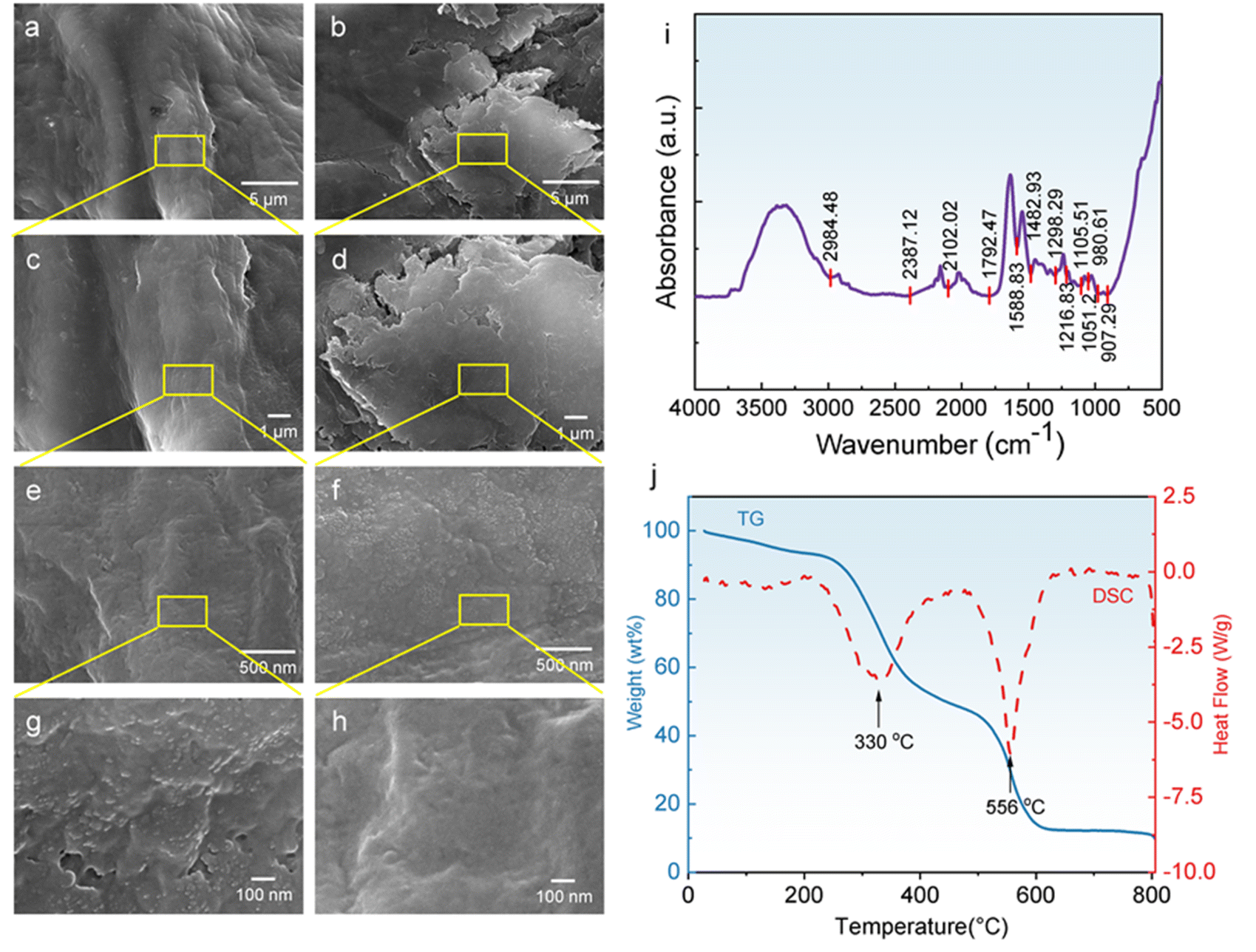 | ||
| Fig. 2 SEM images (a–h), FT-IR spectra (i), and TG-DSC curves (j) of DLHCG material. The execution condition of TG-DSC is heating in air at a heating rate of 10 °C min−1. | ||
To further evaluate the stability of DLHCG material in natural environments, we characterized its thermal properties in air heated from room temperature to 800 degrees celsius using TG-DSC, as shown in the Fig. 2j. At temperatures below 250 °C, DLHCG material has experienced a continuous and slow process of weight loss, which is mainly caused by the evaporation of water. As the heating temperature further increases, DLHCG material begins to undergo significant endothermic reactions, reaching a peak at 330 °C, mainly due to the partial decomposition of chemical bonds in the material. Further increasing the temperature will lead to the disintegration of more chemical bonds in the DLHCG material, reaching a peak at approximately 556 °C. Specifically, the cross-linked structure of DLHCG material endows the material with certain strength and stability. However, the breakage of these bonds at high temperatures will destroy the network structure of the material, and the originally interconnected network will gradually disintegrate. As the cross-linked structure is disrupted, the material will further decompose to produce volatile products, resulting in weight loss. Meanwhile, the breaking process of the bond absorbs heat and shows endothermic characteristics on the DSC curve.
By characterizing the material properties of DLHCG, we have confirmed its safety, stability, and compatibility for implantation in the human body. Next, we will implant DLHCG material into the knee joint of the patient through surgery, and the specific surgical procedures will be presented in the ESI.† The Fig. 3a presents the macroscopic photos of DLHCG material. The Fig. 3b and c show the processed cartilage defect area and the filling of cartilage material during surgery, respectively. The Fig. 3d and g shows the preoperative MRI image of the patient's knee joint, which clearly shows damage to the knee cartilage. The sequence used for the MRI pictures was T2. It mainly reflects the differences in T2 relaxation time of tissues and can display water-containing tissues (such as edema, cerebrospinal fluid, and tumors) through high signals, thereby assisting in the diagnosis of lesions. After 6 months of using DLHCG material for repair, the cartilage damage at the knee joint of the patient significantly improved, as shown in the Fig. 3e and h. According to the relevant rehabilitation procedures, the patient's injury has been basically repaired 12 months after surgery, as shown in the Fig. 3f and i. Although DLHCG material has shown significant advantages and effects in repairing knee cartilage injuries, there are still some shortcomings that need to be overcome.21 For example, the thickness of the cartilage defect area needs to be measured, otherwise the graft is too thin, which can easily cause damage and wrinkles during implantation. If the graft is too thick, it will be higher than the normal joint surface after implantation, and it is prone to wear and tear after surgery, affecting the effectiveness of cartilage repair.22 The transplant is fixed with protein adhesive, which is not as precise as suture fixation, and the postoperative joint movement and weight-bearing time are relatively long. In summary, we use DLHCG material as the repair material for knee cartilage injury, which is different from osteochondral transplantation technology and autologous chondrocyte transplantation technology, providing a new injury repair strategy. Through techniques such as SEM, FT-IR, and TG-DSC, we systematically characterized the structural morphology and thermal stability of DLHCG material, providing a research basis for its practical application in surgery. The MRI images further demonstrate the patient's cartilage repair after surgery. This work provides a research foundation and guidance for the clinical application of DLHCG material.
 | ||
| Fig. 3 Macroscopic morphology photos of DLHCG material (a). The processed cartilage defect area (b) and the filling of cartilage material during surgery (c). The MRI images before surgery (d and g), after surgery 6 months (e and h) and 12 months (f and i). | ||
Summary
By performing SEM, FT-IR, and TG-DSC characterizations, we reveal the good porous gel properties, excellent human compatibility, and chemical stability of DLHCG material. The good physical properties of DLHCG material are the basis for its clinical application, especially its rich chemical bonds and porous gel structure of trans micron–nano scale, which makes it play a huge advantage in the repair of knee cartilage injury. Furthermore, we used MRI technology to characterize the recovery of knee cartilage injuries in patients at 6 and 12 months after repair. This work proves that the DLHCG material can repair cartilage defects in the knee joint without adverse reactions in patients, and has good clinical application value.Ethical statement
All patients signed informed consent forms. This study was approved by the Medical Ethics Committee of Yantai Mountain Hospital in YCantai City (LL-2022-046-K).Data availability
The authors confirm that the data supporting the findings of this study are available within the article and its ESI.†Conflicts of interest
There are no conflicts to declare.Acknowledgements
This work was supported by the Fund of Natural Science Special (Special Post) Research Foundation of Guizhou University (Grant No. 2023-032).References
- H. Zhou, Z. Zhang and Y. Mu, et al., Harnessing Nanomedicine for Cartilage Repair: Design Considerations and Recent Advances in Biomaterials, ACS Nano, 2024, 18(16), 10667–10687 CrossRef CAS PubMed.
- H. Cao, S. Deng and X. Chen, et al., An injectable cartilage-coating composite with long-term protection, effective lubrication and chondrocyte nourishment for osteoarthritis treatment, Acta Biomater., 2024, 179, 95–105 CrossRef CAS PubMed.
- E. Vina and C. Kwoh, Epidemiology of osteoarthritis: literature update, Curr. Opin. Rheumatol., 2018, 30(2), 160–167 CrossRef.
- J. Li, B. Jiang and P. Zhang, et al., Cartilage Decellularized Extracellular Matrix-Based Hydrogel with Enhanced Tissue Adhesion and Promoted Chondrogenesis for Cartilage Tissue Engineering, ACS Appl. Polym. Mater., 2024, 6(8), 4394–4408 CrossRef CAS.
- D. Chen, et al., Osteoarthritis: toward a comprehensive understanding of pathological mechanism, Bone Res., 2017, 5(1), 1–13 Search PubMed.
- W. Dai, X. Gong and C. Wang, et al., Injectable decellularized extracellular matrix hydrogel with cell-adaptable supramolecular network enhances cartilage regeneration by regulating inflammation and facilitating chondrogenesis, Chem. Eng. J., 2024, 498, 155138 CrossRef CAS.
- J. Shi, H. Yao and H. Chong, et al., Tissue-engineered collagen matrix loaded with rat adipose-derived stem cells/human amniotic mesenchymal stem cells for rotator cuff tendon-bone repair, Int. J. Biol. Macromol., 2024, 282, 137144 CrossRef CAS PubMed.
- D. Huey, J. Hu and K. Athanasiou, Unlike Bone, Cartilage Regeneration Remains Elusive, Science, 2012, 338(6109), 917–921 CrossRef CAS PubMed.
- M. Soleymani, E. S. Motiee and S. Karbasi, et al., Evaluation of the effects of decellularized umbilical cord Wharton's Jelly ECM on polyhydroxy butyrate electrospun scaffolds: A new strategy for cartilage tissue engineering, Mater. Today Chem., 2024, 39, 102145 CrossRef CAS.
- H. Kwon, et al., Surgical and tissue engineering strategies for articular cartilage and meniscus repair, Nat. Rev. Rheumatol., 2019, 15(9), 550–570 CrossRef PubMed.
- S. Pina, et al., Scaffolding Strategies for Tissue Engineering and Regenerative Medicine Applications, Materials, 2019, 12(11), 1824 CrossRef CAS PubMed.
- R. Longley, A. Ferreira and P. Gentile, Recent approaches to the manufacturing of biomimetic multi-phasic scaffolds for osteochondral regeneration, Int. J. Mol. Sci., 2018, 19(6), 1755 CrossRef PubMed.
- M. Shive, et al., BST-CarGel® Treatment Maintains Cartilage Repair Superiority over Microfracture at 5 Years in a Multicenter Randomized Controlled Trial, Cartilage, 2015, 6(2), 62–72 CrossRef CAS PubMed.
- J. Dhillon, et al., Third-Generation Autologous Chondrocyte Implantation (Cells Cultured Within Collagen Membrane) Is Superior to Microfracture for Focal Chondral Defects of the Knee Joint: Systematic Review and Meta-analysis, Arthroscopy, 2022, 38(8), 2579–2586 CrossRef.
- L. Fortier, et al., Clinical and Magnetic Resonance Imaging Outcomes After Microfracture Treatment With and Without Augmentation for Focal Chondral Lesions in the Knee: A Systematic Review and Meta-analysis, Am. J. Sports Med., 2023, 51(8), 2193–2206 CrossRef PubMed.
- E. Kon, et al., Combined subchondral and intra-articular injections of bone marrow aspirate concentrate provide stable results up to 24 months. Knee Surgery, Sports Traumatology, Arthroscopy, 2023, 31(6), 2511–2517 Search PubMed.
- C. Li, R. Deng and M. Yang, et al., Advanced hydrogel material for meniscus repair, Adv. Funct. Mater., 2024, 34(16), 2312276 CrossRef CAS.
- K. Lee, K. Chung, D. Nam, M. Jung, S. Kim and H. Kim, Decellularized allogeneic cartilage paste with human costal cartilage and rosslinked hyaluronic acid-carboxymethyl cellulose carrier augments microfracture for improved rticular cartilage repair, Acta Biomater., 2023, 172, 297–308 CrossRef CAS PubMed.
- C. R. Rowland, L. A. Colucci and F. Guilak, Fabrication of anatomically-shaped cartilage constructs using decellularized cartilage-derived matrix scaffolds, Biomaterials, 2016, 91, 57–72 CrossRef CAS PubMed.
- U. Mendibil, Y. Lópiz-Morales and B. Arnaiz, et al., Development of bioactive solid-foam scaffolds from decellularized cartilage with chondrogenic and osteogenic properties, Mater. Today Bio, 2024, 28, 101228 CrossRef CAS PubMed.
- L. Yu, S. Cavelier and B. Hannon, et al., Recent development in multizonal scaffolds for osteochondral regeneration, Bioact. Mater., 2023, 5, 122–159 Search PubMed.
- X. Hu, M. Jin and K. Sun, et al., Type II collagen scaffolds repair critical - -sized osteochondral defects under induced conditions of osteoarthritis in rat knee joints via inhibiting TGF-β-Smad1/5/8 signaling pathway, Bioact. Mater., 2024, 35, 416–428 CAS.
Footnotes |
| † Electronic supplementary information (ESI) available. See DOI: https://doi.org/10.1039/d5ra01186h |
| ‡ These authors contributed equally to this work. |
| This journal is © The Royal Society of Chemistry 2025 |