DOI:
10.1039/D4RA07294D
(Paper)
RSC Adv., 2025,
15, 2023-2033
Design and development of an isatin-1,2,3-triazole hybrid analogue as a potent anti-inflammatory agent with enhanced efficacy and gene expression modulation†
Received
11th October 2024
, Accepted 2nd January 2025
First published on 22nd January 2025
Abstract
Isatin (1H-indole-2,3-dione) and its derivatives have been found to exhibit various biological activities, including anticancer and antidiabetic properties. In this study, a series of nine isatin-1,2,3-triazole conjugates were synthesized and evaluated for their anti-inflammatory potential via in vitro experiments. Their synthesis involved the propargylation of isatin 1 with propargyl bromide to obtain N-propargyl isatin 2, which was subjected to click reactions with different aromatic azides to yield isatin-N-1,2,3-triazoles (3a–i). The structures of all the compounds were confirmed via NMR and HR-MS. The final isatin analogues were tested for their ability to attenuate the production of proinflammatory cytokines in the lipopolysaccharide (LPS)-induced human leukemia monocytic THP-1 cells. Importantly, none of the compounds had any negative effect on THP-1 cell viability at the tested concentrations of 4 mM and 8 mM. LPS induced the production of the cytokines: Tumor necrosis factor-α (TNF-α), interleukin-6 (IL-6) and monocyte chemoattractant protein-1 (MCP-1) by 351.4, 7.9 and 14.3 fold, respectively, in THP-1 cells. However, treatment with compound 3e markedly attenuated the levels of TNF-α (by 6.6 fold and 1.5 fold), IL-6 (by 1.03 fold and 1.41 fold) and MCP-1 (by 3.3 fold and 1.7 fold) by several fold at concentrations of 4 mM and 8 mM, respectively. Furthermore, in the gene expression modulation studies, 3e was found to downregulate the genes responsible for the production of TNF-α (24 and 25 fold), IL-6 (148 and 502 fold) and MCP-1 (50 and 25 fold) at the two tested concentrations compared with their expression in the LPS-induced THP-1 cells (135 fold, 6612 fold, and 68.8 fold, respectively). Thus, 3e markedly attenuated the secretion of TNF-α, IL-6 and MCP-1 from LPS-treated THP-1 cells, and also the expression of the concerned genes. At the lowest dose tested, i.e., 4 mM, 3e had the greatest effect on both gene expression and marker secretion.
1 Introduction
Isatin (1H-indole-2,3-dione) and substituted isatins are heterocyclic molecules found in plants such as Isatis spp,1 Calanthe discolor,2 Couroupita guianensis,3 and Melochia tomentosa,4 bacteria such as Streptomyces albus5 and fungi such as Chaetomium globosum.6 Isatin is a versatile substrate that can be used to prepare a variety of heterocyclic compounds, such as indoles and quinolones.7 Furthermore, Schiff bases and Mannich bases of isatin are known to possess a wide range of pharmacological properties including antibacterial I,8 anticonvulsant,9 anti-HIV II,10 antifungal III,11 antiviral IV12 and anti-TB13 activities. The bis-imine of isatin has shown antimicrobial properties14 and affected cell viability15 (Fig. 1). Isatin is used as an intermediate in the synthesis of drugs such as diclofenac and pirquinozol.
 |
| | Fig. 1 Structures of some reported bioactive isatin derivatives. | |
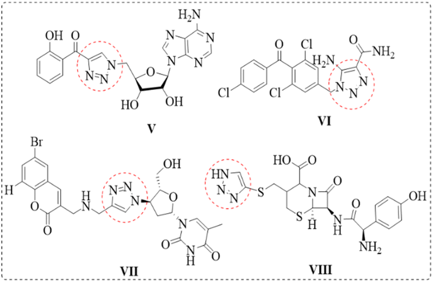
The construction of molecular hybrids of potential bioactives using differently substituted nitrogen-rich heterocyclic pharmacophores such as 1,2,3-triazoles has enabled the production of new and biologically promising drugs.16–18 Chemically, 1,2,3-triazole is stable against acidic/basic hydrolysis and oxidative/reductive conditions, thus reflecting high aromatic stabilization and relative resistance to metabolic degradation.19–21 1,2,3-Triazole is one of the key structural units found in a large variety of bioactive molecules such as antifungal,22 antibacterial,23,24 antiallergic,25 anti-HIV,26,27 antitubercular28,29 and anti-inflammatory30 agents. For example, triazole nucleoside derivative V31 inhibited Mycobacterium tuberculosis, which disrupts siderophore biosynthesis. Carboxyamidotriazole VI is an angiogenesis inhibitor useful in cancer therapy,32 and 1,2,3-triazole-containing compound VII is a potential dual-action HIV-1 protease and nonnucleoside reverse transcriptase inhibitor.33 The cephalosporin analogue cefatrizine VIII is also an antibiotic used to treat bacterial infections of the urinary tract, liver and gallbladder, etc.34,35 (Fig. 2).
 |
| | Fig. 2 Structures of some reported bioactive triazole derivatives. | |
Our earlier research on the synthesis of novel hybrid analogues such as piperonal-thiazolidinedione-triazoles,36 chromanochalcone-triazoles,37 andrographolide-triazoles38 resulted in the production of potential anticancer, hypolipidemic and antidiabetic agents. In continuation, herein, we discuss the synthesis of isatin-1,2,3-triazole hybrid molecules via click chemistry and their anti-inflammatory potential in LPS-induced THP-1 cells.
Inflammation is an immunological response of the body to several factors including dietary lipids, mechanical injury, burns, allergens and noxious stimuli.39 Usually, inflammation is a defence mechanism of the body; however, chronic inflammation may damage normal functioning, especially in the context of autoimmune diseases, atherosclerosis, arthritis, etc. The inflammatory response involves various immune cells, blood vessels, and molecular mediators. Key players include white blood cells (such as neutrophils, macrophages, and lymphocytes), cytokines, chemokines, and other signalling molecules. The synthesized isatin-1,2,3-triazoles 3a–i were treated with LPS-induced human monocytic THP-1 cells in vitro to assess their ability to attenuate the production of three proinflammatory cytokines i.e., TNF-α, IL-6 and MCP-1, and investigate the nuclear factor character of the most potent analogue in modulating the expression of concerned genes in the tested monocytic cells.
2 Results and discussion
2.1 Chemistry
The synthetic strategy of novel isatin N-substituted 1,2,3-triazoles is outlined in Scheme 1. Isatin 1 was N-propargylated with propargyl bromide (1.5 eq.) in the presence of anhydrous K2CO3 (2 eq.) in 10 mL of DMF at room temperature for 4 h, resulting in N-propargyl-isatin 2. Compound 2 was reacted with substituted phenyl azides a and b and benzyl azides c–i (prepared in situ, as previously reported40 under click chemistry reaction conditions (Cu-catalyzed 1,3-dipolar cycloaddition)) in the presence of copper(I) iodide as the catalyst in dry THF at room temperature for 12–14 h, resulting in the production of novel substituted 1-((1-phenyl-1H-1,2,3-triazol-4-yl) methyl) indoline-2,3-diones 3a and b (yield: 90–92%) and 1-((1-benzyl-1H-1,2,3-triazol-4-yl)methyl) indoline-2,3-diones 3c–i (yield: 85–93%), respectively.
 |
| | Scheme 1 General scheme for the synthesis of isatin-1,2,3-triazol derivatives (reaction conditions: step-1: propargyl bromide (1.0 mL), K2CO3, DMF, RT 4 h; step-2: CuI, THF, RT, 14 h). | |
All the synthesized isatin-triazole derivatives 3a–i were characterized by 1H NMR, 13C NMR, IR and HR-MS spectra. In the IR spectra, compounds 3a–i presented IR absorption bands ranging from 1020–1250, 1724–1740 and 3000–3134 cm−1 for C–N, C![[double bond, length as m-dash]](https://www.rsc.org/images/entities/char_e001.gif) O and aromatic C–H stretching, respectively. In the 1H NMR spectra, the characteristic protons of compounds 3a and 3e were assigned as follows. In compound 3a, one singlet at δ 5.57 (2H) confirmed the presence of an N–CH2 linkage. The characteristic triazole CH proton appeared at δ 8.28 (1H) and seven aromatic protons (four aromatic protons of the isatin ring plus three aromatic protons of the phenyl ring) resonated in the range of δ 7.39–8.27. Furthermore, the structure of compound 3a was confirmed by ESI-MS, and a molecular ion peak at m/z 386 (M + 23) was observed for the molecular formula C18H13N5O4 (M = 363). In the 1H NMR spectrum of compound 3e, the presence of two characteristic singlets at δ 5.6 (2H) to 4.9 (2H) confirmed the N–CH2 and Ar–CH2 linkages, respectively. A characteristic singlet peak was observed at δ 8.19 (1H) for the CH proton of the triazole ring. Furthermore, in the 13C-NMR spectrum of compound 3e, three characteristic peaks were observed for N–CH2, Ar–CH2 and CH of the triazole ring at δ 51.01, 35.36 and 123.1 respectively. In the ESI-mass spectrum, a molecular ion peak at m/z 409 (M + 23) was also observed for the molecular formula C18H12Cl2N4O2 (M = 386); thus, the structure of 3e was confirmed.
O and aromatic C–H stretching, respectively. In the 1H NMR spectra, the characteristic protons of compounds 3a and 3e were assigned as follows. In compound 3a, one singlet at δ 5.57 (2H) confirmed the presence of an N–CH2 linkage. The characteristic triazole CH proton appeared at δ 8.28 (1H) and seven aromatic protons (four aromatic protons of the isatin ring plus three aromatic protons of the phenyl ring) resonated in the range of δ 7.39–8.27. Furthermore, the structure of compound 3a was confirmed by ESI-MS, and a molecular ion peak at m/z 386 (M + 23) was observed for the molecular formula C18H13N5O4 (M = 363). In the 1H NMR spectrum of compound 3e, the presence of two characteristic singlets at δ 5.6 (2H) to 4.9 (2H) confirmed the N–CH2 and Ar–CH2 linkages, respectively. A characteristic singlet peak was observed at δ 8.19 (1H) for the CH proton of the triazole ring. Furthermore, in the 13C-NMR spectrum of compound 3e, three characteristic peaks were observed for N–CH2, Ar–CH2 and CH of the triazole ring at δ 51.01, 35.36 and 123.1 respectively. In the ESI-mass spectrum, a molecular ion peak at m/z 409 (M + 23) was also observed for the molecular formula C18H12Cl2N4O2 (M = 386); thus, the structure of 3e was confirmed.
2.2 Anti-inflammatory activity
2.2.2 Effect of isatin-triazole conjugates on the LPS-induced increase in TNF-α in THP-1 cells. In THP-1 cells, LPS has been shown to induce the production of proinflammatory cytokines such as tumor necrosis factor (TNF-α), interleukin-6 (IL-6) and MCP-1 via the TLR4-NFκB signalling pathway.41 The amount of TNF-α secreted into the cell culture supernatants was measured via ELISA; the data are shown in Fig. 4 and Table 2. The uninduced THP-1 cells released basal levels of TNF-α (152.70 pg mL−1), and induction with LPS for 3 h increased the release of TNF-α to 53671.32 pg mL−1. This LPS-dependent increase in TNF-α secretion was significantly inhibited by pretreatment with the synthesized analogues. LPS induced the secretion of TNF-α by 351.46 fold in the THP-1 cells. Among the tested samples (3a–i), compound 3e significantly attenuated the LPS-induced secretion of TNF-α by 6.6 fold and 1.5 fold at all the tested concentrations of 4 mM and 8 mM, followed by 3g, which was significant (152.1 fold) only at 8 mM. Dexamethasone (Dex) attenuated the LPS-induced secretion of TNF-α by 331.68, 38.21, and 123 fold at concentrations of 12 μM, 60 μM, and 120 μM, respectively. After 12 h of pretreatment instead of 30 min with Dex at 12 μM,42 it did not affect the secretion of TNF-α. However, pretreatment with 60 μM Dex under the same conditions significantly attenuated the secretion of TNF-α.
 |
| | Fig. 4 Effects of the anti-inflammatory standard compound Dex, the parent compound isatin, and its derivatives 3a-–i on LPS-induced TNF- α secretion in THP-1 cells. THP-1 cells were pretreated with Dex, isatin and its derivative compounds 3a–i for 12 h, followed by 3 h of LPS treatment. Experiments were performed at least in triplicate, and the results are expressed as mean ± S.D. *p < 0.001 for comparison between LPS-induced cells and LPS-uninduced cells. #p < 0.001 compared between cells treated with LPS in the presence of test compounds vs. control. | |
Table 2 Effects of synthesized samples on LPS-induced TNF-α secretion in THP-1 cells. THP-1 cells were pretreated with the anti-inflammatory standard compound Dex, the parent compound isatin and its derivatives for 12 h, followed by LPS for 3 h
| TNF-α release (fold change) |
| Cells (control) |
1 ± 0.00177 |
|
|
| Cells + LPS (3 h) |
351.46 ± 0.0248 |
|
|
![[thin space (1/6-em)]](https://www.rsc.org/images/entities/char_2009.gif) |
| Pretreatment with dexamethasone (μM) for 12 h |
| |
12 |
60 |
120 |
| Cells + Dex + LPS |
331.6891 ± 0.05 |
38.21044 ± 9.97 |
123.3 ± 29.83 |
|
| Isatin (mM) |
| |
5 |
|
10 |
| Cells + isatin + LPS |
418.10 ± 0.06 |
|
324.2066 ± 0.09 |
|
| Isatin derivatives (mM) |
| |
4 |
|
8 |
| Cells + 3a + LPS |
332.72 ± 0.01 |
|
336.55 ± 0.09 |
| Cells + 3b + LPS |
318.06 ± 0.24 |
|
303.58 ± 0.07 |
| Cells + 3c + LPS |
342.84 ± 0.06 |
|
341.04 ± 0.02 |
| Cells + 3d + LPS |
306.9061 ± 0.08 |
|
313.6059 ± 0.14 |
| Cells + 3e + LPS |
6.65 ± 0.01 |
|
1.50 ± 0.004 |
| Cells + 3f + LPS |
328.15 ± 0.12 |
|
338.54 ± 0.05 |
| Cells + 3g + LPS |
300.87 ± 0.06 |
|
152.19 ± 0.11 |
| Cells + 3h + LPS |
335.6804 ± 0.11 |
|
276.31 ± 0.18 |
| Cells + 3i + LPS |
353.60 ± 0.07 |
|
334.40 ± 0.08 |
2.2.3 Effect of 3e on LPS-induced IL-6 in THP-1 cells. IL-6 is a pleiotropic cytokine that initiates the acute phase response of innate immunity. The uninduced THP-1 cells released basal levels of IL-6 (96.8 pg mL−1), and induction with LPS for 3 h increased the release of IL-6 to 770.5 pg mL−1. LPS induced the secretion of IL-6 by 7.95 fold in the THP-1 cells. Pre-treatment of induced THP-1 cells was performed with standard dexamethasone, parent isatin or compound 3e (given that only 3e could significantly reduce IL-6). Pre-treatment of induced THP-1 cells with the standard dexamethasone (Dex) at three different concentrations (12 μM, 60 μM, and 120 μM) attenuated IL-6 secretion by 5.74, 6.11, and 5.53 fold, respectively. Isatin (parent compound) markedly attenuated the LPS-induced secretion of IL-6 by 4.08 fold at a concentration of 5 mM. Sample 3e (Fig. 5 and Table 3) markedly attenuated the LPS-induced secretion of IL-6 by 1.03 fold and 1.41 fold at concentrations of 4 mM and 8 mM, respectively.
 |
| | Fig. 5 Effects of the anti-inflammatory standard compound dexamethasone, the parent compound isatin, and 3e on LPS-induced IL-6 secretion in THP-1 cells. THP-1 cells were pretreated with Dex, isatin or its derivative compound 3e for 12 h, followed by 3 h of LPS treatment. Experiments were performed in at least triplicate, and the results are expressed as mean ± S.D. *p < 0.001 for comparison between LPS-induced cells and LPS-uninduced cells. #p < 0.001 compared between cells treated with LPS in the presence of test compounds vs. control. | |
Table 3 Effects of isatin derivative 3e on LPS-induced IL-6 secretion in THP-1 cells. THP-1 cells were pretreated with anti-inflammatory standard compound Dex, the parent compound isatin and 3e for 12 h, followed by LPS for 3 h
| IL-6 release (fold change) |
| Cells (control) |
1 ± 0.0062 |
| Cells + LPS (3 h) |
7.95 ± 1.67 |
|
| Pretreatment with dexamethasone (μM) for 12 h |
| |
12 |
60 |
120 |
| Cells + Dex + LPS |
5.74 ± 0.1 |
6.11 ± 0.36 |
5.53 ± 0.82 |
|
| Isatin (mM) |
| |
5 |
|
10 |
| Cells + isatin + LPS |
4.08 ± 0.01 |
|
4.86 ± 0.15 |
|
| 3e (mM) |
| |
4 |
|
8 |
| Cells + 3e + LPS |
1.03 ± 0.20 |
|
1.41 ± 0.53 |
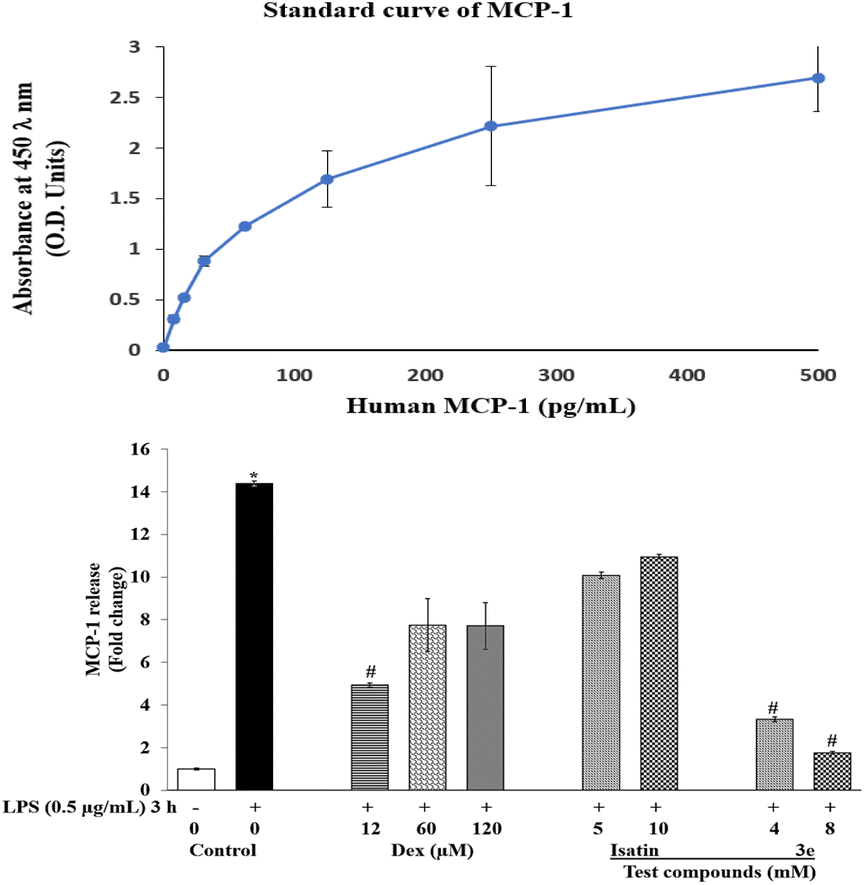
2.2.4 Effect of 3e on LPS-induced MCP-1 in THP-1 cells. Monocyte chemoattractant protein-1 (MCP-1) is a key chemokine involved in the regulation, migration and infiltration of monocytes to the site of inflammation. Uninduced THP-1 cells released basal levels of MCP-1 (204 pg mL−1), and induction with LPS for 3 h increased the release of MCP-1 to 2934.6 pg mL−1. In lipopolysaccharide (LPS)-induced THP-1 cells, MCP-1 production was significantly upregulated (by 14.37 fold) as part of the inflammatory response (Fig. 6 and Table 4). Treatment of these differentiated THP-1 cells with standard dexamethasone markedly attenuated MCP-1 production by 4.93, 7.75, and 7.71 fold at concentrations of 12 μM, 60 μM, and 120 μM, respectively. Similarly, compound 3e markedly attenuated the secretion of MCP-1 by 3.32 fold and 1.75 fold at concentrations of 4 mM and 8 mM, respectively.
 |
| | Fig. 6 Effects of the anti-inflammatory standard compound dexamethasone, the parent compound isatin, and 3e on LPS-induced MCP-1 secretion in THP-1 cells. THP-1 cells were pretreated with Dex, isatin or its derivative compound 3e for 12 h, followed by 3 h of LPS treatment. Experiments were performed at least in triplicate, and the results are expressed as mean ± S.D. *p < 0.001 for comparison between LPS-induced cells and LPS-uninduced cells. #p < 0.001 compared between cells treated with LPS in the presence of test compounds vs. control. | |
Table 4 Effects of isatin derivative 3e on LPS-induced MCP-1 secretion in THP-1 cells. THP-1 cells were pretreated with the anti-inflammatory standard compound Dex, the parent compound isatin and 3e for 12 h, followed by LPS for 3 h
| MCP-1 release (fold change) |
| Cells (control) |
1 ± 0.04 |
| Cells + LPS (3 h) |
14.37 ± 0.12 |
|
| Pretreatment with dexamethasone (μM) for 12 h |
| |
12 |
60 |
120 |
| Cells + Dex + LPS |
4.93 ± 0.08 |
7.75 ± 1.23 |
7.71 ± 1.1 |
|
| Isatin (mM) |
| |
5 |
|
10 |
| Cells + isatin + LPS |
10.07 ± 0.15 |
|
10.95 ± 0.097 |
|
| 3e (mM) |
| |
4 |
|
8 |
| Cells + 3e + LPS |
3.32 ± 0.11 |
|
1.75 ± 0.08 |
2.2.5 Effect of the isatin-triazole analogue 3e on TNF-α, IL-6 and MCP-1 transcript levels in LPS stimulated THP-1 cells. Gene expression regulation is a critical aspect of understanding the cellular response to various stimuli. The exposure of THP-1 monocytes to LPS strongly induced TNF-α, IL-6 and MCP-1 gene expression with maximal expression occurring after 3 h of stimulation. To assess the nuclear factor characteristic of compound 3e, the mRNA expression levels of these cytokine genes were determined via qRT-PCR. Although the expression of all the above-mentioned cytokine genes was upregulated in stimulated THP-1 monocytes, the IL-6 gene presented the highest fold change in expression (6612.6), followed by the TNF- α (135.2) and MCP-1 genes (68.8).
2.2.6 Effect of 3e on TNF-α genes. Tumour necrosis factor-alpha (TNF-α) is a proinflammatory cytokine that plays a pivotal role in inflammation and immune responses. The expression of TNF-α is tightly regulated at the transcriptional level, and its dysregulation is associated with numerous pathological conditions, including sepsis, autoimmune diseases, and cancer. Lipopolysaccharides (LPS) are known to induce the expression of TNF-α, making them a useful model for studying inflammatory responses. The gene expression regulation of TNF-α demonstrated that the TNF-α gene transcript was markedly upregulated in the LPS-treated cells compared with the uninduced control cells (135 fold). Pretreatment with isatin significantly inhibited TNF-α expression by 55.9 and 31.4 fold at concentrations of 5 mM and 10 mM, respectively. In contrast, pretreatment with sample 3e significantly inhibited TNF-α expression by 24 and 25 fold at concentrations of 4 mM and 8 mM, respectively (Fig. 7 and Table 5).
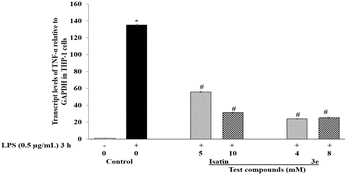 |
| | Fig. 7 Effects of the parent compound isatin and its derivative 3e on LPS-induced TNF-α gene expression, as determined via quantitative real time PCR. THP-1 cells were pretreated with isatin or 3e for 12 h, followed by 3 h of LPS induction. At the end of the treatment, the transcript levels of TNF-α were quantified via real-time PCR. Experiments were performed at least in triplicate, and the results were expressed as mean ± S.D. *p < 0.001 for comparison between LPS-induced cells vs. LPS-uninduced cells. #p < 0.001 compared between cells treated with LPS in the presence of test compounds vs. control. | |
Table 5 Transcript levels of TNF- α, IL-6 and MCP-1 relative to that of GAPDH in the THP-1 cells (in fold changes)
| Genes |
Cells (control) |
Cells + LPS |
Cells + isatin + LPS |
Cells + 3e + LPS |
| 5 mM |
10 mM |
4 mM |
8 mM |
| TNF-α |
1 ± 0.31 |
135.2 ± 0.81 |
55.9 ± 0.50 |
31.4 ± 0.39 |
23.9 ± 0.41 |
25.3 ± 0.59 |
| IL-6 |
1 ± 0.18 |
6612.6 ± 0.21 |
1145.8 ± 1.01 |
596.8 ± 0.83 |
148.5 ± 0.53 |
502.5 ± 2.2 |
| MCP-1 |
1 ± 0.19 |
68.8 ± 0.70 |
6.21 ± 0.67 |
1.73 ± 0.54 |
49.9 ± 0.21 |
24.9 ± 0.49 |
2.2.7 Effect of 3e on IL-6 genes. Interleukin-6 (IL-6) is a cytokine that plays a vital role in the immune response, inflammation, and haematopoiesis. It is involved in various physiological processes and pathological conditions, including autoimmune diseases, chronic inflammation, and cancer. The expression of IL-6 is tightly regulated, and its dysregulation can lead to severe inflammatory responses. The gene expression regulation of IL-6 demonstrated that the IL-6 gene transcript was significantly upregulated in the LPS-treated cells compared with the uninduced control cells (6612 fold). Pretreatment with isatin (parent compound) significantly inhibited IL-6 expression by 1145 and 596 fold at concentrations of 5 mM and 10 mM, respectively. In contrast, pretreatment with sample 3e significantly inhibited IL-6 expression by 148 and 502 fold at concentrations of 4 mM and 8 mM, respectively (Fig. 8 and Table 5).
 |
| | Fig. 8 Effects of the parent compound isatin and its derivative 3e on LPS-induced IL-6 gene expression, as determined via quantitative real-time PCR. THP-1 cells were pretreated with isatin or 3e for 12 h, followed by 3 h of LPS induction. At the end of the treatment, the transcript levels of IL-6 were quantified via real-time PCR. Experiments were performed at least in triplicate, and the results were expressed as mean ± S.D. *p < 0.001 for comparison between LPS-induced cells vs. LPS-uninduced cells. #p < 0.001 compared between cells treated with LPS in the presence of test compounds vs. control. | |
2.2.8 Effect of 3e on MCP-1 genes. Monocyte chemoattractant protein-1 (MCP-1) is a key chemokine involved in the recruitment of monocytes to sites of injury and infection. It plays a crucial role in inflammatory responses and has been implicated in various pathological conditions, including atherosclerosis, rheumatoid arthritis, and other inflammatory diseases. The regulation of MCP-1 expression is tightly controlled at the genetic level, and its overexpression can lead to excessive inflammatory responses, contributing to disease progression. The gene expression regulation of MCP-1 demonstrated that the MCP-1 gene transcript was markedly upregulated in LPS-treated cells compared with the uninduced control cells (69 fold). Pretreatment with isatin (parent compound) significantly inhibited MCP-1 expression by 6.21, and 1.73 fold at concentrations of 5 mM and 10 mM, respectively. Pretreatment with sample 3e significantly inhibited MCP-1 expression by 50 and 25 fold at concentrations of 4 mM and 8 mM, respectively (Fig. 9).
 |
| | Fig. 9 Effects of the parent compound isatin and its derivative 3e on LPS-induced MCP-1 gene expression, as determined via quantitative real-time PCR. THP-1 cells were pretreated with isatin or 3e for 12 h, followed by 3 h of LPS induction. At the end of the treatment, the transcript levels of MCP-1 were quantified via real-time PCR. The data were presented as mean ± S.D. *p < 0.001 for comparison between LPS-induced and LPS-uninduced cells, and the experiments were conducted at least three times. #p < 0.001 when comparing cells treated with LPS and test compounds to the control. | |
3 Experimental
3.1 Chemistry
The melting points of all the compounds were recorded on a Casia-Siamia (VMP-AM) melting point apparatus and uncorrected. IR spectra were recorded on a PerkinElmer FT-IR 240 C spectrometer using KBr optics. NMR spectra were recorded on a Bruker Avance 400/500 MHz in DMSO-d6 using TMS as an internal standard. All reactions were monitored on silica gel percolated TLC plates from Merck, and the spots were visualized with UV light. The silica gel (100–200 mesh) used for column chromatography was procured from Merck.
3.1.1 General experimental procedure for compound 2. Compound 1 (1.0 mmol) and potassium carbonate (1.5 mmol) were dissolved in 10 mL of dry DMF and propargyl bromide (1.0 mmol) was subsequently slowly added to the mixture while stirring. The reaction mixture was stirred at room temperature for 4 h. To the reaction mixture, excess ethyl acetate was added, and the mixture was washed with water (70 mL × 3). The organic layer was dried over Na2SO4, filtered, concentrated under reduced pressure and washed with hexane to obtain the final analytically pure compound 2. Red solid, yield: 95%.
3.1.1.1 1-(Prop-2-ynyl)indoline-2,3-dione (2). Orange solid, yield: 98%, m.p.156–157 °C, 1H-NMR (DMSO-d6, 400 MHz) δ (ppm): 6.74–6.79 (t, 1H), 6.64–6.66 (d, J = 7.4 Hz, 1H), 6.22–6.30 (m, 2H), 3.62 (d, J = 2.4 Hz, 2H), 2.24 (t, 1H). ESI-MS found MNa+, 208 calculated for C11H7NO2: M, 185.1820.
3.1.2 General experimental procedure for compounds 3a–i. Compound 2 (1.0 mmol) was dissolved in dry THF (Na 10 mL) and a catalytic amount of copper iodide was added. To this end, substituted aromatic azides (phenyl and benzyl) (1.0 mmol) in dry THF were added slowly while stirring at room temperature under a nitrogen atmosphere for 14 h. Later, excess CHCl3 was added, and the mixture was filtered, and washed with CHCl3. The solvent was removed under reduced pressure to obtain the crude product. The crude product was purified through silica gel (100–200 mesh) column chromatography (in ethyl acetate–hexane) to afford the desired products 3a–i.
3.1.2.1 1-((1-(2-Methyl-3-nitrophenyl)-1H-1,2,3-triazol-4-yl)methyl)indoline-2,3-dione (3a). Orange solid, yield: 92%, m.p. 198–199 °C, IR (KBr) 3145, 3093, 1739, 1612, 1527, 1467, 1296, 1175, 1097 cm−1; 1H-NMR (DMSO-d6, 500 MHz) δ (ppm): 8.26–8.28 (dd, J = 8.1, 1.3 Hz, 1H), 8.12 (s, 1H), 7.85–7.90 (m, 2H), 7.79–7.81 (dd, J = 7.9, 1.3 Hz, 1H), 7.73–7.76 (t, J = 8.0 Hz, 1H), 7.61 (t, J = 7.62 Hz, 1H), 7.50 (s, 1H), 7.36–7.43 (m, 1H), 5.37 (s, 2H), 1.84 (s, 3H). HR-MS found MH+, 364.1036. Calculated for C18H14N5O4: MH, 364.1046 [m/z: 364.10 (100.0%), 386.08.10 (30.2%), 146.06 (15%), 308.10 (12%), 158.06 (7%)].
3.1.2.2 1-((1-(4-Acetylphenyl)-1H-1,2,3-triazol-4-yl)methyl)indoline-2,3-dione (3b). Orange solid, yield: 90%, m.p. 252–253 °C, IR (KBr) 3153, 2959, 2924, 1737, 1687, 1608, 1468, 1263, 1158, 1098 cm−1; 1H-NMR (DMSO-d6, 500 MHz) δ (ppm): 8.05 (s, 1H), 7.19–7.22 (d, J = 8.7 Hz, 2H), 7.07–7.10 (d, J = 8.7 Hz, 2H), 6.65–6.71 (m, 2H), 6.21–6.28 (m, 2H), 4.15 (s, 2H), 1.68 (s, 3H). HR-MS found: MH+, 347.1154. Calculated for C19H15N4O3: MH, 347.1164 [m/z: 347.11 (100.0%), 348.11 (20%), 349.12 (4%), 345.13 (2%)].
3.1.2.3 1-((1-(4-Nitrobenzyl)-1H-1,2,3-triazol-4-yl)methyl)indoline-2,3-dione (3c). Orange solid, yield: 86%, m.p. 232–234 °C, IR (KBr) 3126, 2954, 1788, 1731, 1610, 1519, 1467, 1269, 1184, 1033 cm−1; 1H-NMR (DMSO-d6, 500 MHz) δ (ppm): 7.33 (s, 1H), 7.25–7.27 (d, J = 8.7 Hz, 2H), 6.66–6.71 (t, J = 7.8 Hz, 1H), 6.60–6.62 (d, J = 7.2 Hz, 1H), 6.53–6.56 (d, J = 8.6 Hz, 2H), 6.16–6.22 (m, 2H), 4.79 (s, 2H), 4.03 (s, 2H). ESI-MS found: MNa+, 386. Calculated for C15H14N5O4: MH, 364.3330 [m/z: 386 (100.0%), 235 (22.6%), 118 (10%), 166 (8%), 402 (6%)].
3.1.2.4 1-((1-(3-Chlorobenzyl)-1H-1,2,3-triazol-4-yl)methyl)indoline-2,3-dione (3d). Orange solid, yield: 89%, m.p. 145–146 °C, IR (KBr) 3130, 3069, 1732, 1609, 1468, 1322, 1217, 1124, 1049 cm−1; 1H-NMR (DMSO-d6, 400 MHz) δ (ppm): 9.01 (s, 1H), 8.03–8.15 (m, 4H), 7.60–7.64 (d, J = 15.8 Hz, 2H), 7.14–7.22 (m, 2H), 5.09 (s, 2H), 3.35 (s, 3H). 13C-NMR (CDCl3, 75 Hz) δ (ppm): 196.89, 182.97, 149.98, 143.20, 139.38, 138.09, 136.34, 130.06, 124.48, 123.40, 121.91, 119.56, 117.68, 111.07, 34.99, 26.81. HR-MS found: MH+, 353.0813. Calculated for C18H14ClN4O2: MH, 353.0823 [m/z: 353.08 (100.0%), 355.07 (31.2%), 356.08 (8%), 357.08 (1.8%)].
3.1.2.5 1-((1-(2,4-Dichlorobenzyl)-1H-1,2,3-triazol-4-yl)methyl)indoline-2,3-dione (3e). Orange solid, yield: 85%, m.p. 175–176 °C, IR (KBr) 3123, 3080, 2924, 2855, 1786, 1731, 1610, 1559, 1432, 1229, 1120, 1031 cm−1; 1H-NMR (DMSO-d6, 400 MHz) δ (ppm): 4.970 (s, 2H), 5.657 (s, 2H), 7.108–7.147 (m, 2H), 7.236–7.257 (d, 1H, J = 8.533 Hz), 7.552–7.575 (dd, 1H, J = 1.506 Hz), 7.608–7.651 (m, 1H), 7.688–7.694 (d, 1H, J = 2.259 Hz), 8.199 (s, 1H). 13C-NMR (CDCl3, 75 Hz) δ (ppm): 183.02, 157.92, 150.18, 142.12, 138.62, 135.88, 134.38, 131.42, 130.53, 129.93, 128.02, 125.34, 124.06, 123.17, 117.51, 111.50, 51.01, 35.36. HR-MS found: MH+, 387.0415. Found for C18H13C12N4O2: MH, 387.0425 [m/z: 387.04 (100.0%), 389.03 (63.5%), 359.04 (7%), 331.04 (4.2%)].
3.1.2.6 1-((1-(4-Fluorobenzyl)-1H-1,2,3-triazol-4-yl)methyl)indoline-2,3-dione (3f). Orange solid, yield: 93%, m.p. 186–187 °C, IR (KBr) 3150, 2924, 1786, 1726, 1607, 1509, 1466, 1354, 1266, 1051 cm−1; 1H-NMR (DMSO-d6, 500 MHz) δ (ppm): 7.27 (s, 1H), 6.61–6.68 (m, 2H), 6.39–6.43 (dd, J = 8.6, 5.6 Hz, 2H), 6.19–6.27 (dd, J = 17.5, 8.4 Hz, 4H), 4.61 (s, 2H), 4.01 (s, 2H). HR-MS found: MH+, 337.1114. Calculated for C18H14FN4O2: MH, 337.1124 [m/z: 337.11 (100.0%), 338.11 (21%), 281.11 (11.1%), 309.11 (8.6%), 282.11 (1.9%)].
3.1.2.7 1-((1-(2-Fluorobenzyl)-1H-1,2,3-triazol-4-yl)methyl)indoline-2,3-dione (3g). Orange solid, yield: 90%, m.p. 137–138 °C, IR (KBr) 3126, 3080, 2936, 1740, 1611, 1466, 1326, 1236, 1145, 1039 cm−1; 1H-NMR (DMSO-d6, 500 MHz) δ (ppm): 7.25 (s, 1H), 6.60–6.68 (dd, J = 19.1, 3.9 Hz, 2H), 6.25–6.45 (m, 4H), 6.18–6.20 (d, J = 7.8 Hz, 3H), 4.67 (s, 2H), 4.01 (s, 2H). HR-MS found: MH+, 337.1115. Calculated for C18H14FN4O2: MH, 337.1125 [m/z: 337.11 (100.0%), 338.11 (21%), 309.11 (18%), 310.11 (4%)].
3.1.2.8 2-((4-((2,3-Dioxoindolin-1-yl)methyl)-1H-1,2,3-triazol-1-yl)methyl)benzonitrile (3h). Orange solid, yield: 86%, m.p. 166–167 °C, IR (KBr) 3081, 3032, 2960, 2230, 1862, 1724, 1608, 1467, 1327, 1120, 1024 cm−1; 1H-NMR (DMSO-d6, 500 MHz) δ (ppm): 7.31 (s, 1H), 6.93–6.96 (dd, J = 7.7, 0.9 Hz, 1H), 6.6.-6.76 (m, 4H), 6.38–6.41 (d, J = 7.8 Hz, 1H), 6.18–6.22 (dd, J = 7.8, 1.6 Hz, 2H), 4.83 (s, 2H), 4.03 (s, 2H). HR-MS found: MH+, 344.1190. Calculated for C19H14N5O2: MH, 344.1200 [m/z: 344.11 (100.0%), 366.09 (60%), 345.11 (38%), 288.11 (37%), 316.11 (7.1%), 289.11 (7.6%)].
3.1.2.9 4′-((4-((2,3-Dioxoindolin-1-yl)methyl)-1H-1,2,3-triazol-1-yl)methyl)biphenyl-2-carbonitrile (3i). Orange solid, yield: 85%, m.p. 170–171 °C, IR (KBr) 3143, 2950, 2221, 1854, 1738, 1612, 1469, 1327, 1122, 1098 cm−1; 1H-NMR (DMSO-d6, 400 MHz) δ (ppm): δ 7.37 (s, 1H), 7.00–7.03 (d, J = 7.7 Hz, 1H), 6.83–6.86 (d, J = 6.8 Hz, 1H), 6.63–6.71 (dd, J = 15.5, 7.6 Hz, 6H), 6.47–6.50 (d, J = 8.1 Hz, 2H), 6.20–6.25 (m, 2H), 4.73 (s, 2H), 4.05 (s, 2H). HR-MS found: MH+, 420.1482. Calculated for C25H18N5O2: MH, 420.1492. [m/z: 420.14 (100.0%), 421.14 (18%), 442.12 (45%), 443.13 (8%)]
3.2 Anti-inflammatory activity
3.2.1 Chemicals. The chemicals used in this study included lipopolysaccharide (LPS), trypan blue, MTT, isopropanol and chloroform from Sigma-Aldrich. ELISA kits (for human-TNF-α, IL-6 and MCP-1) were purchased from BD Biosciences (San Diego, CA, USA). The iScript cDNA synthesis kit from Bio-Rad (Hercules, CA, USA) was used. Power SYBR green PCR Master Mix was procured from Applied Biosystems (Warrington, UK). Fetal bovine serum (FBS), Pen Strep, and RPMI 1640 media were procured from Gibco (USA). All other reagents were of analytical grade.
3.2.2 Cell cultures and treatments. THP-1 cells were obtained from NCCS (Pune, India) and grown in RPMI-1640 with 10% (v/v) fetal bovine serum (FBS) and 1% (v/v) pen strep (Penicillin streptomycin). The cells were kept in an incubator at 37 °C and 5% CO2.43 The trials were carried out at a cell density of 5 × 10⁵. The trypan vital staining exclusion technique was used to determine the viability of the cultured cells. The cells were treated with isatin triazoles (3a–i) at two different concentrations (4 mM and 8 mM) for anti-inflammatory drug purposes, and their viability was tested via the trypan vital staining exclusion method. For the trypan blue vital assay, the treated cell suspension was mixed with trypan blue solution in at a 1:1 ratio. Colourless and blue cells were recorded based on morphology through an inverted microscope while being watched with a haemocytometer.44
3.2.3 Cell viability (MTT assay). The effects of various concentrations of the selected pure compounds and isatin triazoles on THP-1 cells were tested via 3-(4,5-dimethylthiazol-2-yl)-2,5-diphenyltetrezolium bromide (MTT) assays, as described previously by Reddi & Tetali (2019).44 THP-1 cells were distributed at a density of 5 × 10⁵ cells/mL in 24-well plates, treated with different concentrations of test compounds and incubated at 37 °C overnight. After overnight incubation, the cells were harvested and the cell pellet was washed three times with RPMI medium. Then, 20 μL of MTT (5 mg mL−1) dissolved in media was added, and the mixture was incubated for 4 h. The cells were pelleted via low-speed centrifugation and incubated for 15 min with 100 μL of DMSO to dissolve the insoluble purple colored formazan crystals. The absorbance of MTT formazan was determined at λ of 570 nm with the reference λ of 690 nm using a 96-well plate reader (Thermo Fisher Scientific). Incomplete media with the tested compounds were used as blanks. The percentage of viable cells from each well after incubation with the test compounds was obtained using following equation:
| % viability = (O.D. of treated cells//O.D. of untreated cells) × 100. |
3.2.4 Treatment of Dex. The anti-inflammatory effect of Dex (0.01 μM to 1 μM) was reported in THP-1 cells,42 and therefore we included it using higher concentrations (12 μM, 60 μM, and 120 μM) as a reference standard compound.
3.2.5 Measurement of secreted TNF-α, MCP-1, and IL-6. Secreted TNF-α, IL-6 and MCP-1 are proinflammatory cytokines and chemokines secreted by THP-1 cells and were measured with BD OptEIA-™ Set Human ELISA kits (San Diego, CA, USA). THP-1 cells at a density of 5 × 10⁵ cells per mL were incubated with and without different concentrations of test compounds overnight and induced with LPS (0.5 μg mL−1) on the following day for 3 h.41 After induction, the cells were spun, and the supernatants were collected for quantification of the above-mentioned marker levels via ELISA kits (BD Biosciences). The reaction mixture was assayed by measuring the absorbance at a wavelength of 450 nm using a microplate reader with the wavelength correction set at 570 nm. The secretory levels in the culture supernatants were determined using a standard curve, which was constructed via serial dilutions of the above-mentioned markers, as provided with the respective kits.
3.2.6 Quantitative real time PCR. For the real-time quantitative PCR assay, two microliters of cDNA were used along with 1XFG, power SYBR green PCR Master Mix (Applied Bio Systems). By generating an amplicon melting curve by plotting fluorescence as a function of temperature, the specificity of SYBR green fluorescence was tested. The transcript levels of the marker genes were normalized to that of GAPDH mRNA.44
3.2.7 Quantitation of transcripts of TNF- α, IL-6 and MCP-1. Total RNA was isolated from the THP-1 cells after the indicated treatments using TRIzol reagent according to the manufacturer's (Invitrogen) protocol. cDNA was synthesized from the prepared RNA via an iScript™ cDNA synthesis kit. The gene expression levels of the TNF-α, MCP-1, and IL-6 genes were analysed by using 1× SYBR green PCR reagent and gene–specific primers.43–45
3.2.8 Statistical analysis. The entire data in the manuscript represents the mean ± standard deviation (SD) of triplicates. Data were analysed by one way analysis of variance (ANOVA).
4 Conclusion
The synthesized isatin-1,2,3-triazole hybrid analogues demonstrated significant anti-inflammatory activity, with compound 3e showing the most potent effects. Specifically, compound 3e significantly attenuated the secretion of TNF-α by 6.65 fold and 1.50 fold; that of IL-6 by 1.03 and 1.41 fold; and that of MCP-1 by 3.32 and 1.75 fold at concentrations of 4 mM and 8 mM, respectively. The gene expression analysis further revealed that 3e downregulated TNF-α by 24 fold and 25 fold; IL-6 by 148 fold and 502 fold; and MCP-1 by 50 fold and 25 fold at the same concentrations. Importantly, none of the synthesized analogues, including 3e, exhibited cytotoxicity toward THP-1 cells at the tested concentrations, confirming their safety. These findings highlight the potential of isatin-1,2,3-triazole hybrids, particularly compound 3e, as nontoxic and effective anti-inflammatory agents.
Abbreviations
| C | Control |
| V-ctrl | Vehicle control |
| LPS | Lipopolysaccharides |
| TNF-α | Tumor necrosis factor-alpha |
| IL-6 | Interleukin-6 |
| MCP-1 | Monocyte chemoattractant protein-1 |
| MTT | 3-(4,5-Dimethylthiazol-2-yl)-2,5-diphenyltetrezolium bromide |
| Dex | Dexamethasone |
| TC | Test compound |
Data availability
The data supporting the findings of this study are available from the corresponding author JKK upon reasonable request.
Author contributions
A. N. K. performed the synthetic protocol and wrote the first draft of the manuscript; J. K. K. and K. V. N. S. S. edited the manuscript and formatted the last version; S. R. D. and S. D. T. performed the anti-inflammatory activity.
Conflicts of interest
Authors declare no conflict of interest.
Acknowledgements
The authors thank Director, CSIR-CIMAP, Lucknow, India, and Scientist in-charge, CSIR-CIMAP-RC, Hyderabad, India for their constant encouragement and support. Author Smruti expresses gratitude to CSIR for granting SRF and acknowledges UoH-IoE for their financial support towards consumables. CIMAP Publication Communication Number: CIMAP/PUB/2024/136.
References
- Y. Guo and F. Chen, TLC-UV-spectrophotometric and TLC-scanning determination of isatin in leaf of Isatis, Zhongcaoyao, 1986, 17, 8–11 CAS.
- M. Yoshikawa, T. Murakami, A. Kishi, T. Sakurama, H. Matsuda, M. Nomura, H. Matsuda and M. Kubo, Novel indole S,O-bisdesmoside, calanthoside, the precursor glycoside of tryptanthrin, indirubin, and isatin, with increasing skin blood flow promoting effects, from two Calanthe species (Orchidaceae), Chem. Pharm. Bull., 1998, 46, 886–888, DOI:10.1248/cpb.46.886.
- J. Bergman, J. O. Lindström and U. L. F. Tilstam, The structure and properties of some indolic constituents in Couroupita guianensis aubl, Tetrahedron, 1985, 41(14), 2879–2881, DOI:10.1016/S0040-4020(01)96609-8.
- G. J. Kapadia, B. K. Shukla, S. P. Chowdhury, H. M. Basak and E. A. Falesm, Phenylpelisatins: a novel class of alkaloids from Melochia tomentosa, J. Chem. Soc., Chem. Commun., 1997, 535–536, 10.1039/C39770000535.
- U. D. O. Grafe and L. Rasics, Isolation and structure elucidation of 6-(3′-methylbuten-2′-yl) isatin, an unusual metabolite from Streptomyces albus, J. Antibiot., 1986, 39(1), 162–163, DOI:10.7164/antibiotics.39.162.
- J. Breinholt, H. Demuth, M. Heide, G. W. Jensen, I. L. Moeller, R. I. Nielsen, C. E. Olsen and C. N. Rosendahl, Prenisatin (5-(3-Methyl-2-butenyl)-indole-2,3-dione): An antifungal isatin derivative from Chaetomium globosum, Acta Chem. Scand., 1996, 50(5), 443–445, DOI:10.3891/acta.chem.scand.50-0443.
- J. F. M. Da Silva, S. J. Garden and A. C. Pinto, The Chemistry of Isatins: a Review from 1975 to 1999, J. Braz. Chem. Soc., 2001, 12(3), 273–324 CrossRef CAS . https://ssrn.com/abstract=2969403.
- S. N. Pandeya and D. Sriram, Synthesis and screening for antibacterial activity of Schiff's and Mannich bases of Isatin and its derivatives, Acta. Pharm. Turc., 1998, 40, 33–38 CAS.
- M. Verma, S. N. Pandeya, K. N. Singh and J. P. Stables, Anticonvulsant Activity of Schiff Bases of Isatin Derivatives, Acta Pharm., 2004, 54, 49–56 CAS.
- S. N. Pandeya, D. Sriram, G. Nath and E. De Clercq, Synthesis, antibacterial, antifungal and anti-HIV evaluation of Schiff and Mannich bases of isatin and its derivatives with triazole, Arzneim.-Forsch., Drug Res., 2000, 50(2), 55–59, DOI:10.1055/s-0031-1300164.
- S. N. Pandeya, D. Sriram, G. Nath and E. De Clercq, Synthesis, antibacterial, antifungal and anti-HIV activities of Schiff and Mannich bases derived from isatin derivatives and N-[4-(4′-chlorophenyl)thiazol-2-yl] thiosemicarbazide, Eur. J. Pharm. Sci., 1999, 9(1), 25–31, DOI:10.1016/s0928-0987(99)00038-x.
- S. P. Singh, S. K. Shukla and L. P. Awasthi, Synthesis of some 3-(4′-nitrobenzoylhydrazono)-2-indolinones as a potential antiviral agents, Curr. Sci., 1983, 52, 766–769 CAS.
- N. Karah, N. Terzioglu and A. Gursoy, Synthesis and Structure-Activity Relationships of 3-Hydrazono-1H-2-indolinones with Antituberculosis Activity, Arzneim.-Forsch., Drug Res., 1998, 48(7), 758–763, DOI:10.1002/chin.199844123.
- A. Bacchi, M. Carcelli, P. Pelagatti, G. Pelizzi, M. C. Rodriguez-Arguelles, D. Rogolino, C. Solinas and F. Zani, Antimicrobial and mutagenic properties of organotin(IV) complexes with isatin and N-alkylisatin bisthiocarbonohydrazones, J. Inorg. Biochem., 2005, 99(2), 397–408, DOI:10.1016/j.jinorgbio.2004.10.008.
- G. Cerchiaro, K. Aquilano, G. Filomeni, G. Rotilio, M. R. Ciriolo and A. M. D. C. Ferriera, Isatin-Schiff base copper(II) complexes and their influence on cellular viability, J. Inorg. Biochem., 2005, 99(7), 1433–1440, DOI:10.1016/j.jinorgbio.2005.03.013.
- N. G. Aher, V. S. Pore, N. N. Mishra, A. Kumar, P. K. Shukla, A. Sharma and M. K. Bhat, Synthesis and antifungal activity of 1,2,3-triazole containing fluconazole analogues, Bioorg. Med. Chem. Lett., 2009, 19(3), 759–763, DOI:10.1016/j.bmcl.2008.12.026.
- J. A. Demaray, J. E. Thuener, M. N. Dawson and S. J. Sucheck, Synthesis of triazole-oxazolidinones via a one-pot reaction and evaluation of their antimicrobial activity, Bioorg. Med. Chem. Lett., 2008, 18(17), 4868–4871, DOI:10.1016/j.bmcl.2008.07.087.
- X. L. Wang, K. Wan and C. H. Zhou, Synthesis of novel sulfanilamide-derived 1,2,3-triazoles and their evaluation for antibacterial and antifungal activities, Eur. J. Med. Chem., 2010, 45(10), 4631–4639, DOI:10.1016/j.ejmech.2010.07.031.
- S. B. Ferreira, A. C. Sodero, M. F. Cardoso, E. S. Lima, C. R. Kaiser, F. P. Silva and V. F. Ferreira, Synthesis, biological activity, and molecular modelling studies of 1H-1,2,3-triazole derivatives of carbohydrates as alpha-glucosidases inhibitors, J. Med. Chem., 2010, 53(6), 2364–2375, DOI:10.1021/jm901265h.
- Y. Bourne, H. C. Kolb, Z. Radic, K. B. Sharpless, P. Taylor and P. Marchot, Freeze-frame inhibitor captures acetylcholinesterase in a unique conformation, Proc. Natl. Acad. Sci. U. S. A., 2004, 101(6), 1449–1454, DOI:10.1073/pnas.0308206100.
- M. Whiting, J. Muldoon, Y. C. Lin, S. M. Silverman, W. Lindstron, A. J. Olson, H. C. Kolb, M. G. Finn, K. B. Sharpless, J. H. Elder and V. V. Fokin, Inhibitors of HIV-1 protease by using in situ click chemistry, Angew. Chem., Int. Ed., 2006, 45(9), 1435–1439, DOI:10.1002/anie.200502161.
- M. S. Costa, N. Boechat, E. A. Rangel, F. D. C. D. Silva, A. M. T. D. Souza, C. R. Rodrigues, H. C. Castro, I. N. Junior, M. C. S. Lourenc, S. M. S. V. Wardell and V. F. Ferreirab, Synthesis, tuberculosis inhibitory activity, and SAR study of N-substituted-phenyl-1,2,3-triazole derivatives, Bioorg. Med. Chem., 2006, 14(24), 8644–8653, DOI:10.1016/j.bmc.2006.08.019.
- R. Patpi, L. Pulipati, P. Yogeeswari, D. Sriram, N. Jain, B. Sridhar, R. Murthy, D. T. Anjana, S. V. Kalivendi and S. Kantevar, Design, Synthesis, and Structure–Activity Correlations of Novel Dibenzo[b,d]furan, Dibenzo[b,d]thiophene, and N-Methylcarbazole Clubbed 1,2,3-Triazoles as Potent Inhibitors of Mycobacterium tuberculosis, J. Med. Chem., 2012, 55, 3911–3922, DOI:10.1021/jm300125e.
- R. D. Simone, M. G. Chini, I. Bruno, R. Riccio, D. Mueller, O. Werz and G. Bifulco, Structure-Based Discovery of Inhibitors of Microsomal Prostaglandin E2 Synthase−1,5-Lipoxygenase and 5-Lipoxygenase-Activating Protein: Promising Hits for the Development of New Anti-inflammatory Agents, J. Med. Chem., 2011, 54(6), 1565–1575 CrossRef PubMed . doi.org/10.1021/jm101238d.
- I. S. Bennet, G. Brooks, N. J. P. Broom, S. H. Calvert, K. Coleman and I. Francois, 6-(Substituted methylene)penems, potent broad spectrum inhibitors of bacterial beta-lactamase. V. Chiral 1,2,3-triazolyl derivatives, J. Antibiot., 1991, 44(9), 969–978, DOI:10.7164/antibiotics.44.969.
- M. J. Soltis, H. J. Yeh, K. A. Cole, N. Whittaker, R. P. Wersto and E. C. Kohn, Identification and characterization of human metabolites of CAI [5-amino-1-1(4′-chlorobenzoyl-3,5-dichlorobenzyl)-1,2,3-triazole- 4-carboxamide], Drug Metab. Dispos., 1996, 24(7), 799–806 CAS.
- M. J. Fray, D. J. Bull, C. L. Carr, E. C. L. Gautier, C. E. Mowbray and A. Stobie, Structure−Activity Relationships of 1,4-Dihydro-(1H,4H)-quinoxaline-2,3-diones as N-Methyl-d-aspartate (Glycine Site) Receptor Antagonists. 1. Heterocyclic Substituted 5-Alkyl Derivatives, J. Med. Chem., 2001, 44(12), 1951–1962, DOI:10.1021/jm001124p.
- D. Imperio, T. Pirali, U. Galli, F. Pagliai, L. Cafici, P. L. Canonico, G. Sorba, A. A. Genazzani and G. C. Tron, Replacement of the lactone moiety on podophyllotoxin and steganacin analogues with a 1,5-disubstituted 1,2,3-triazole via ruthenium-catalyzed click chemistry, Bioorg. Med. Chem., 2007, 15(21), 6748–6757, DOI:10.1016/j.bmc.2007.08.020.
- L. S. K. Q. Lu, W. Chen, T. Tomaszek, G. Yang, D. Tew, T. D. Meek, G. A. Hofmann, C. K. S. Pritchard, W. W. Smith, T. F. Ho, P. W. Fisher, M. R. Mattern, R. K. Johnson, M. J. Hansbury, J. D. Winkler, K. W. Ward, D. F. Veber and S. K. Thompson, 4-Aryl-1,2,3-triazole: a novel template for a reversible methionine aminopeptidase 2 inhibitor, optimized to inhibit angiogenesis in vivo, J. Med. Chem., 2005, 48(18), 5644–5647, DOI:10.1021/jm050408c.
- F. Pagliai, T. Pirali, E. D. Grosso, R. D. Brisco, G. C. Tron, G. Sorba and A. A. Genazzani, Rapid synthesis of triazole-modified resveratrol analogues via click chemistry, J. Med. Chem., 2006, 49(2), 467–470, DOI:10.1021/jm051118z.
- R. V. Somu, H. Boshoff, C. Qiao, E. M. Bennett, C. E. Barry and C. C. Aldrich, Rationally designed nucleoside antibiotics that inhibit siderophore biosynthesis of Mycobacterium tuberculosis, J. Med. Chem., 2006, 49(1), 31–34, DOI:10.1021/jm051060o.
- J. K. Luzzi, J. H. Varghese, C. I. MacDonald, E. E. Schmidt, C. Kohn, L. V. Morris, E. K. Marshall, F. A. Chamber and C. A. Groom, Inhibition of angiogenesis in liver metastases by carboxyamidotriazole (CAI), Angiogenesis, 1998, 2, 373–379, DOI:10.1023/a:1009259521092.
- T. O. Olomola, R. Klein, K. A. Lobb, Y. Sayed and P. T. Kaye, Towards the synthesis of coumarin derivatives as potential dual-action HIV1 protease and reverse transcriptase inhibitors, Tetrahedron Lett., 2010, 51(48), 6325–6328, DOI:10.1016/j.tetlet.2010.09.121.
- M. Kume, T. Kubota, Y. Kimura, K. Nakashimizu, M. Motokawa and M. Nakano, Synthesis and structure-activity relationships of new 7β-[(z)-2-(2-aminothiazol-4-yl)-2-hydroxyiminoacetamido]-cephalosporins with 1,2,3-triazole in c-3 side chain, J. Antibiot., 1993, 46, 177–192, DOI:10.7164/antibiotics.46.177.
- C. H. Neu and P. K. Fu, Cefatrizine Activity Compared with That of Other Cephalosporins, Antimicrob. Agents Chemother., 1979, 15, 209–212, DOI:10.1128/aac.15.2.209.
- Ch. Yakaiah, D. A. Kumar, A. Sarfaraz, S. P. Singh, A. N. Kumar, N. Gupta, K. V. N. S. Srinivas, J. K. Kumar, F. Khan, A. K. Tiwari and P. Grover, Synthesis, biological evaluation and molecular modelling studies of some novel thiazolidi-nediones with triazole ring, Eur. J. Med. Chem., 2013, 70, 308–314, DOI:10.1016/j.ejmech.2013.10.005.
- Ch. Yakaiah, T. Sneha, T. Shalini, S. Chinde, D. A. Kumar, A. N. Kumar, K. V. N. S. Srinivas, S. Alam, J. K. Kumar, F. Khan, A. Tiwari and P. Grover, Synthesis, docking and ADMET studies of novel chalcone triazoles for anticancer and anti-diabetic activity, Eur. J. Med. Chem., 2015, 93, 564–573, DOI:10.1016/j.ejmech.2015.02.027.
- Ch. Yakaiah, K. Manjulatha, P. Sharma, K. V. N. S. Srinivas, J. K. Kumar, A. N. Kumar, F. Khan and O. H. Setty, Synthesis and Cytotoxicity Evaluation of Novel Andrographolide-1,2,3-Triazole Derivatives, J. Heterocycl. Chem., 2016, 53(6), 1902–1910, DOI:10.1002/jhet.2505.
- S. D. Silab, B. Kiskan, M. Antonietti and Y. Yagci, Mesoporous graphitic carbon nitride as a heterogeneous catalyst for photoinduced copper(I)-catalyzed azide–alkyne cycloaddition, RSC Adv., 2014, 4, 52170–52173, 10.1039/c4ra09954k.
- H. Bosshart and M. Heinzelmann, THP-1 cells as a model for human monocytes, Ann. Transl. Med., 2016, 4, 438, DOI:10.21037/atm.2016.08.53.
- S. S. Choudhury, L. Bashyam, N. Manthapuram, P. Bitla, P. Kollipara and S. D. Tetali, Ocimum sanctum leaf extracts attenuate human monocytic (THP-1) cell activation, J. Ethnopharmacol., 2014, 154(1), 148–155, DOI:10.1016/j.jep.2014.03.049.
- K. V. Reddy, G. Bhattacharjee, G. Schabbauer, A. Hollis, K. Kempf, M. Tencati and N. Mackman, Dexamethasone enhances LPS induction of tissue factor expression in human monocytic cells by increasing tissue factor mRNA stability, J. Leukocyte Biol., 2004, 76(1), 145–151 CrossRef CAS PubMed.
- K. K. Reddi and S. D. Tetali, Dry leaf extracts of Tinospora cordifolia (Willd.) Miers attenuate oxidative stress and inflammatory condition in human monocytic (THP-1) cells, Phytomedicine, 2019, 61, 152831–185, DOI:10.1016/j.phymed.2019.152831.
- R. K. Kumar, M. Heuchel, K. Kratz, A. Lendlein, J. Jankowski and S. D. Tetali, Effects of extracts prepared from modified porous poly (ether imide) microparticulate absorbers on cytotoxicity, macrophage differentiation and proinflammatory behavior of human monocytic (THP-1) cells, Clin. Hemorheol. Microcirc., 2018, 69(1–2), 175–185, DOI:10.3233/CH-189112.
- P. K. Kokkiripati, R. V. Kamsala, L. Bashyam, N. Manthapuram, P. Bitla, V. Peddada, A. S. Rafgavendra and S. D. Tetali, Stem-bark of Terminalia arjuna attenuates human monocytic (THP-1) and aortic endothelial cell activation, J. Ethnopharmacol., 2013, 146(2), 456–464, DOI:10.1016/j.jep.2012.12.050.
Footnote |
| † Electronic supplementary information (ESI) available: The supplementary material includes experimental data and spectroscopy data of all the synthesized compounds such as IR, NMR and mass spectra. See DOI: https://doi.org/10.1039/d4ra07294d |
|
| This journal is © The Royal Society of Chemistry 2025 |
Click here to see how this site uses Cookies. View our privacy policy here. 
 Open Access Article
 This Open Access Article is licensed under a Creative Commons Attribution-Non Commercial 3.0 Unported Licence
 *a,
K. V. N. S. Srinivas*a and
Sarada D. Tetalib
*a,
K. V. N. S. Srinivas*a and
Sarada D. Tetalib