
Fluorescence lifetime imaging-guided photodynamic therapy over two-photon responsive metal–organic frameworks†
Bo
Li
,
Xin
Lu
*,
Xianshun
Sun
,
Hongping
Zhou
 ,
Yupeng
Tian
,
Zijuan
Hai
* and
Dandan
Li
*
,
Yupeng
Tian
,
Zijuan
Hai
* and
Dandan
Li
*
Institutes of Physical Science and Information Technology, Faculty of Materials Science and Engineering, School of Chemistry and Chemical Engineering, Key Laboratory of Structure and Functional Regulation of Hybrid Materials, Ministry of Education, Anhui University, Hefei 230601, P. R. China. E-mail: chemlidd@163.com; zijuan@ahu.edu.cn; luxin1105@163.com
First published on 11th January 2025
Abstract
In the realm of photodynamic therapy (PDT), the incorporation of real-time feedback through two-photon fluorescence lifetime imaging poses a significant challenge, primarily due to the intricate nature of photosensitizer design. In our investigation, we have effectively constructed a versatile platform labeled as ZTBH using a post-ligand modification approach, resulting in enhanced two-photon fluorescence capabilities and notable responsiveness of fluorescence lifetime to variations in the cellular microenvironment. The distinctive synergy between intersystem crossing and linker-to-cluster charge transfer within ZTBH empowers the generation of ample reactive oxygen species (1O2 and O2˙−), thereby yielding remarkable efficiency in PDT. Moreover, the capping of hyaluronic acid (HA) through the coordination method confers cancer-specific targeting properties on ZTBH. Subsequently, with the aid of a two-photon fluorescence lifetime imaging microscope (TP-FLIM), ZTBH not only achieves successful two-photon photodynamic therapy but also enables real-time visualization of cellular microenvironment changes throughout the apoptosis process. This investigation underscores a viable approach for the creation of two-photon fluorescence lifetime photosensitizers for visualizing the PDT procedure.
Introduction
Photodynamic therapy (PDT) has emerged as a highly promising non-invasive option, offering notable selectivity and efficacy in treating localized cancer.1–6 Real-time feedback is pivotal in this treatment modality, with fluorescence image-guided PDT garnering significant attention in recent years.7–12 However, traditional fluorescence imaging, reliant on fluorescence intensity variations, often produces unreliable and inaccurate outcomes due to issues like concentration fluctuations, autofluorescence, and photobleaching, leading to potential under- or overexposure during PDT.13 To overcome these challenges, one effective approach involves leveraging fluorescence lifetime imaging microscopy (FLIM) to capture images based on the decay time of fluorescent probes.14–16 The inherent characteristic of fluorescence lifetime remains unaffected by changes in the concentration or the surrounding environment. Presently, FLIM finds widespread application in diagnosing and monitoring subtle alterations in the cellular microenvironment, including factors like polarity, pH, temperature, and viscosity.14 Consequently, FLIM has emerged as a promising alternative for imaging, offering real-time feedback capabilities during the PDT process.In comparison with visible light, near-infrared (NIR) light offers several advantages, such as reduced phototoxicity, minimal background fluorescence, and enhanced tissue penetration, making it a powerful tool for effective deep-seated oncological therapy.17–20 Therefore, the development of FLIM photosensitizers with NIR light excitation properties holds significant importance. In this regard, numerous studies have proposed a strategy involving the construction of two-photon-excited fluorescence materials to achieve NIR absorption.21–23 Bipyrromethene boron difluoride (BODIPY) represents a class of exceptional luminescent materials known for their high modifiability, positioning them as promising candidates for FLIM photosensitizers.24–26 To circumvent the fluorescence quenching phenomenon associated with BODIPY probes, metal–organic frameworks (MOFs) have been selected as carriers due to their substantial specific surface area, versatility, and pronounced porosity.27–33 Furthermore, tailored MOFs offer the advantage of regulating their physical and chemical properties through processes like electron transfer and energy transfer.34–39 Consequently, the integration of multifunctional fluorescent units within MOFs through design and assembly can pave the way for fluorescence lifetime imaging-guided photodynamic therapy, enhancing both selectivity and sensitivity in the process.
Taking into consideration the aforementioned aspects, a construction strategy has been devised to develop a FLIM-responsive platform, termed ZTBH, through post-synthetic modification, building upon our prior work involving a two-photon-responsive MOF (ZrTc).40 The expended conjugated system and enhanced charge transfer capabilities of ZrTc@BODIPY have resulted in exceptional two-photon excited fluorescence characteristics when exposed to NIR light irradiation. Furthermore, owing to processes like intersystem crossing (ISC) and linker-to-cluster charge transfer (LCCT), ZrTc@BODIPY can efficiently generate toxic reactive oxygen species (ROS) under NIR light exposure, leading to remarkable PDT efficacy. Throughout this procedure, changes in fluorescence lifetime can be tracked via FLIM imaging to gauge the extent of treatment. Additionally, the colloidal stability and cancer cell-specific targeting capabilities of ZrTc@BODIPY can be implemented through wrapping of hyaluronic acid (HA) (termed as ZTBH). Leveraging these distinctive features, ZTBH not only induces apoptosis under NIR light irradiation but also furnishes real-time feedback through alterations in fluorescence lifetime. This study presents a straightforward and practical approach for real-time feedback during the PDT process.
Results and discussion
ZTBH was synthesized following the procedures outlined in Fig. 1A. In the synthesis pathway, nanoscale ZrTc was initially obtained through a hydrothermal method, following our prior work.40 Subsequently, synthesized BODIPY (Fig. S1–S3†) was loaded onto ZrTcvia a click reaction involving azide–alkyne coupling. Lastly, hyaluronic acid (HA) was applied to coat the surface of ZrTc@BODIPY, utilizing the unsaturated coordination site of Zr. This coating aimed to enhance dispersion and stability in aqueous solution while imparting cancer cell-specific targeting properties. The MOF composite, as indicated by powder X-ray diffraction (PXRD) analysis in Fig. 1B, exhibited no discernible changes compared to ZrTc, showcasing its excellent crystallinity. Additionally, scanning electron microscope and transmission electron microscope (TEM) images, as depicted in Fig. S4,† revealed that the morphology of ZrTc was maintained well after the modification of BODIPY and HA. Element mapping analysis of ZrTc@BODIPY confirmed the successful assembly of BODIPY, which could be further supported by Fourier-transform infrared spectroscopy (Fig. 1C and S5†). The zeta potential of ZTBH in deionized water, assessed through dynamic light scattering (DLS) experiments, exhibited a significant shift from 33.23 to −40.03 mV, verifying the successful HA modification (Fig. 1D). Lastly, the stability and dispersity of ZTBH under physiological conditions were confirmed from the consistent particle size in phosphate buffer solution (PBS) and in serum-containing medium over a 7-day period (Fig. S6†). Collectively, the aforementioned details highlight the successful fabrication of ZTBH. | ||
| Fig. 1 (A) Synthesis route of ZTBH. (B) PXRD patterns of ZrTc, ZrTc@BODIPY, and ZTBH. (C) TEM images of ZTBH and elemental mapping of the Zr K edge, O K edge, N K edge, S K edge, and F K edge signals (scale bar: 100 nm). (D) Zeta potentials of ZrTc, ZrTc@BODIPY, and ZTBH in deionized water. | ||
Encouraged by the characterization results mentioned above, we embarked on investigating the photophysical properties of ZTBH. In the absorption spectrum of ZTBH, both the characteristic absorption bands of ZrTc and BODIPY are evident, providing further evidence of the successful modification of BODIPY (Fig. 2A). Notably, the fluorescence intensity of ZTBH exhibited a significant enhancement compared to ZrTc and BODIPY, accompanied by a noticeable red shift attributed to the enlarged conjugated structure of ZTBH (Fig. 2B). The extended conjugated system of ZTBH plays a crucial role in its multiphoton activity. Obviously, both ZrTc@HA and ZTBH demonstrated distinct reverse saturable absorption signals using an open-aperture Z-scan method (Fig. S7†), indicating their multiphoton absorption activity under 800 nm laser irradiation. Moreover, the calculated value of the imaginary part of the third-order susceptibility (Imχ(3)) for ZTBH (5 × 10⁻¹⁴ esu) exceeded that of ZrTc (3.23 × 10⁻¹⁴ esu), underscoring its superior multiphoton absorption activity. Furthermore, as depicted in Fig. 2C, it is noteworthy that ZTBH exhibited enhanced multiphoton excited fluorescence performance under NIR laser excitation (700–900 nm). The slope of log![[thin space (1/6-em)]](https://www.rsc.org/images/entities/char_2009.gif) Iinvs. logIout for ZTBH was calculated as 2.01 under 850 nm irradiation, demonstrating its two-photon excited fluorescence (TPEF) behavior (Fig. S8†). The two-photon action cross-section (Φδ2PA, a product of the fluorescence quantum yield (Φ, 13.04%) and the two-photon absorption cross-section (δ)) of ZTBH (Fig. 2D) was calculated to be 740 GM at 850 nm, significantly surpassing the commonly employed criterion of 50 GM in TPEF imaging (50 GM).41 Moreover, ZTBH exhibited exceptional stability under NIR light irradiation, as depicted in Fig. S9.† This photostability characteristic positions it as a promising candidate for TPEF imaging applications.
Iinvs. logIout for ZTBH was calculated as 2.01 under 850 nm irradiation, demonstrating its two-photon excited fluorescence (TPEF) behavior (Fig. S8†). The two-photon action cross-section (Φδ2PA, a product of the fluorescence quantum yield (Φ, 13.04%) and the two-photon absorption cross-section (δ)) of ZTBH (Fig. 2D) was calculated to be 740 GM at 850 nm, significantly surpassing the commonly employed criterion of 50 GM in TPEF imaging (50 GM).41 Moreover, ZTBH exhibited exceptional stability under NIR light irradiation, as depicted in Fig. S9.† This photostability characteristic positions it as a promising candidate for TPEF imaging applications.
 | ||
| Fig. 2 (A) Ultraviolet–visible (UV-vis) absorption spectra of ZrTc@HA, BODIPY, and ZTBH (50 μg mL−1). (B) Fluorescence spectra of ZrTc@HA, BODIPY, and ZTBH (50 μg mL−1). (C) TPEF spectra of ZrTc@HA (500 μg mL−1, fixed power: 1 W cm−2) and ZTBH (500 μg mL−1, fixed power: 1 W cm−2) in deionized water. (D) Two-photon action cross section of ZrTc@HA and ZTBH. (E) Photocurrent response of ZrTc@HA and ZTBH. (F) EIS Nyquist plots of ZrTc@HA and ZTBH. | ||
In order to grasp the underlying reasons for the observed differences in TPEF activity between ZrTc@HA and ZTBH, photoelectronic experiments and density functional theory (DFT) calculations were conducted. The photocurrent density of ZTBH, as presented in Fig. 2E, exhibited a substantial enhancement compared to that of ZrTc@HA, suggesting a heightened charge transfer efficiency. This trend aligns with the decrease observed in the electrochemical impedance spectroscopy (EIS) spectra, as illustrated in Fig. 2F. Furthermore, DFT calculations were carried out based on an optimized model using Gaussian 16. As shown in Fig. S10,†ZrTc@BODIPY exhibited optimized charge transfer properties. The calculated dipole moment of ZTBH (213.6) was notably larger than that of ZrTc (0.07), a characteristic that is advantageous for improving two-photon activity (Fig. S11†). These experimental and theoretical findings collectively confirm that the modification of BODIPY enhances the charge transfer ability and dipole moment, thereby effectively optimizing the two-photon activity.42–44
In the assessment of ROS generation activity and production efficiency within ZTBH, electron spin resonance (ESR) and spectroscopy experiments were conducted. Initially, ESR assays were employed to determine the type of ROS produced, utilizing 5,5-dimethyl-1-pyrroline-N-oxide (DMPO) and 2,2,6,6-tetramethylpiperidine (TEMP) as the O2˙− and 1O2 trapped agents, respectively. Characteristic peaks were observed under NIR light irradiation (Fig. S12†), confirming the effective generation of O2˙− and 1O2 by ZTBH due to the LCCT and ISC processes.45–47 Subsequently, the ROS generation ability of ZTBH under NIR light excitation (850 nm) was further confirmed using 9,10-anthracenediyl-bis(methylene)dimalonic (ABDA, a 1O2 probe) and dihydrohodamine 123 (DHR123, an O2˙− probe). As shown in Fig. S13,† the reduction in the absorption signal of ABDA and the enhancement in the fluorescence signal of DHR123 provided evidence of the effective generation of O2˙− and 1O2 by ZTBH under NIR light irradiation. Overall, the above results demonstrated the generation of toxic agents under NIR light excitation, underscoring the potential of ZTBH as a promising agent for PDT.
Owing to the modification of hyaluronic acid (HA) enabling interaction with CD44, which is commonly overexpressed in various cancer cells, ZTBH exhibits selective targeting of HepG2 cells (human hepatoma cells, CD44 positive) over HEK 293T cells (human embryonic kidney cells, CD44 negative) (Fig. S14†).48,49 Then we supplemented a cell uptake experiment in which free HA occupies the receptor position of CD44 positive cells in advance to further verify the cancer-specific targeting of ZTBH. The NIR light-activated ROS generation and PDT efficacy of ZTBH were subsequently evaluated using confocal laser scanning microscopy (CLSM). As shown in Fig. 3A and B, the distinct emission signals from singlet oxygen sensor green (SOSG, a 1O2 indicator) and dihydroethidium (DHE, an O2˙− indicator) in ZTBH-treated HepG2 cells confirmed significant intracellular ROS production.50,51 Furthermore, the in vitro PDT efficacy was assessed by staining HepG2 cells with live/dead cell indicators (calcein acetoxymethyl ester, calcein AM/propidium iodide, PI). The red fluorescent signal of PI from dead cells was effectively detected under light irradiation, indicating the noteworthy PDT efficacy of ZTBH (Fig. 3C and S15†). This efficacy was further supported by the results of the standard methyl-thiazole diphenyl tetrazolium bromide assay (Fig. S16†). To investigate the mechanism of cell death induced by ZTBH, HepG2 cells were stained with annexin V-FITC and PI. Following irradiation for 15 minutes, obvious fluorescence signals of annexin V-FITC (green) and PI (red) were observed, characteristic of cell apoptosis (Fig. S17†). These findings collectively suggest that ZTBH can induce cell apoptosis through ROS production under NIR light irradiation.
 | ||
| Fig. 3 (A) CLSM images of ZTBH incubated HepG2 cells treated with SOSG under NIR light irradiated for different time durations (50 μg mL−1, λex = 850 nm, scale bar: 20 μm). (B) CLSM images of ZTBH incubated HepG2 cells treated with DHE under NIR light irradiated for different time durations (50 μg mL−1, λex = 850 nm, scale bar: 20 μm). (C) CLSM images of ZTBH incubated HepG2 cells after different irradiation time durations, stained with calcein-AM/PI (50 μg mL−1, λex = 850 nm, scale bar: 20 μm). | ||
In addition, ZTBH, serving as a TPEF agent, boasts advantages such as high resolution (Fig. 4A) and deep tissue penetration in bioimaging (Fig. 4B and S18†). It emerges as a promising tool in fluorescence imaging-guided PDT. Importantly, its remarkable sensitivity of the TPEF lifetime to changes in the cellular microenvironment equips it with considerable potential for precise PDT. As shown in Fig. 4C and D, the fluorescence intensity of ZTBH exhibited minimal variation with the extension of illumination time. Conversely, the fluorescence lifetime of ZTBH experienced a significant shift from 2.34 to 2.73 ns. These findings signify that ZTBH can offer real-time feedback during the PDT process through subtle alterations in the cellular microenvironment, thereby preventing excessive PDT. To further investigate the applicability of ZTBH for real-time monitoring of dynamic changes in HepG2 cells during the PDT process, TP-FLIM images were captured every 5 minutes under 850 nm laser irradiation. The results demonstrated an increase in the fluorescence lifetime with prolonged irradiation time, facilitating the dynamic monitoring of cell death during PDT (Fig. S19†).
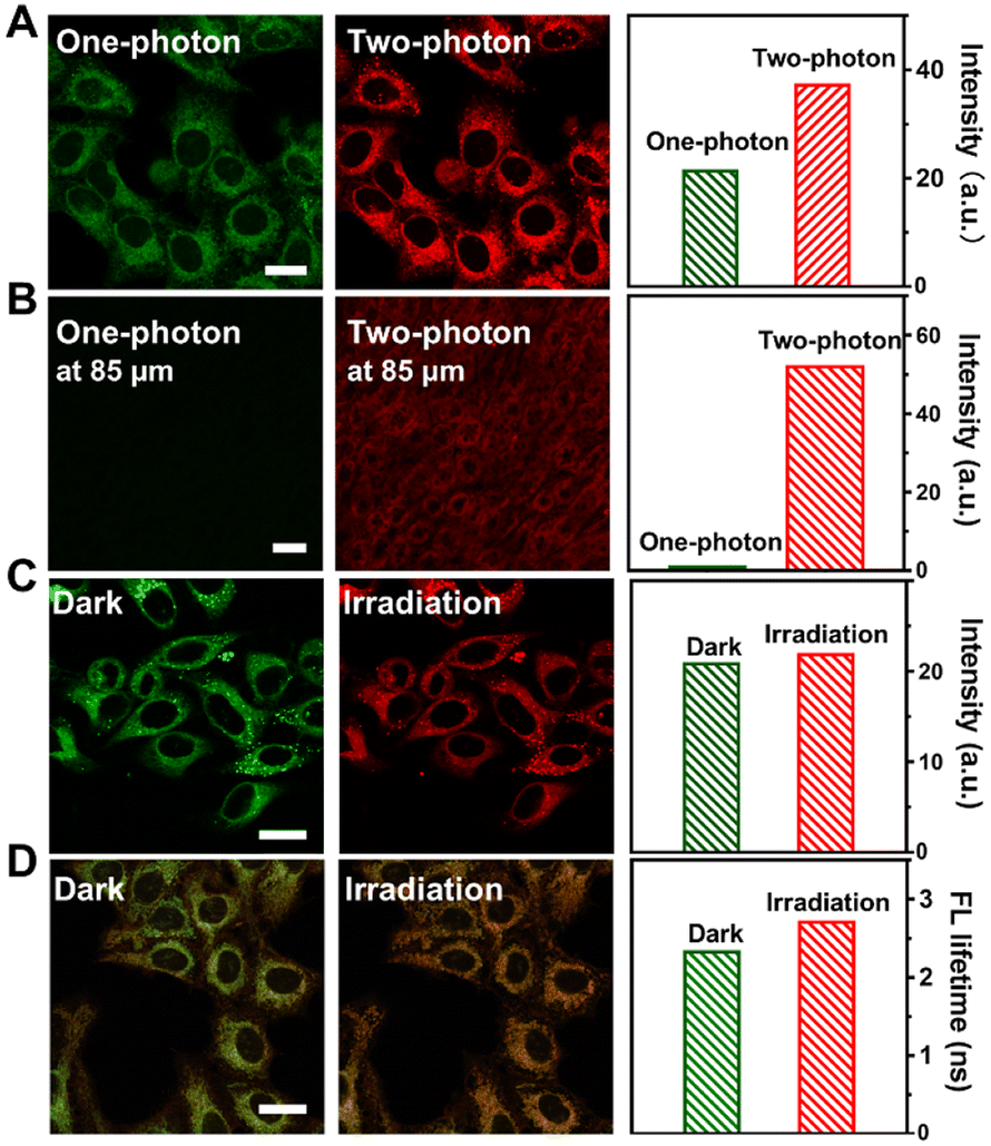 | ||
| Fig. 4 (A) One-/two-photon CLSM images of HepG2 cells treated with ZTBH (50 μg mL−1) upon irradiation of 500 nm and 850 nm (input laser power: 0.1 W cm−2), respectively (scale bar: 20 μm). (B) One-/two-photon CLSM fluorescence images of kidney tissue treated with ZTBH (50 μg mL−1) at 85 μm (λex: 500 nm, 850 nm; input laser power: 0.1 W cm−2; scale bar: 200 μm). (C) Two-photon CLSM images of HepG2 cells under dark and laser irradiation (λex: 850 nm; input laser power: 0.1 W cm−2; scale bar: 20 μm). (D) TP-FLIM images of HepG2 cells under dark and laser irradiation (λex: 850 nm; input laser power: 0.1 W cm−2; scale bar: 20 μm). | ||
Moreover, TP-FLIM guided real-time monitoring PDT was subsequently conducted using HepG2 tumor-bearing zebrafish embryos as integrated models (Fig. 5A). In comparison with one-photon fluorescence imaging, two-photon fluorescence imaging showed stronger fluorescence intensity and higher resolution (Fig. 5B). As depicted in Fig. 5C, the fluorescence lifetime exhibited a gradual increase with prolonged irradiation time, showcasing its potential for monitoring the PDT process in vivo. Collectively, the aforementioned experiments collectively demonstrated that ZTBH functions effectively as a TP-FLIM photosensitizer, providing precise real-time feedback during the PDT process.
 | ||
| Fig. 5 (A) Schematic diagram of a TP-FLIM guided PDT process of xenograft zebrafish embryo tumor model. (B) One- and two-photon fluorescence images of zebrafish incubated with ZTBH (50 μg mL−1; λex = 500/850 nm, 0.1 W cm−2; scale bar: 200 μm). (C) TP-FLIM images of zebrafish incubated with ZTBH (50 μg mL−1) with different irradiation time durations (λex = 850 nm, 0.1 W cm−2; scale bar: 200 μm). | ||
Conclusions
In conclusion, we have successfully developed a two-photon photosensitizer based on metal–organic frameworks, allowing for real-time feedback during photodynamic therapy through two-photon fluorescence lifetime imaging. Thanks to its excellent two-photon activity and ROS generation ability, ZTBH proves to be an efficient two-photon photosensitizer, triggering apoptosis under NIR laser irradiation. Moreover, the fluorescence lifetime of ZTBH undergoes significant changes in response to alterations in the cellular microenvironment during the PDT process, positioning it as a potential indicator for fluorescence lifetime imaging-guided PDT. This work establishes a promising design strategy for NIR-responsive two-photon fluorescence lifetime photosensitizer materials and provides their proof-of-concept applications in real-time feedback PDT.Author contributions
Bo Li: data curation, methodology, investigation, data curation and writing – original draft; Xin Lu: formal analysis, software, investigation, data curation and writing – original draft; Xianshun Sun: investigation, methodology and formal analysis; Hongping Zhou: formal analysis; Yupeng Tian: formal analysis; Zijuan Hai: formal analysis and writing – review and editing; Dandan Li: conceptualization, project administration, supervision and writing – review and editing.Data availability
The data supporting this article have been included as part of the ESI.†Conflicts of interest
The authors declare no competing financial interest.Acknowledgements
This work was supported by the National Natural Science Foundation of China (22171001, 22475001, 22305001 and 52372073), the Natural Science Foundation of Anhui Province (2408085Y010), and the China Postdoctoral Science Foundation (2024M760009).References
- T. C. Pham, V.-N. Nguyen, Y. Choi, S. Lee and J. Yoon, Recent strategies to develop innovative photosensitizers for enhanced photodynamic therapy, Chem. Rev., 2021, 121, 13454–13619 CrossRef CAS PubMed.
- J. Gao, Y. Tian, Y. Li, F. Hu and W. Wu, Design strategies for aggregation-induced emission photosensitizers with enhanced safety in photodynamic therapy, Coord. Chem. Rev., 2024, 507, 215756 CrossRef CAS.
- X. Zhao, J. Liu, J. Fan, H. Chao and X. Peng, Recent progress in photosensitizers for overcoming the challenges of photodynamic therapy: From molecular design to application, Chem. Soc. Rev., 2021, 50, 4185–4219 RSC.
- H. Chen, J. Yang, L. n Sun, H. Zhang, Y. Guo, J. Qu, W. Jiang, W. Chen, J. Ji, Y.-W. Yang and B. Wang, Synergistic chemotherapy and photodynamic therapy of endophthalmitis mediated by zeolitic imidazolate framework-based drug delivery systems, Small, 2019, 15, 1903880 CrossRef CAS PubMed.
- X.-Y. Lou, G. Zhang, N. Song and Y.-W. Yang, Supramolecular materials based on AIEgens for photo-assisted therapy, Biomaterials, 2022, 286, 121595 CrossRef CAS PubMed.
- S. Liang, M.-H. Li, M.-L. Qi, H. Hui, H.-P. Zhang, J. Zhou, L. Wang and Y.-W. Yang, Reactive oxygen species-responsive pillararene-embedded covalent organic frameworks with amplified antimicrobial photodynamic therapy for the targeted elimination of periodontitis pathogens, Nano Lett., 2024, 24, 13708–13717 CrossRef CAS PubMed.
- E. Kamya, Z. Lu, Y. Cao and R. Pei, Effective design of organic luminogens for near-infrared-II fluorescence imaging and photo-mediated therapy, J. Mater. Chem. B, 2022, 10, 9770–9788 RSC.
- L. Ma, Y. Wang, X. Wang, Q. Zhu, Y. Wang, L. Li, H.-B. Cheng, J. Zhang and X.-J. Liang, Transition metal complex-based smart AIEgens explored for cancer diagnosis and theranostics, Coord. Chem. Rev., 2022, 473, 214822 CrossRef CAS.
- J.-F. Yu, Y. Wen and M. Li, Sterically twisted mitochondria-targeting photosensitizers for augmented near-infrared II fluorescence–guided photodynamic immunotherapy, Adv. Funct. Mater., 2024, 38, 2402663 CrossRef.
- X. Zhang, Y. Dou, S. Liu, P. Chen, Y. Wen, J. Li, Y. Sun and R. Zhang, Adv. Healthcare Mater., 2024, 13, 2303842 CrossRef CAS PubMed.
- X. Gu, J. Shen, Z. Xu, W. Wang, Y. Wu, W. Zhou, C. Xie and Q. Fan, Rational design of biodegradable semiconducting polymer nanoparticles for NIR-II fluorescence imaging-guided photodynamic therapy, Nano Res., 2024, 17, 5399–5408 CrossRef CAS.
- N. Song, Z. Zhang, P. Liu, D. Dai, C. Chen, Y. Li, L. Wang, T. Han, Y.-W. Yang, D. Wang and B. Z. Tang, Pillar[5]arene-modified gold nanorods as nanocarriers for multi-modal imaging-guided synergistic photodynamic-photothermal therapy, Adv. Funct. Mater., 2021, 31, 2009924 CrossRef CAS.
- J. Sha, W. Liu, X. Zheng, Y. Guo, X. Li, H. Ren, Y. Qin, J. Wu, W. Zhang, C.-S. Lee and P. Wang, Polarity-sensitive probe for two-photon fluorescence lifetime imaging of lipid droplets in vitro and in vivo, Anal. Chem., 2023, 95, 15350–15356 CrossRef CAS PubMed.
- M. Y. Berezin and S. Achilefu, Fluorescence lifetime measurements and biological imaging, Chem. Rev., 2010, 110, 2641–2684 CrossRef CAS PubMed.
- Y. Ouyang, Y. Liu, Z. M. Wang, Z. Liu and M. Wu, FLIM as a promising tool for cancer diagnosis and treatment monitoring, Nano–Micro Lett., 2021, 13, 133 CrossRef CAS PubMed.
- R. Datta, T. M. Heaster, J. T. Sharick, A. A. Gillette and M. C. Skala, Fluorescence lifetime imaging microscopy: fundamentals and advances in instrumentation, analysis, and applications, J. Biomed. Opt., 2020, 25, 07120 Search PubMed.
- W. Liang, D. Chen, H. Guan, H.-C. Park, K. Li, A. Li, M.-J. Li, I. Gannot and X. Li, Label-free metabolic imaging in vivo by two-photon fluorescence lifetime endomicroscopy, ACS Photonics, 2022, 9, 4017–40296 CrossRef CAS PubMed.
- Y. Song, H. Zhang, X. Wang, X. Geng, Y. Sun, J. Liu and Z. Li, One stonethree birds: pH triggered transformation of aminopyronine and iminopyronine based lysosome targeting viscosity probe for cancer visualization, Anal. Chem., 2021, 93, 1786–1791 CrossRef CAS PubMed.
- Z. Wu, M. Liu, Z. Liu and Y. Tian, Real-time imaging and simultaneous quantification of mitochondrial H2O2 and ATP in neurons with a single two-photon fluorescence lifetime-based probe, J. Am. Chem. Soc., 2020, 142, 7532–7541 CrossRef CAS PubMed.
- W. Li, X. Li, C. Zhang, H. Wang, Y. Zhu, Y. Wang, W. Yan, L. Liu and J. Qu, Research of pulmonary fibrosis lesions based on FLIM and SHG imaging microscopy, Anal. Chem., 2024, 96, 12286–12295 CAS.
- L.-L. Li, K. Li, M.-Y. Li, L. Shi, Y.-H. Liu, H. Zhang, S.-L. Pan, N. Wang, Q. Zhou and X.-Q. Yu, BODIPY-based two-photon fluorescent probe for real-time monitoring of lysosomal viscosity with fluorescence lifetime imaging microscopy, Anal. Chem., 2018, 90, 5873–5878 CrossRef CAS PubMed.
- X. Wang, G. Shi, S. Xu, Y. Sun, H. Qiu, Q. Wang, X. Han, Q. Zhang, T. Zhang and H.-Y. Hu, Unravelling immune-inflammatory responses and lysosomal adaptation: insights from two-photon excited delayed fluorescence imaging, Adv. Healthcare Mater., 2024, 13, 2304223 CrossRef CAS PubMed.
- B. Li, S. Yu, R. Feng, Z. Qian, K. He, G.-J. Mao, Y. Cao, K. Tang, N. Gan and Y.-X. Wu, Dual-mode gold nanocluster-based nanoprobe platform for two-photon fluorescence imaging and fluorescence lifetime imaging of intracellular endogenous miRNA, Anal. Chem., 2023, 95, 14925–14933 CrossRef CAS PubMed.
- T. Kowada, H. Maeda and K. Kikuchi, BODIPY-based probes for the fluorescence imaging of biomolecules in living cells, Chem. Soc. Rev., 2015, 44, 4953 RSC.
- A. Kamkaew, S. H. Lim, H. B. Lee, L. V. Kiew, L. Y. Chung and K. Burgess, BODIPY dyes in photodynamic therapy, Chem. Soc. Rev., 2013, 42, 77 RSC.
- A. Loudet and K. Burgess, BODIPY dyes and their derivatives: Syntheses and spectroscopic properties, Chem. Rev., 2007, 107, 4891–4932 CrossRef CAS PubMed.
- H.-G. Jin, P.-C. Zhao, Y. Qian, J.-D. Xiao, Z.-S. Chao and H.-L. Jiang, Metal–organic frameworks for organic transformations by photocatalysis and photothermal catalysis, Chem. Soc. Rev., 2024, 53, 9378–9418 RSC.
- H.-Y. X.-J. Kong, S.-D. Han, J. Pang, T. He, G.-M. Wang and X.-H. Bu, Metalation of metal–organic frameworks: fundamentals and applications, Chem. Soc. Rev., 2024, 53, 5626–5676 RSC.
- G. Zhang, Y. Shen, J. Phipps, L. Sun and S. Ma, Metal–organic frameworks for the diagnosis and treatment of Alzheimer's disease: Current status and perspectives, Coord. Chem. Rev., 2024, 518, 216059 CrossRef CAS.
- C. Zhang, Z. Lin, L. Jiao and H.-L. Jiang, Metal–organic frameworks for electrocatalytic CO2 reduction: From catalytic site design to microenvironment modulation, Angew. Chem., Int. Ed., 2024, 63, e202414506 CrossRef CAS PubMed.
- L. Jiao, J. Wang and H.-L. Jiang, Microenvironment modulation in metal–organic framework-based catalysis, Acc. Mater. Res., 2021, 2, 327–339 CrossRef CAS.
- J. Yang and Y.-W. Yang, Metal–organic frameworks for biomedical applications, Small, 2020, 16, 1906846 CrossRef CAS PubMed.
- J. Yang, D. Dai, X. Zhang, L. Teng, L. Ma and Y.-W. Yang, Multifunctional metal–organic framework (MOF)-based nanoplatforms for cancer therapy: from single to combination therapy, Theranostics, 2023, 13, 295–323 CrossRef CAS PubMed.
- D. Wang, H. Yao, J. Ye, Y. Gao, H. Cong and B. Yu, Metal–organic frameworks (MOFs): Classification, synthesis, modification, and biomedical applications, Small, 2024, 20, 2404350 CrossRef CAS PubMed.
- A. E. Amooghin, H. Sanaeepur, M. Ghomi, R. Luque, H. Garcia and B. Chen, Flexible-robust MOFs/HOFs for challenging gas separations, Coord. Chem. Rev., 2024, 505, 215660 CrossRef.
- B. Mohanty, S. Kumari, P. Yadav, P. Kanoo and A. Chakraborty, Metal–organic frameworks (MOFs) and MOF composites based biosensors, Coord. Chem. Rev., 2024, 519, 216102 CrossRef CAS.
- L. Zhu, B. Zhu, J. Luo and B. Liu, Design and property modulation of metal–organic frameworks with aggregation-induced emission, ACS Mater. Lett., 2021, 3, 77–89 CrossRef CAS.
- M. D. Allendorf, C. A. Bauer, R. K. Bhakta and R. J. T. Houk, Luminescent metal–organic frameworks, Chem. Soc. Rev., 2009, 38, 1330–1352 RSC.
- Y. Cui, J. Zhang, H. He and G. Qian, Photonic functional metal–organic frameworks, Chem. Soc. Rev., 2018, 47, 5740–5785 RSC.
- B. Li, X. Lu, Y. Tian and D. Li, Embedding multiphoton active units within metal–organic frameworks for turning on high-order multiphoton excited fluorescence for bioimaging, Angew. Chem., Int. Ed., 2022, 61, e202206755 CrossRef CAS PubMed.
- H. M. Kim and B. R. Cho, Small-molecule two-photon probes for bioimaging applications, Chem. Rev., 2015, 115, 5014–5055 CrossRef CAS PubMed.
- Y.-B. Tian, Q.-H. Li, Z. Wang, Z.-G. Gu and J. Zhang, Coordination-induced symmetry breaking on metal–porphyrinic framework thin films for enhanced nonlinear optical limiting, Nano Lett., 2023, 23, 3062–3069 CrossRef CAS PubMed.
- S. Yang, Y. Sun, K. Geng, Y. Cui, J. Huang, X. Meng and H. Hou, Investigating the third-order NLO performance of a three-phase core–shell PANI@MIL-101(Cr)@CeO2 with internal and external synergies, Adv. Opt. Mater., 2024, 12, 2400475 CrossRef CAS.
- X. Jiang, L. Zhang, S. Liu, Y. Zhang, Z. He, W. Li, F. Zhang, Y. Shi, W. Lü, Y. Li, Q. Wen, J. Li, J. Feng, S. Ruan, Y.-J. Zeng, X. Zhu, Y. Lu and H. Zhang, Ultrathin metal–organic framework: an emerging broadband nonlinear optical material for ultrafast photonics, Adv. Opt. Mater., 2018, 6, 1800561 CrossRef.
- C. Xu, H. Liu, D. Li, J.-H. Su and H.-L. Jiang, Direct evidence of charge separation in a metal–organic framework: efficient and selective photocatalytic oxidative coupling of amines via charge and energy transfer, Chem. Sci., 2018, 9, 3152 RSC.
- J. Karges, U. Basu, O. Lacque, H. Chao and G. Gasser, Polymeric encapsulation of novel homoleptic bis(dipyrrinato) Zinc(II) complexes with long lifetimes for applications as photodynamic therapy photosensitisers, Angew. Chem., Int. Ed., 2019, 58, 14334–14340 CrossRef CAS PubMed.
- Y. Cai, P. Liang, Q. Tang, X. Yang, W. Si, W. Huang, O. Zhang and X. Dong, Diketopyrrolopyrrole–triphenylamine organic nanoparticles as multifunctional reagents for photoacoustic imaging-guided photodynamic/photothermal synergistic tumor therapy, ACS Nano, 2017, 11, 1054–1063 CrossRef CAS PubMed.
- W. Cai, H. Gao, C. Chu, X. Wang, J. Wang, P. Zhang, G. Lin, W. Li, G. Liu and X. Chen, Engineering phototheranostic nanoscale metal–organic frameworks for multimodal imaging-guided cancer therapy, ACS Appl. Mater. Interfaces, 2017, 9, 2040–2051 CrossRef CAS PubMed.
- K. Kim, S. Lee, E. Jin, L. Palanikumar, J. H. Lee, J. C. Kim, J. S. Nam, B. Jana, T.-H. Kwon, S. K. Kwak, W. Choe and J.-H. Ryu, MOF × biopolymer: Collaborative combination of metal–organic framework and biopolymer for advanced anticancer therapy, ACS Appl. Mater. Interfaces, 2019, 11, 27512–27520 CrossRef CAS PubMed.
- B. Li, H. Cao, J. Zheng, B. Ni, X. Lu, X. Tian, Y. Tian and D. Li, Click modification of a metal–organic framework for two-photon photodynamic therapy with near-infrared excitation, ACS Appl. Mater. Interfaces, 2021, 13, 9739–9747 CrossRef CAS PubMed.
- B. Li, X. Yu, J. Wang, H. Tang, X. Sun, L. Cheng, H. Zhou, Y. Tian and D. Li, Unlocking efficient high-order multiphoton-excited fluorescence of metal–organic framework via octupolar module in situ formation, Adv. Funct. Mater., 2023, 33, 2305391 CrossRef CAS.
Footnote |
| † Electronic supplementary information (ESI) available. See DOI: https://doi.org/10.1039/d4qi03014a |
| This journal is © the Partner Organisations 2025 |