
More than a delivery system: the evolving role of lipid-based nanoparticles
Senjuti
Karmaker†
,
Plinio D.
Rosales†
,
Barath
Tirumuruhan†
 ,
Amartya
Viravalli†
and
Natalie
Boehnke
*
,
Amartya
Viravalli†
and
Natalie
Boehnke
*
Department of Chemical Engineering and Materials Science, University of Minnesota, Twin Cities Minneapolis, MN 55455, USA. E-mail: nboehnke@umn.edu
First published on 2nd April 2025
Abstract
Lipid-based nanoparticles, including liposomes and lipid nanoparticles (LNPs), make up an important class of drug delivery systems. Their modularity enables encapsulation of a wide range of therapeutic cargoes, their ease of functionalization allows for incorporation of targeting motifs and anti-fouling coatings, and their scalability facilitates rapid translation to the clinic. While the discovery and early understanding of lipid-based nanoparticles is heavily rooted in biology, formulation development has largely focused on materials properties, such as how liposome and lipid nanoparticle composition can be altered to maximize drug loading, stability and circulation. To achieve targeted delivery and enable improved accumulation of therapeutics at target tissues or disease sites, emphasis is typically placed on the use of external modifications, such as peptide, protein, and polymer motifs. However, these approaches can increase the complexity of the nanocarrier and complicate scale up. In this review, we focus on how our understanding of lipid structure and function in biological contexts can be used to design intrinsically functional and targeted nanocarriers. We highlight formulation-based strategies, such as the incorporation of bioactive lipids, that have been used to modulate liposome and lipid nanoparticle properties and improve their functionality while retaining simple nanocarrier designs. We also highlight classes of naturally occurring lipids, their functions, and how they have been incorporated into lipid-based nanoparticles. We will additionally position these approaches into the historical context of both liposome and LNP development.
 Natalie Boehnke | Natalie Boehnke received her PhD in organic chemistry from UCLA in 2017. After completing a postdoc at the Koch Institute for Integrative Cancer Research at MIT, which focused on engineering targeted nanomaterials for drug delivery applications, she joined the Chemical Engineering and Materials Science Department at the University of Minnesota as an assistant professor in 2022. Natalie's current research interests are inspired by her multidisciplinary background, focusing on next-generation tools to advance nanomedicine. Specifically, her group takes a data driven approach to nanocarrier design, developing barcoded and pooled screening approaches and leveraging bioactive building blocks to develop intrinsically therapeutic drug delivery systems. |
Introduction
Lipid-based nanoparticles have emerged as a groundbreaking platform for drug delivery over the past 60 years.1 As the name suggests, lipid-based nanoparticles include nanoparticle constructs that are composed of lipids, including liposomes, lipid nanoparticles (LNPs), solid lipid nanoparticles (SLNs), and nanostructured lipid carriers (NLCs).2,3 In this review, we will limit our focus to liposomes and LNPs. The evolution of lipid-based nanoparticles began with the discovery of the liposome structure in the 1960s4 and has progressed to their development into sophisticated therapeutic nanocarriers. The modularity, biodegradability and biocompatibility of lipid-based nanoparticles have made them the gold standard for drug delivery. Lipid-based nanocarriers currently make the largest proportion of nanocarriers in clinical trials.5Researchers in the field have primarily focused on the use of external modifications, such as peptides, proteins, and polymers, to impart lipid-based nanoparticles with additional functionality, including the use of targeting and immunomodulatory motifs.6–8 This additive approach to designing functional nanocarriers can increase nanocarrier complexity and result in scale up and manufacturing challenges, hindering translational efforts.9,10 Moreover, from an ‘atom economy’ perspective, the majority of the nanocarrier is composed of lipids that serve only a carrier function, while the therapeutic itself constitutes an additional, and often smaller, portion.11 This prompted the question whether an alternative approach to synthesizing functional lipid-based nanoparticles that leverages the inherent functionality of the lipid building blocks could address the limitations of additive nanocarrier design.
In this review, we focus on recent advances in lipid-based nanoparticle design which leverage existing understanding of lipid structure and function to engineer structurally simple yet intrinsically multifunctional carriers. The review begins by providing brief overviews of liposomes and LNPs in addition to a historical context of the development of these nanocarriers (Fig. 1). We then categorize recent formulation strategies into two key approaches – (1) incorporation of bioactive lipids and (2) modifying nanoparticle physical properties. These strategies enable functionalities beyond delivery such as targeting, immunomodulation, and antimicrobial activity. Specifically, we further classify bioactive lipid incorporation into two sub-categories, naturally occurring lipids and synthetically modified lipids. We highlight the vastness of naturally occurring lipids that are underexplored and can be leveraged to design functional nanoparticles. Ultimately, we hope this review inspires a multi-disciplinary effort in paving the way for the next generation of lipid-based nanoparticles.
 | ||
| Fig. 1 Timeline of key discoveries and advancements in lipid-based nanoparticle development. | ||
General overview of liposomes
Liposomes consist of amphiphilic phospholipid molecules that organize into bilayers in aqueous environments.12,13 Due to their amphiphilic nature, liposomes serve as versatile nanocarriers capable of transporting both hydrophobic and hydrophilic molecules, including small molecules, nucleic acids, proteins, and imaging agents, with hydrophobic drugs integrating into the lipid bilayer and hydrophilic drugs encapsulated within the aqueous core.14,15Liposome composition and structure
Anti-fouling coatings
Despite their advantages, liposomes face challenges like short circulation times and limited stability due to rapid clearance by the mononuclear phagocyte system (MPS). To overcome this, anti-fouling polymers, like polyethylene glycol (PEG), can be added to create “stealth” liposomes, providing steric protection that prolongs circulation by evading opsonization.12,38–40 However, in recent years, PEG and PEG-lipid conjugates have been associated with drawbacks, such as hypersensitivity reactions with repeated administration and complement system activation, which can amplify the clearance of these nanocarriers.41–44 To mitigate these issues, alternative materials, such as polysialic acids, glycoproteins, zwitterionic polymers, and polysaccharides, are being explored to enhance the circulation time of liposomes.45–47Targeting motifs
Liposomes can be functionalized with small molecule ligands, carbohydrates, proteins and nucleic acid aptamers to enhance the targeting of liposomes, thereby improving drug efficacy and reducing side effects.14,48 Current efforts are focused on decorating the surface of liposomes with ligands that can target and bind to receptors overexpressed on the surfaces of cancer cells. Common targets include transferrin receptor (TfR), folate receptor (FR), and the epidermal growth factor receptor (EGFR).14 Additionally, stimuli-responsive liposomes have been designed to release their cargo in response to specific environmental triggers such as pH, temperature, enzymes, or redox conditions within the tumor microenvironment, reducing premature degradation of the cargo and enhancing treatment efficacy.49,50Despite extensive efforts to enhance liposome functionality through external modifications, challenges remain in evading premature clearance, addressing receptor expression heterogeneity, and minimizing ‘on-target, off-site’ effects, where liposomes bind to the intended receptor but accumulate in tissues other than the intended target.
As an alternative approach to targeted and controlled delivery, this review will focus on how lipid-based nanoparticle formulation components can be strategically selected, designed, and incorporated to modulate nanocarrier properties. This, in turn, can improve targeting efficacy, immune response, stability in blood circulation, antimicrobial activity and additional functions such as antitumor effects, transfection efficiency and stimuli-responsiveness.
General overview of LNPs
In contrast to liposomes, LNPs are considered to have a less ordered structure. The primary focus of LNP development has been on nucleic acid encapsulation and delivery, with current efforts largely focused on synthesizing LNP formulations for delivery beyond the liver.LNP composition and structure
Targeting strategies
Similar to liposomes, LNPs can be functionalized externally using ligands like antibodies, proteins, and aptamers to enhance LNP targeting.73–76 For instance, Palanki et al. were able to actively target the CD45 receptor for in-utero delivery of targeted ionizable lipid nanoparticles.77 Aptamers have gained significant interest due to their size (6–30 kDa), which enables them to bind to smaller targets or hidden binding domains which are inaccessible for antibodies.78,79Despite various efforts to incorporate external modifications using ligands, receptors, or aptamers to lipid-based nanoparticles to improve functionality, challenges remain in targeted delivery, extrahepatic delivery in the context of LNPs, and immunomodulation and evasion. This review will emphasize how lipid-based nanoparticle formulations can be leveraged to modulate biological responses and functions using both natural and synthetic lipids as building blocks. This, in turn, can improve targeting efficacy, immune response, stability in blood circulation, antimicrobial activity and additional functions such as transfection efficiency and stimuli-responsiveness.
Timeline of lipid-based nanoparticle development
Early exploration
The early development of lipid-based nanoparticles finds its roots in the study of biological membranes and the fundamental inquiry of “what holds biological membranes together”. In 1964, Alec Bangham published the first electron microscopy image of vesicles formed by ovolecithin, a natural phospholipid extracted from egg, following an accidental discovery made while studying the role of phospholipids in blood clotting.4 Initially referred to as “multi-lamellar smectic mesophases” or “banghosomes”, these vesicles were later termed “liposomes” by Gerald Weissmann.15 Bangham's pioneering work on ion sequestration and transport in these vesicles sparked interest in using phospholipid liposome models for biological membranes and as potential drug delivery vehicles.80,81,82–87The progression from liposome discovery to their application in drug delivery unfolded at a remarkable pace. In 1970, Gerald Weissmann provided the first experimental evidence of therapeutic loading into nanoparticles by encapsulating lysozyme into liposomes, positing their use for enzyme replacement therapy.88 Subsequently, Brenda Ryman and Gregory Gregoriadis’ pioneering work reported the delivery of Aspergillus niger amyloglucosidase and radiolabeled albumin encapsulated in liposomes to liver and spleen—the main organs of the reticuloendothelial system (RES), marking the first nanoparticle biodistribution study.89 Notably, the observed challenges associated with RES clearance of nanoparticles still impede clinical translation to this day. Subsequent studies rapidly explored the encapsulation of a range of therapeutics, including proteins, enzymes, hormones, and small molecules, targeting diseases like cancer, microbial infections, and metabolic conditions.90,91
In parallel, several early investigations also aimed at interpreting liposome interactions with the biological environment. In vitro studies investigated the effect of physicochemical properties such as composition, fluidity, charge, and size on liposome uptake mechanisms.92–95 Building on these insights, in vivo studies revealed how these properties modulate pharmacokinetics and biodistribution of encapsulated therapeutic cargo.96–98 This prompted the development of several liposome synthesis techniques to enhance these physicochemical properties including ethanol dilution, thin lipid membrane hydration, and high-pressure extrusion.99–106 Around the same time, the concept of using liposomes for immune modulation was introduced by Gregoriadis and Allison, who found that an inactivated form of diphtheria toxoid loaded into negatively charged liposomes elicited higher antibody responses compared to free antigens or antigen loaded into positively charged liposomes.107,108
Formulation refinement
In the 1980s and 1990s, efforts to resolve key challenges in liposome technology, including drug leakage97,109–111 and immune detection, to enhance intracellular delivery,112 and to develop scalable synthesis protocols paved the way for the founding of companies like The Liposome Company, Liposome Technology Inc., Vical, and Acuitas, and the first clinical translation of lipid-based nanotherapeutics.112–114Research efforts focused on optimizing formulations to enhance drug loading and retention, including the incorporation of cholesterol to modulate the nanoparticle's membrane fluidity and bilayer thickness.115,116 Another change was the shift from using lipids that were solids in physiological temperatures instead of fluids, which reduced drug leakage and liposome degradation.117 Remote loading was also introduced as an approach that uses transmembrane pH or ion gradients to achieve high drug-to-lipid ratio loading and up to 100% encapsulation efficiency.118–125
There was also a focus to address the premature clearance of administered liposomes, including pre-dosing with ‘empty’ liposomes to block the RES system126–128 and size modifications.129,130 Interestingly, the former concept was revisited in Chan et al.'s recent work identifying a nanoparticle dose threshold for blocking liver's Kupffer cells.131 Concurrently, in late-1970s, it was hypothesized that liposome opsonization by serum proteins causes accumulation in the RES organs.132–134 Early reports of increased liposome circulation half-life involved surface modifications with monoasialoglycoprotein GM1 and substituting egg PC with sphingomyelin.135 These ‘stealth’ liposomes avoided RES clearance. In 1990, Klibanov et al. reported in their seminal work that grafting polyethylene glycol (PEG) chains onto the liposome surface enhanced circulation,136 using hydrophilic interactions to repel opsonins.137–141
The cumulative outcome of addressing drug leakage via remote loading and clearance via PEGylation led to the development of Doxil, doxorubicin-loaded PEGylated liposomes, the first FDA-approved nanoparticle-based therapeutic in the US in 1995 after successful trials demonstrating prolonged circulation and improved safety.142,143 The rapid translation of liposomes from discovery to FDA approval in just thirty years underscores the potential of nanoparticles as drug delivery vehicles.
Intracellular and targeted delivery
The use of nanocarriers to serve as ‘magic bullets’144,145 and improve accumulation of therapeutic cargo at target disease sites has been a long-standing challenge in the field. Targeted nanocarriers have traditionally been designed with external targeting motifs. This concept originated from attempts to improve intracellular delivery of liposomes, which were substantially larger and more complex compared to biomolecules typically endocytosed by cells. Initial attempts focused on the use of antibodies to enhance the homing of nanoparticles and utilize receptor-mediated endocytosis for uptake.146–150 This concept, which is referred to as active or ligand-based targeting, was expanded to include targeting of cell surface receptors via the addition of ligands to nanoparticle surfaces to facilitate localization to target tissues, disease sites, and cell populations. Many classes of molecules capable of targeting cell surface receptors, including vitamins, peptides, and carbohydrates, have been since explored. Lipid-based nanoparticles modified with tissue and cell-penetrating peptides, polysaccharides, and proteins are a few of the notable strategies that have been explored for clinical use.151–167 However, clinical translation remains elusive because of factors including toxicity, difficulty navigating biological barriers, serum protein interaction, and disease specific heterogeneities.168–170Lipid-based nanoparticles for nucleic acid delivery
In addition to small molecule delivery and targeting efforts, the field quickly recognized the potential of lipid-based nanocarriers to deliver a different class of therapeutics: nucleic acids. Early efforts to encapsulate nucleic acids in nanoparticles showed promise, but challenges including low encapsulation efficiency and charge-based repulsion between anionic nucleic acids and liposomes presented early barriers to translation.171–176 In 1987, Phil Felgner created lipoplexes that achieved 100% DNA encapsulation by complexing DNA with the permanent cationic lipid DOTMA.177 This process was refined to create Lipofectin, a commercial carrier with excellent in vitro DNA and RNA transfection.178,179 However, the in vivo success of cationic liposomes was hindered due to immunogenicity imparted by the overall positive charge of the nanocarrier.180–182Stable plasmid lipid particles (SPLPs) represent an early attempt at mitigating immunogenicity. SPLPs are formed by detergent dialysis to incorporate small amounts of a permanent cationic lipid, DODAC, along with PC or PE, cholesterol, and PEG-lipids. SPLPs formed particles with a plasmid-encapsulated lipid complex enclosed by a bilayer.183 SPLPs achieved 70% plasmid trapping efficiency while maintaining charge neutrality and demonstrated effective transfection at tumor sites in several murine models. However, the detergent dialysis process presented challenges for scale up, preventing its clinical translation. Despite this, the promising encapsulation efficiency and transfection outcomes of SPLPs helped evolve LNPs as specialized nucleic acid carriers, making SPLPs precursors to modern LNPs.
An alternative to permanent cationic lipids was the incorporation of ionizable lipids into LNPs. These lipids have apparent pKa values slightly below physiological pH. This enables efficient nucleic acid complexation and retention of neutral nanoparticle surface charge while facilitating endosomal escape. The first ionizable lipid, 1,2-dioleoyl-3-dimethylammonium propane (DODAP),184 was incorporated into LNPs for the delivery of antisense oligonucleotides, which achieved approximately 90% nucleotide encapsulation and a circulation half-life of around six hours in ICR mice.185 These LNPs were termed stabilized antisense lipid particles (SALPs). The pH-dependent modulation of lipid charge continues to be leveraged to induce endosomal escape and enhance intracellular nucleic acid delivery, resulting in particles referred to as stable nucleic acid-lipid particles (SNALP),186,187 optimized for encapsulating and delivering siRNA. DODAP used in SALPs evolved into the “DLinDMA” lipid family, from which 4-(N,N-dimethylamino)butyric acid (dilinoleyl) methyl ester, DLinMC3DMA (MC3), emerged as the present “gold standard” for siRNA delivery.52
In addition to nucleic acid delivery, current efforts are on the use of LNPs for the encapsulation and delivery of small molecules and protein therapeutics, including for CRISPR/Cas9 delivery.188 Challenges in CRISPR/Cas9 delivery include the development of optimized delivery vectors, ensuring biocompatibility, and achieving efficient and targeted delivery to target cells.189,190 LNPs have been successfully used to incorporate and deliver CRISPR/Cas9 technology,188,191–193 including through the encapsulation of plasmid DNA (pDNA) encoding both Cas9 protein and gRNA or pDNA encoding Cas9 protein in combination with gRNA oligos, or Cas9 mRNA and gRNA.
Clinical translation
Several liposomes were successfully translated into the clinic following Doxil® (1995) for a broad range of applications, including VYXEOS for acute myeloid leukemia, and Onivyde for metastatic pancreatic cancer.5,194,195 In recent years, there has been growing interest in using liposomes in combination therapy,196 such as liposomal irinotecan and nituzumab for nasopharyngeal carcinoma, currently in phase 2 trials (NCT06414577), and liposomal nemvaleukin combined with pembrolizumab for platinum resistant ovarian cancer, currently in phase 3 (ARTISTRY-7, NCT05092360) trials.On the LNP front, research showed that DLinMC3DMA-based LNPs preferentially accumulated in the liver due to Apolipoprotein B adsorption, which facilitates receptor-mediated endocytosis by hepatocytes.197 Additionally, these LNPs were two orders of magnitude more potent than DLinDMA-containing LNPs in silencing transthyretin (TTR) protein in the liver.52 Together, these factors resulted in the FDA approval of the first RNAi therapeutic, Onpattro®, in 2018.66
The COVID-19 pandemic spurred the unparalleled development and subsequent FDA approval of mRNA vaccines, Pfizer/BioNTech's Comirnaty®61 and Moderna's Spikevax®,67 using ionizable lipids ALC-0315 and SM-102, respectively.198–203 Post-pandemic, LNP research has expanded to include cholesterol variants, biodegradable ionizable lipids, SORT lipids, and CRISPR/Cas9. These advances will be discussed in detail in the upcoming sections. Several LNP formulations that are currently in clinical trials have been reviewed previously.14,30,204–206 Notable LNP formulations in clinical trials include Pfizer-BioNTech's BNT122-01 for colorectal cancer, Moderna's mRNA-4157 for melanoma, CureVac's CV7202 for rabies, and Moderna's mRNA-1345 for respiratory syncytial virus.
Evolving role of lipid-based nanoparticle systems
It is interesting to note that, while the discovery and use of lipid-based nanoparticles originated from biological studies where researchers observed that lipid membranes enable cells to segregate their internal components from the external environment,207 efforts to optimize lipid-based nanoparticles quickly shifted toward optimization of non-biological parameters, such as drug loading, stability and release, and the use of external modifications to add stealth and targeting capabilities. Lipids, as essential components of biological systems, serve a variety of functions, from modulating cell membrane fluidity to acting as signaling molecules that control cellular processes. The versatility and modularity of both lipid structure and function has inspired the creation of intrinsically functional nanocarriers, which we define as lipid-based nanoparticles in which a formulation component has been leveraged to serve a purpose beyond cargo encapsulation and stabilization. One main approach is through the use of bioactive lipids, which we define as both natural and synthetic lipids that serve a biological role or function. These formulations consist of relatively simple designs, helping to circumvent challenges associated with the complexity of multicomponent nanoparticles that rely on exogenous stealth and targeting modalities.In this section we will explore how bioactive lipids have been incorporated into lipid-based nanoparticles as well as their subsequent uses. We will additionally provide examples on how formulation components can be used to modulate mechanical properties of lipid-based nanoparticles for added functionality, including tumor penetration, enhanced blood circulation, and immune cell targeting (Table 1).
| Type of lipid | Lipid replaced or formulation modified | NP form | Function(s) altered | Ref. |
|---|---|---|---|---|
| Natural | N-Octanoylglucosylceramide | Lipid nanovesicle | Biodistribution | 274 |
| Monophosphoryl lipid A | Liposome | Immune response | 290 | |
| αGalCer-containing oligomannose-ooated liposomes (Mannotriose-DPPEconj) | Liposome | Immune response | 294 | |
| Saponin monophosphoryl lipid A (SMNP) | SMNP | Immune response | 296 and 297 | |
| Cholesteryl oleate, 7α-hydroxycholesterol, 7β-hydroxycholesterol, and 4β-hydroxycholesterol | LNP | Nucleic acid delivery, transfection | 406 | |
| 7α-hydroxycholesterol | LNP | Nucleic acid delivery, transfection | 57 | |
| Sphingomyelin | Liposome | Sequester bacterial endotoxins | 248 | |
| Egg PC | Liposome | Therapeutic efficacy | 233 | |
| Ceramide | Liposome | Therapeutic efficacy | 260 | |
| N-Octanoylglucosylceramide | Liposome | Therapeutic efficacy | 274 | |
| Cholesterol | Liposome | Therapeutic efficacy, tumor penetration | 22 | |
| Archaeal ether lipids | LNP | Transfection, tumor accumulation | 277 | |
| Cholesterol | Liposome | Tumor accumulation | 21 | |
| Synthetic | Polyglycerol | Liposome | Circulation time | 329 |
| Polyglycerol fatty acid esters | Liposome | Circulation time | 331 | |
| Folic-acid conjugated to DSPE-PEG | Liposome | Circulation time | 332 | |
| Polycarboxybetaine (PCB) variants | Liposome | Circulation time | 334 | |
| Polysialic acid-octadecyl dimethyl betaine (PSA-BS18) | Liposome | Circulation time | 335 | |
| CD47 mimicry self-peptide | Liposome | Circulation time | 338 | |
| Dimethyldioctadecylammonium (DDA) | Liposome | Immune response | 400 | |
| Cetyltrimethylammonium bromide (CTAB) and DDAB | Liposome | Targeting | 323 | |
| pSar BA12-50 | LNP | Transfection, nucleic acid delivery, immune response, immune evasion | 301 | |
| DDAB | LNP | Transfection, uptake, biodistribution, tumor accumulation | 315 | |
| Natural & synthetic | DOPE, DOPS, DOTAP | LNP | Biodistribution, transfection, nucleic acid delivery | 300 |
| DODAP, DOTAP, 18PA | LNP | Transfection. therapeutic efficacy, biodistribution | 362 | |
Use of natural bioactive lipids
The inherent biocompatibility and cellular recognition of naturally occurring lipids make them appealing building blocks for the design of intrinsically therapeutic nanocarriers (Fig. 2). In this section, we highlight how naturally occurring bioactive lipids are being used to create lipid-based nanoparticles for a range of biomedical applications. Beyond their role in maintaining structural integrity of cell membranes,207–210 lipids are involved in energy metabolism,211–214 signaling pathways,46,215,216 protein–lipid interactions217–221 and immune modulation.222,223 Although not a direct function, the biosynthesis of lipids is a vital metabolic process essential for survival224–228 (Table 2). Here we highlight key examples of how these natural lipids have been used to impart their functionality into nanocarriers.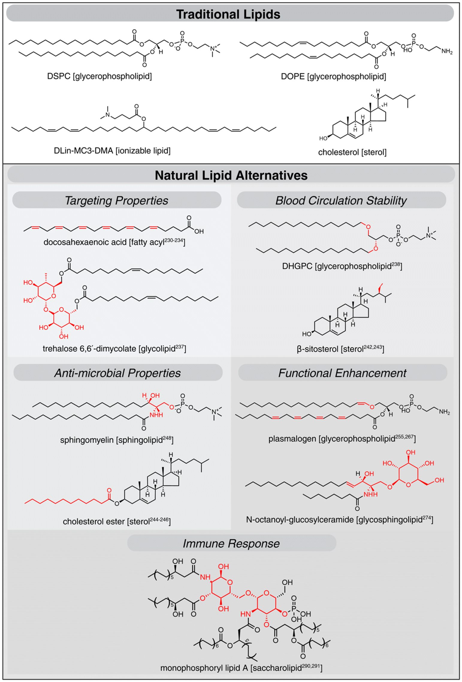 | ||
| Fig. 2 Representative structures of natural lipids used for a range of drug delivery applications, with specific references to studies where they have been utilized. Structural variations compared to traditional components are highlighted in red. | ||
| Function | Description | Examples | Ref. |
|---|---|---|---|
| Structural role | Lipids maintain structural integrity, fluidity, and functionality of cellular membranes providing a dynamic platform for cellular processes | Glycerophospholipids (PC, PE, PS, PI, PA), sphingolipids (sphingomyelin, glycosphingolipids,) sterols (cholesterol) | 207–210 |
| Energy source | Lipid molecules act as energy substrates, stored by the body and systematically broken down to release energy in the form of ATP as needed | Triglycerides, fatty acids | 211–214 |
| Signaling molecules | Lipids mediate signal transduction, regulate immune responses, and influence cell survival and proliferation | Phosphatidylinositols (PIs), sphingolipids (ceramides, S1P), eicosanoids (prostaglandins, leukotrienes), steroid hormones (cortisol, testosterone, estrogens) | 46, 215 and 216 |
| Protein-lipid interactions | Lipids enable peripheral, integral, and lipid- anchored protein interactions, facilitating cellular signaling, transport, and structural integrity | GPI-anchored proteins, actin-binding proteins, integral membrane proteins (e.g., EGFR) | 217–221 |
| Immune regulation | Lipids act as antigens, modulating immune responses and inflammation | Eicosanoids (prostaglandins, leukotrienes), glycosphingolipids (β-GlcCer, β-GalCer), phospholipids (LPC, eLPA), polyunsaturated fatty acids (Omega-3, Omega-6) | 222–223 |
| Lipid biosynthesis | Dysregulation of biosynthesis pathways can lead to diseases like cancer, cardiovascular diseases, and neurodegenerative conditions | Ether phospholipids (plasmalogens), cholesterol, fatty acid derivatives | 224–228 |
Apart from PUFAs, glycolipids can be used to achieve tissue specific targeting by leveraging the specific interactions between the carbohydrate moieties of glycolipids and lectins or other carbohydrate-binding proteins on the surface of target cells.235,236 For instance, Bae et al. incorporated a trehalose glycolipid into an LNP formulation by substituting half of the ionizable lipid and observed improved accumulation in spleen and lymph. The platform also demonstrated lower immunogenicity and biotoxicity compared to traditional LNP formulations in BALB/c mice. This was attributed to the reduction in ionizable lipid content, while the trehalose glycolipid stabilized the mRNA through hydrogen bonding, maintaining delivery efficacy.237
As mentioned previously, cholesterol is included in many clinically approved nanoparticle formulations due to its ability to enhance membrane stability.2,240 Aside from cholesterol, other cholesterol analogues like stigmasterol and β-sitosterol (plant sterols) can be used to modulate properties such as membrane fluidity and nanocarrier stability over a wider temperature range. For instance, stigmasterol is bulkier than cholesterol and has been found to reduce overall lipid packing, whereas incorporation of β-sitosterol has led to formation of liposomes with improved long-term stability compared to cholesterol-containing formulations.241 Incorporation of ergosterol, a major sterol found in fungi, was found to broaden the temperature range over which the membranes undergo phase transition, which enables the membranes to maintain fluidity and stability over wider temperature ranges.242,243 More about how incorporation of cholesterol analogues into LNP formulations affects the transfection efficiency is included in the Enhanced Functionality subsection.
Another strategy to develop antimicrobial liposomes includes the incorporation of lipids such as cholesterol and sphingomyelin, which are specific to animal cell membranes; these lipids are primary targets for bacterial toxins and have the ability to neutralize bacterial toxins and protect host cells from infection. Drug delivery systems composed of these lipids have been shown to sequester toxins, preventing mammalian cell lysis. For instance, Henry et al. demonstrated that liposomes composed of sphingomyelin and cholesterol (66 mol%), combined with antibiotics like vancomycin, effectively competed with host cells for toxin binding during active infections. The authors observed that the administered liposomes effectively rescued pneumonic mice (caused by S. aureus and S. pneumoniae) within 10 hours.248
Antitumor activity. PUFAs and PUFA-containing phospholipids (PUFA-PLs) are highly susceptible to peroxidation249 and play key roles in free radical reactions and lipid peroxidation processes. They also contribute to ferroptosis, an iron-dependent, non-apoptotic form of cell death characterized by the accumulation of lipid peroxides.250–252 For instance, Huang et al. demonstrated that liposomes enriched with linoleic acid, oleic acid, or α-linolenic acid (C18:2) elevated cellular reactive oxygen species (ROS), inducing lipid peroxidation and triggering ferroptosis in B16 tumor cells in vitro and in B16 tumor-bearing models in vivo.253 Induction of ferroptosis is a promising strategy that can be used to tackle cancers and a significant body of research has been conducted to highlight the pivotal role of PUFA-PLs in increasing the susceptibility of cells to ferroptosis. For instance, Zou et al., through genome-wide CRISPR/Cas9 screens identified the role of peroxisome synthesized PUFA-containing ether phospholipids (PUFA-ePLs), predominantly plasmalogens, in ferroptosis induction. The authors showed exogenous treatment with PUFA-ePLs liposomes resulted in increased susceptibility to ferroptosis in high grade human serous ovarian cancer cells, with an additional potential for modulating cell membranes.250,254,255 Qiu et al., through systematic studies across 20 different cell lines, identified a specific class of phospholipids containing PUFA tails at both the sn-1 and sn-2 positions as critical drivers of ferroptosis. These phospholipids were shown to initiate ROS production in mitochondria and trigger lipid peroxidation within the endoplasmic reticulum, guiding the ferroptosis process.256 Building on these studies, researchers have begun incorporating PUFA-PLs into liposomal formulations to enable ferroptosis, often encapsulating ferroptosis inducing compounds to effectively kill cancer cells.253 For instance, Gao et al. synthesized surface-engineered liposomes with fucose ligands and incorporated phospholipids with two C20:4 tails, encapsulating immunostimulatory CpG oligodeoxynucleotides to enhance the synergy between ferroptosis and immunotherapy. These liposomes exhibited improved anti-tumor activity in 4T1 tumor-bearing mice, where ROS triggered the peroxidation of the incorporated phospholipid, which led to the release of immunostimulatory cargo, boosting immune response and promoting ferroptosis.257
In addition, other lipids like ceramides, which are central intermediates of sphingolipid metabolism, exhibit tumor suppressive properties and are key mediators of cellular death programs like apoptosis and autophagy.258,259 Ceramides have been incorporated into nanoparticle formulations to achieve anti-tumor effects. For example, Zhai et al., synthesized ceramide (C6) containing liposomes to increase the apoptotic sensitivity induced by methotrexate and doxorubicin by twofold in U2OS human osteosarcoma xenograft model.260
Treatment against neurodegenerative conditions. The involvement of plasmalogens in various neurodegenerative conditions makes them an appealing therapeutic strategy.254,261–263 Plasmalogens are essential for proper neuronal function and therefore are enriched in synaptic vesicles, where plasmalogens support release of neurotransmitters and vesicular fusion.264 Their unique properties make them promising candidates for the treatment of disorders associated with neuroinflammation, oxidative stress, mitochondrial dysfunction, and perturbations in lipid metabolism. In a recent study, Wu et al. synthesized plasmalogen-containing nanoparticles for treating neurodegenerative diseases such as Parkinson's disease (PD), which is linked to reduced plasmalogen levels.265,266 In this study, the authors ameliorated the behavioral PD symptoms in transgenic PD mouse models by treatment with nanoparticles containing scallop-derived plasmalogens intranasally.267
Stimuli-responsive cargo release. Unsaturated phospholipids have been explored for light-triggered cargo release in liposomal formulations.268 For instance, a study by Miranda and Lovell demonstrated that increasing 1,2-dioleoyl-sn-glycero-3-phosphocholine (DOPC) content in doxorubicin-loaded liposomes enhanced drug release rates up to tenfold under 665 nm NIR light due to membrane destabilization and permeabilization.269 Apart from this, plasmalogens have also been incorporated into liposomes to enable stimuli-responsive drug release through the photoinduced cleavage of the labile sn-1 vinyl ether linkage.270,271 Synthetic approaches to develop stimuli responsive platforms are discussed in the Use of Synthetic Bioactive Lipids section.
Improved efficacy. The trans-membrane diffusion of amphiphilic drugs can be improved by treating cells with liposomes enriched with short-chain sphingolipids, which cause lipid packing imperfections in cell membranes.272,273 In a study by Lummel et al., the authors observed almost twofold reduction in tumor growth rate when dosed with N-octanoyl-glucosylceramide-enriched doxorubicin-containing nanoparticles compared to only doxorubicin-containing nanoparticles in an A431 xenograft model, with no reported systemic toxicity.274
Ether lipids from archaea, owing to their fusogenic and lyotropic properties that enhance endosomal escape, have been incorporated into LNP systems, enhancing transfection efficiency for pDNA and mRNA.275,276 For example, Sedlmayr et al. demonstrated that incorporating archaeal ether lipids into mRNA LNPs improved internalization and increased mRNA expression up to tenfold in primary (HSMM) and established (Caco-2, C2C12) cell lines.277 Additionally, incorporation of cholesterol analogues instead of cholesterol into LNP systems can impact particle surface morphology, which indirectly affects gene transfection. For example, in a study by Eygeris et al., the authors compared the transfection efficiencies of LNPs containing either cholesterol or its analogs in HeLa cells. The authors observed that LNPs containing β-sitosterol had the highest transfection efficiency and the authors attribute this to reduced membrane ordering, resulting in polymorphic or faceted morphologies, which contribute to improved fusogenic properties.278,279
Researchers have also designed lipid-based nanoparticles composed of multi-functional lipids that are immunogenic in addition to serving as critical structural components in nanoparticle assemblies.282,295 For instance, Irvine and coworkers reported the self-assembly of saponin and monophosphoryl lipid A (MPLA), resulting in caged nanoparticles, referred to as saponin/MPLA nanoparticles (SMNPs), which have substantial adjuvanticity in non-human primate models. This formulation has been fast-tracked to phase I human-clinical trials for use as an HIV vaccine (NCT06033209).296,297
As research advances, new lipid species and their functions will continue to be discovered, and the potential for developing functional lipid-based nanoparticle systems will continue to expand. As discussed in this section, naturally occurring lipids can be incorporated into lipid-based nanoparticles for a wide range of biomedical applications. By taking inspiration from the diverse array of lipids present in the body and their functions, it is possible to strategically select and incorporate bioactive lipids into nanoparticle systems tailored to meet specific functional requirements.
Use of synthetic bioactive lipids
While naturally-occurring lipids provide the field with a comprehensive toolkit of building blocks for designing functional nanocarriers, synthetic approaches have also been taken to design lipids and nanocarriers with specific and systematically tailored properties (Fig. 3). As LNPs continue to garner interest in the field, a large part of our discussion in this section will center around LNP development in the context of synthetic bioactive lipids. | ||
| Fig. 3 Synthetic bioactive lipids and their respective functions. In grey, the traditional lipid-based nanoparticles are highlighted. In shades of orange, the synthetic bioactive lipids used to enhance carrier functions are indicated. | ||
Modifications to traditional formulation components: cholesterol and PEG-lipids. Systematic alterations of the structure of cholesterol can be used to modulate lipid-based particle functionality. Various studies have synthesized cholesterol variants to improve delivery. For example, Dahlman and coworkers investigated the impact of esterified cholesterol variants, including cholesteryl stearate and cholesteryl oleate, and oxidized cholesterol variants, like 7α-hydroxycholesterol, 7β-hydroxycholesterol, and 4β-hydroxycholesterol, on LNP function. They concluded that LNPs formulated with esterified cholesterol delivered DNA barcodes to hepatic endothelial cells in mice with deficiency in the low-density lipoprotein receptor (LDLR−/−) and mice with deficiency in the very low-density lipoprotein receptor (VLDLR−/−) more efficiently than LNPs formulated with regular or oxidized cholesterol, likely due to differential interactions with serum proteins and the protein corona.57 Patel et al. used a hydroxycholesterol substitution in LNPs for mRNA delivery to T-cells and found that the substituting cholesterol with 7α-hydroxycholesterol LNP-mediated mRNA delivery and targeting to primary human T cells ex vivo by 1.8-fold and 2.0-fold in protein expression.58 In a recent study, Radmand et al. explored the tropism of charge-dependent lipid-based nanoparticles by investigating the substitution of cholesterol with positively charged cholesterol variants including DC cholesterol, a cholesterol derivative with an ionizable tertiary amine and GL67, a spermine-based cholesterol variant. They observed a 25–75% increase in tdTomato + expression across all liver cell lines in Ai14 and BL/6 mice—including endothelial cells, dendritic cells, hepatocytes, and T-cells—when using cationic cholesterol compared to LNPs with cationic helper lipids and neutral cholesterol.59 Overall, the addition of modified cholesterol variants has been shown to serve as a useful tool for modulating lipid-based nanoparticle delivery and targeting.
While PEG-lipids have been shown to control the size and stability of both liposomes and LNPs,55,64,65 chemical modifications to the PEG end groups can also be used to modulate LNP targeting. In a recent study by Sahay and coworkers, lipid-based particles with three different surface modifications were designed to deliver mRNA into the retina using 661w cell line model. The LNP surfaces were modified by amine-, carboxy-, and carboxyl ester-modified PEG lipids. They showed that incorporating carboxy and carboxyl ester-modified PEG lipids in LNPs increased mRNA delivery by 20 and 35% relative to LNPs containing unmodified PEG lipids.301
SORT lipids. Selective organ targeting (SORT) LNPs were developed by Siegwart and coworkers as a means to create LNPs capable of targeting organs other than the liver.302–305 Although not the first example of LNPs for extrahepatic delivery,306–308 SORT technology shows an alternative to circumvent liver tropism. SORT-LNP formulations consist of traditional components (ionizable lipid, helper phospholipid, cholesterol, and PEG lipid) with the addition of a SORT lipid, which can be a cationic, ionizable or negatively charged lipid. A recent study showed that the addition of unsaturated lipids to SORT LNPs can promote transfection efficiency relative to other traditional saturated LNPs in vivo.309 SORT lipids have been shown to alter the apparent pKa of LNPs, leading to changes in transfection efficacy and tropism.300 In addition, it was shown that the interaction of quaternary ammonium-containing lipids, used as SORT lipids, with serum proteins can influence protein corona formation and impact targeting. Dilliard et al. characterized a panel of quaternary ammonium lipids to assess how chemical structure affects the organ-targeting of SORT LNPs and found that chemical structure of both the lipid alkyl tail and headgroup impact the potency and specificity of mRNA delivery to the lungs due to alterations in protein corona identity.310 This work and other studies300,304,311 have shown that the chemical structure of the SORT lipid impacts plasma protein interactions, leading to organ-specific delivery. In a more recent study, SORT technology with crosslinked lipids has been leveraged for LNP delivery for bone marrow gene editing.312 Bone targeting is a growing area of interest to treat osteoporosis, bone cancer, and osteoarthritis. As bone marrow is the primary site of blood cell production, including red blood cells, white blood cells, and platelets, it is an important target for leukemia and other genetic disorders.313,314 Another study by Alvarez-Benedicto et al. has demonstrated that splenic T cells and macrophages can be separately transfected by LNPs containing an anionic SORT lipid.311 In addition to these advantages, the change in apparent pKA as a result of incorporating SORT lipids have been shown to lead to changes in transfection efficacy.370
Overall, SORT technology has been shown to efficiently deliver LNPs to the lungs, spleen, kidney, heart and bone marrow. Some generalizable rules that have been discovered in these studies include the use of permanently cationic SORT lipids (e.g. DOTAP) to increase lung tropism and permanently anionic SORT lipids (e.g. 18PA) to increase spleen tropism. These modifications to LNPs change the apparent pKa of LNPs to be greater than 11 for lung tropism and less than 6 to increase spleen tropism, respectively.304,305,310
Replacement of helper lipids with charged lipids. As charged lipids can be used to alter the homing capabilities of lipid-based nanoparticles, replacing the helper lipid with charged lipids can be used to modulate targeting and trafficking. A study from Whitehead and coworkers demonstrated that replacing the helper phospholipid with a cationic or anionic lipid enhanced extrahepatic targeting properties. They achieved both lung and spleen homing based on only four lipid components by changing the apparent pKa of the particle replacing the neutral helper phospholipid with a permanent cationic or anionic lipid, respectively.315 Consistent with other studies and SORT technology, adding an anionic helper lipid containing PS achieved spleen tropism, while adding a cationic helper lipid like DOTAP significantly improved lung tropism. More specifically, substituting the neutral phospholipid with PS altered the ratio of liver to spleen protein expression from 8
![[thin space (1/6-em)]](https://www.rsc.org/images/entities/char_2009.gif) :1 to 1:3, while substituting the neutral phospholipid with DOTAP changed the protein the ratio of liver to lung protein expression from 36:1 to 1:56. In a more recent study, Kuzminich et al. used a similar strategy using the cationic lipid dimethyldioctadecylammonium bromide (DDAB) to replace the helper phospholipid in an ionizable LNP formulation to target the lungs in vivo via intravenous administration. This study shows that extrahepatic lung tropism of these cationic lipid-based nanoparticles was enhanced even compared to SORT formulation and also showed central nervous system (CNS) tropism in these particles in Ai9 and Ai14 mice models, in addition to showing a 30% increase in protein expression in endothelial cells relative to LNP formulation without the cationic lipid.316 In addition, Melamed et al. have shown that LNPs containing cationic lipids may benefit pancreatic mRNA delivery in C57BL/6 mice in combination with intraperitoneal delivery. A 40% luminescence increase was observed in the pancreas in formulations containing DOTAP and the ionizable lipid 306Oi10 relative to formulations containing DOPE and PS, indicating that the presence of the cationic lipid can improve specificity for the pancreas.317 A limitation of this approach that needs to be highlighted lies in previously reported toxicity concerns associated with positively charged lipids.318 Nonetheless, these studies show decreased immune response either due to LNPs being significantly smaller than liposomes315 or due to intraperitoneal delivery which causes peritoneal macrophage extracellular vesicle (EV) transfer, which refers to the process by which macrophages in the peritoneal cavity release extracellular vesicles that can transfer various bioactive molecules to other cells, thereby reducing overall immunogenicity.317 Overall, incorporation of cationic helper lipids has been shown to increase extrahepatic delivery, while maintaining lower immunogenicity relative to cationic lipoplexes.
:1 to 1:3, while substituting the neutral phospholipid with DOTAP changed the protein the ratio of liver to lung protein expression from 36:1 to 1:56. In a more recent study, Kuzminich et al. used a similar strategy using the cationic lipid dimethyldioctadecylammonium bromide (DDAB) to replace the helper phospholipid in an ionizable LNP formulation to target the lungs in vivo via intravenous administration. This study shows that extrahepatic lung tropism of these cationic lipid-based nanoparticles was enhanced even compared to SORT formulation and also showed central nervous system (CNS) tropism in these particles in Ai9 and Ai14 mice models, in addition to showing a 30% increase in protein expression in endothelial cells relative to LNP formulation without the cationic lipid.316 In addition, Melamed et al. have shown that LNPs containing cationic lipids may benefit pancreatic mRNA delivery in C57BL/6 mice in combination with intraperitoneal delivery. A 40% luminescence increase was observed in the pancreas in formulations containing DOTAP and the ionizable lipid 306Oi10 relative to formulations containing DOPE and PS, indicating that the presence of the cationic lipid can improve specificity for the pancreas.317 A limitation of this approach that needs to be highlighted lies in previously reported toxicity concerns associated with positively charged lipids.318 Nonetheless, these studies show decreased immune response either due to LNPs being significantly smaller than liposomes315 or due to intraperitoneal delivery which causes peritoneal macrophage extracellular vesicle (EV) transfer, which refers to the process by which macrophages in the peritoneal cavity release extracellular vesicles that can transfer various bioactive molecules to other cells, thereby reducing overall immunogenicity.317 Overall, incorporation of cationic helper lipids has been shown to increase extrahepatic delivery, while maintaining lower immunogenicity relative to cationic lipoplexes.
The incorporation of charged lipids can also enhance targeting and delivery in liposomal formulations. Perrie and coworkers demonstrated that upon intramuscular injection anionic formulations, including dimyristoyl-sn-glycero-3-phospho-(1′-rac-glycerol) (DMPG) and PS, facilitated lymphatic targeting.319 Among these, PS-containing liposomes exhibited greater accumulation in draining lymph nodes, likely due to preferential uptake by macrophages, highlighting their potential for lymphatic-targeted therapy. The same study also reported that cationic DOTAP-containing liposomes showed retention at the injection site. Sun et al. synthesized a cationic lipid, HC-Y2, and incorporated it into sialic acid (SA)-modified cationic liposomes loaded with Paclitaxel to assess their antitumor efficacy.320 Their study showed that increasing the proportion of cationic lipids enhanced uptake in tumor-associated immune cells (RAW264.7) and tumor cells (4T1). In vivo, these cationic liposomes reduced lung metastases in a 4T1 metastatic tumor-bearing BALB/c mouse model of breast cancer, likely due to their ability to target tumor-associated macrophages. Another study reported that cationic liposomes containing stearyl amine loaded with resveratrol, an anticancer drug, enhanced targeting of hepatocellular carcinoma.321 This was attributed to electrostatic interactions with anionic PS on liver cancer cell surfaces. In addition, Mooney and coworkers revealed that encapsulating 2′3′ cyclic guanosine monophosphate-adenosine monophosphate (cGAMP) in cationic liposome containing DOTAP lipid resulted in targeted delivery to metastatic melanoma tumors in the lung, leading to antitumor activity.322 Lin et al. demonstrated the potential of cationic liposomes made of cetyltrimethylammonium bromide (CTAB) and DDAB encapsulating doxorubicin for targeted glioma treatment with focused ultrasound in C6 glioma model.323 These studies highlight that lipids can enhance the targeting of liposomes to desired tissues, offering a promising strategy for improving the specificity and efficacy of treatments.
Although many advancements have been made in the design and use of lipid-based nanoparticles for targeted delivery, immunogenicity remains a significant challenge.283 We will next discuss the immune response of lipid-based nanoparticles, different strategies to circumvent these, as well as how immunogenicity has been leveraged for immune-based therapies.
A main approach is surface modification of lipid-based nanoparticles with hydrophilic polymers, including pSar,326–328 polyglycerol,329–331 folic acid,332 zwitter-ionic polymers,333,334 and polysaccharides.332,335 For example, Ito and coworkers investigated the effect of replacing PEG with pSar using identical HSPC-based liposomes and found that that pSar-liposomes elicited significantly lower antipolymer antibodies’ levels compared to PEG-liposomes.327 In a separate study, replacing PEG with polyglycerol in liposomes made of hydrogenated egg PC and cholesterol, loaded with doxorubicin, resulted in reduced ABC and antibody responses. This formulation also enhanced anti-tumor activity in a mouse model of Lewis lung carcinoma.329
Instead of replacing PEG, chemical structure modification of PEG-lipids with moieties like CD47-derived self-peptides can also enhance stealth behavior.336–339 These self-peptides transmit a “don't eat me” signal to macrophages constituting the MPS and thus mask the liposomes against ABC during circulation. One study demonstrated enhanced antitumor efficacy of self-peptide incorporated liposomal doxorubicin in subcutaneous tumor of B cell lymphoma-A20 in Balb/c mice.338
Another challenge for PEG lipids is their detachment from the nanoparticles during circulation, which affects its stability. One approach for tackling this was demonstrated by Pasut et al., who used β-glutamic acid as an anchor to conjugate multiple DSPE molecules to PEG to form dendrite-like structures.340 This enabled more stable interactions of the PEG lipids during circulation, resulting in a 6.2 fold increase in distribution half-life of doxorubicin-loaded liposomes when compared to conventional PEGylation.
Enhanced functionality. Apart from incorporating synthetic lipids into lipid-based nanoparticle formulations for the applications discussed above, we can also enhance carrier functionality such as enabling stimuli-responsive cargo release and improving transfection efficiency by leveraging the bioactivity provided by synthetic lipids.
Stimuli response. To establish stimuli-responsive liposomes, researchers have developed synthetic lipids which undergo conformational changes in the presence of specific stimuli. When these lipids are incorporated into liposomes, their structural alterations can disrupt lipid packing within the membrane, triggering the release of encapsulated contents.349 For instance, in a study by Lou and Best, the authors developed a ROS-responsive liposome platform by incorporating a synthetic lipid bearing a DOPE lipid scaffold with a quinone-methide linker and boronate ester headgroup, which oxidatively breaks down, resulting in membrane disruption and release of both hydrophobic and hydrophilic cargo.350 Another example includes a study by Chander et al., where the authors developed light triggerable liposomal release systems by incorporating small amounts of (∼10 mol%) photoswitchable DSPC analogs which have azobenzenes incorporated into their lipid tails. The authors demonstrated that upon irradiation with UV-A light source (365 nm) the azobenzene group in the lipid tails undergoes photoisomerization (from the trans to the cis form) inducing cargo release up to 80% in 24 hours.351 Apart from using ROS as stimuli, researchers have developed liposomal platforms which use metal cations, ATP, pH, electric fields, magnetic fields, ultrasound waves or enzymes as triggers for cargo release.352–357
Transfection. Synthetic lipids have also been used to improve transfection of lipid-based nanoparticles. A platform for synthesizing a class of branched ionizable lipids called Branched Endosomal Disruptors (BEND) lipids that improve endosomal escape was reported. BEND lipids incorporate terminally branched groups that increase hepatic mRNA delivery and gene editing efficiency, in vitro in HeLa cells and in vivo in C57BL/6J female mice, as well as T cell transfection compared to non-branched lipids via Apo-E mediated mechanism.358 In the context of liposomes, Best and coworkers recently developed ROS-triggered liposomes by incorporating both anionic and cationic synthetic lipids. Initially, the cationic DOTAP positively charge is balanced through the inclusion of anionic lipid containing a boronate headgroup to produce approximately neutral liposomes. These anionic boronate lipids interact with ROS, which leads to clevage of the group unveiling the neutral lipid DOPE. They found that ROS-driven cleavage of anionic lipids was effective at unveiling the positively charged liposomes, which increased the transfection efficiency relative to PC liposomes.359
As mentioned above, pSar has shown promising results as an excipient, providing stealth properties, together with enhanced nanoparticle stability and reduced immunogenicity relative to PEG-containing formulations.360–362 pSar has also been used to increase transfection in lipid-based nanoparticles. Recently, Langguth and coworkers developed a pSar containing LNP platform tailored to avoid the immune response elicit by PEG lipids and found that pSar-containing particles generally have higher transfection efficiency in human peripheral blood mononuclear cells in vitro relative to traditional formulations containing PEG lipids.362
Various strategies have been employed to combat these responses. As mentioned previously, the development of DODAP, and, by extension, the DLinDMA family and other engineered ionizable lipids were engineered to have a neutral charge in blood (avoiding immune response) but a positive charge in endosome (facilitating endosomal escape).66,368,369
Biodegradable Ionizable Lipids. Biodegradable ionizable lipids, such as ester and disulfide-containing lipids, are thought to be one solution to mitigating the immune response to lipid-based nanoparticles.53,370–372 For example, introducing ester bonds into MC3 resulted in a biodegradable variant, L319, which has been shown to increase delivery efficacy and elimination from the liver in 6 to 8 weeks old male C57Bl/6 mice.370 Another example is the lipid 304O13, which showed increased potency relative to its non-biodegradable variant c12–200 in vitro in HeLa cells and in vivo in Female C57BL/6 mice.53
To increase potency of biodegradable ionizable lipids, branched-tail ionizable lipids can be employed. In a recent study by Whitehead and coworkers, a library of 580 ionizable lipids, including branched-tail ionizable lipids for extrahepatic targeting, was developed to screen delivery to extrahepatic targets. This study identified a new ionizable branched-tail lipid with a unique ability to selectively transfect lung natural killer and dendritic cells when incorporated to LNP formulations, without the need for cationic helper lipids in vitro in HeLa cells and in vivo in 6–8 weeks old C57BL/6 female mice.373 Separately, Chaudhary et al. identified potent branched-tail ionizable lipids for LNPs that deliver mRNA selectively to both the placenta and non-reproductive maternal organs without fetal delivery due to biodegradable ionizable lipids maintaining immune homeostasis in order to treat pregnancy disorders in Pr CD-1 mice and Pr Ai9 mice.374
Charged lipids & immune stimulation. Synthetic bioactive lipids can also be leveraged to elicit intrinsic immunomodulatory effects. For instance, a study evaluated the intrinsic immunostimulatory effect of liposome synthesized from two types of cationic lipids, Arg-C3-Clu2C14 (R3C14) or Arg-C5-Clu2C14 (R5C14), revealing the liposomes composed of R5C14 resulted in activation and differentiation of T cells.375
A recent study evaluated liposomes containing various cationic lipids and their effects on immune responses, and it was found that liposomes containing 1,2-Dioleoyl-sn-glycero-3-ethylphosphocholine (EPC), cholesterol, DOPC promoted greater immunomodulation than formulations with other lipid compositions, such as DOTAP, DODMA, and DDA, as indicated by increased expression of cell surface activation markers, enhanced cellular uptake, antigen-specific T-cell activation, cytokine production, and improved cellular viability in an in vitro model using human monocyte-derived dendritic cells (MDDCs).376 Kranz et al. synthesized lipoplexes composed of RNA encoding the firefly luciferase gene (luc-RNA) with lipids such as DOTMA, DOTAP, and DOPE and found that controlling the lipid composition influenced targeting, with negatively charged liposomes accumulating in the spleen and lymph nodes.377 This resulted in dendritic cell targeting in vivo, resulting in immunostimulatory and antitumor effects. Münter et al. developed a cationic liposomal platform by incorporating lipopeptide (cholesterol-anchored tri-arginine peptide) into liposomes378 and demonstrated that monocyte targeting was dependent on lipopeptide content.
Efforts to design synthetic lipids and nanocarriers with intrinsic adjuvanticity are gradually increasing. For instance, Xia and coworkers screened cationic lipid-like materials generated via epoxide ring-opening by poly(-amidoamine) (PAMAM) dendrimers for developing mRNA cancer nanovaccines. They identified C1, a 12-carbon tail molecule, incorporated into lipid-based nanoparticles, was capable of activating TLR-4 signaling in mouse bone marrow dendritic cells and effectively deliver antigen encoding mRNA for OVA expressing tumor cell lines, resulting in significant tumor growth inhibition.379
Specifically, designing amino ionizable lipids for LNP formulations with self-adjuvanticity has garnered considerable interest.380–383 For instance, Anderson and co-workers studied tested 1000 lipids for melanoma and human papillomavirus mRNA vaccines and found that LNPs made with lipid A18, containing a heterocyclic piperidinyl-based head group, triggered 10-fold greater IFN-γ secretion than LNPs with MC3 (used in Onpattro). Other LNP components included cholesterol, DOPE, and C14-PEG 2000. Further, A18-LNP selectively activated adaptive immunity the STING pathway over TLR pathway to enhance antitumor efficacy.384 Similarly, Zhang and co-workers developed STING agonist-derived amino lipids (SALs) SARS-CoV-2 vaccine carriers. LNPs made of SAL, DOPE, cholesterol, and DMG-PEG demonstrated significantly higher IFN levels bone marrow dendritic cells in vitro and STING related cytokine levels in mouse plasma in vivo.383
An alternate design strategy: use of nanoparticle mechanical properties to modulate function
In this section, we will discuss an additional design parameter, the mechanical properties of lipid-based nanocarriers. There are directly linked to the types and combinations of lipids used. Here, we will explore how mechanical properties influence nanocarrier function and biological responses (Fig. 4). | ||
| Fig. 4 Nanoparticle mechanical properties can influence their biological functions, including tumor targeting, accumulation, cellular uptake and immune cell interactions. | ||
The effects of different physicochemical properties of nanoparticles, such as shape, size, and surface properties, in regulating biological performances have been extensively investigated.6,7,385 However, there have been limited studies on how mechanical properties, such as stiffness and elasticity, govern biological interactions, such as blood circulation, immune cell interactions, tumor accumulation, and cellular uptake and trafficking of lipid-based nanocarriers.386
Atomic force microscopy is commonly used to characterize the mechanical properties of nanoparticles.389 As discussed in other review articles, various strategies, including physical, chemical, and biological methods, can be employed to tailor the mechanical properties of nanoparticles.386,388 The influence of mechanical properties of various nanoparticles such as hydrogel particles, layer-by-layer templated capsules, hybrid polymer-lipid nanoparticles, and silica nanoparticles on their biological processes have been covered.386 However, this review will specifically highlight the role the mechanical properties of lipid-based nanoparticles play in key biological processes such as tumor penetration and accumulation, immune cell interactions, and cellular uptake.
The influence of LNP mechanical properties on biological responses is an emerging field, with limited studies effectively characterizing these properties due to challenges in measuring mechanical compliance, which stem from their small size, structural complexity, and soft, deformable nature.390 However, a study found that soft LNPs, with elasticity modulated by the calcium ion density in alginate gel, exhibited enhanced permeation through endothelial layers.390 Additionally, several studies have investigated the impact of LNP rigidity on endosomal escape, including the role of lipid rafts,391 the fusogenicity of lipids,392 and the packing and shape of lipids in facilitating the escape of mRNA-loaded LNPs from endosomes.393 Given the emerging nature of this field, our review primarily focuses on liposomal mechanical properties.
Tumor penetration and accumulation. The mechanical properties of nanoparticles impact cellular uptake and tumor accumulation. Additionally, they help overcome biological barriers like epithelial and mucosal layers, improving the delivery of therapeutic agents to tumor sites.394,395 There are discrepancies between studies regarding whether less rigid or more rigid particles accumulate more effectively in tumors, which can be attributed to differences in tumor models and the use of different nanoparticle systems across studies. However, elasticity-dependent uptake is well-documented in the literature.395–397
Elasticity-mediated tumor accumulation of a nanolipogel (NLG) system has been reported.398 Hybrid NLGs with an alginate hydrogel core and lipid bilayer shell were synthesized to evaluate the effect of nanoparticle elasticity on tumor accumulation. NLG elasticity was modulated by varying the degree of cross-linking of the alginate core. Additionally, nanoliposomes (NLPs) without a hydrogel core were also synthesized, exhibiting Young's modulus of 45 ± 9 kPa. The Young's Modulus of the NLGs ranged from 1.6 ± 0.6 MPa to 19 ± 5 MPa. In vitro studies in MDA-MB-231 and MCF7 breast cancer cell lines revealed that the softer NLPs exhibited greater (5-fold increase) cellular internalization in breast cancer cell lines compared to the NLGs, which was hypothesized to be due to different uptake mechanisms for the soft and stiff nanoparticles. The enhanced internalization of soft NLPs was attributed to internalization through both energy-independent fusion and endocytosis processes compared to the stiffer nanoparticles, which were shown to depend only on energy-dependent clathrin-mediated endocytosis. Furthermore, the soft NLPs demonstrated about 2.6-fold increase in tumor accumulation in an orthotopic mouse 4T1 breast cancer model, which can be attributed to their lower elastic modulus, leading to increased tumor penetration in vivo.
Another study showed the effect of liposome stiffness on nanoparticle trafficking, pharmacokinetics and tumor accumulation.21 This research reported that the stiffness of targeted layer-by-layer (LbL) modified liposomes can be modulated by varying the cholesterol content of the liposomal core. While the core composition was varied, the hyaluronic acid (HA) surface chemistry remained unchanged to ensure consistent targeting across formulations. The influence of their mechanical properties on in vivo trafficking in subcutaneous mouse model of OVCAR8 tumors expressing GFP/Luc2 was analyzed, revealing that soft liposomes resulted in longer elimination half-life (3.5 times higher than stiff counterparts), enhanced tumor accumulation, and exhibited higher penetration of particles in subcutaneous tumors in vivo. Wu et al. used a similar strategy of changing cholesterol content in liposomes to alter rigidity, determining that liposomes with moderate membrane (21.7 to 33.9 MPa) rigidity resulted in enhanced tumor penetration in vitro and in vivo compared to lower (11.5 MPa) or higher rigidities (53.3 MPa).22 Montizaan et al. also reported that softer liposomes were taken up faster and to a greater extent in HeLa and A549 cells compared to harder liposome-coated silica nanoparticles.399
Altering the membrane fluidity and stiffness of liposomes can enhance their tumor penetration. Liposomes with increased fluidity can exhibit improved fusion capabilities, which can enhance the accumulation in tumor cells.400 Bompard et al. engineered liposomes by modulating their phospholipid composition, specifically by varying acyl chain lengths and degrees of saturation of lipid tails and assessed their uptake in various non-tumor and tumor cell lines in vitro.400 The study found that less rigid liposomes, comprised of DMPC, DOPC and 1-palmitoyl-2-oleoyl-sn-glycero-3-phosphocholine (POPC) lipids, had increased uptake in cancer cells and favored membrane fusion, while stiffer liposomes, comprised of DSPC, preferentially targeted healthy (WPMY-1) cells than cancer (LNCaP, C4-2B, and PC-3) cells. In contrast to these findings, when researchers compared hard polymeric PLGA core and lipid shell nanoparticles with soft liposomes in a separate study, the soft liposomes exhibited lower cellular internalization in HeLa cells,401 highlighting the need for the field to gain a more comprehensive understanding of the influence of biological heterogeneity on nanocarrier uptake.
Research has focused on the effect on nanoparticle mechanical property on penetrating biological barriers to enhance tumor accumulation. Gao and coworkers highlighted the importance of nanoparticle rigidity in penetrating biological barriers and enhancing tumor accumulation by developing liposomes with varying stiffness.395 In this work, core–shell nanoparticles with poly(lactic-co-glycolic acid) (PLGA) cores surrounded by lipid bilayer shells and hollow liposomes were separately engineered to evaluate how nanoparticle stiffness impacts diffusion through mucosal barrier. They observed that nanoparticles with intermediate rigidity (50 MPa) demonstrated enhanced diffusion through mucosal barriers and showed increased tumor penetration and accumulation compared to hard core–shell nanoparticles (110 MPa) and soft liposomes (5 MPa) in vitro in HT29-MTX-E12 cell line, human colon carcinoma cell line (Caco-2), in situ in pancreatic adenocarcinoma cell (BxPC-3), and human pancreatic star cells (HPSC).395 To understand the difference in cellular interaction patterns of nanoparticles with varying rigidities, super-resolution microscopy and MD simulations were performed. The study showed that semi-elastic (50 MPa) nanoparticles can deform into ellipsoids, which allows rotation-facilitated diffusion, and enhanced penetration capabilities. It was reasoned that hard nanoparticles (110 MPa) cannot deform, and soft nanoparticles (5MPa) irregularly deform, causing them to exhibit the lowest permeation of the tested formulations.
In another study by Dai et al., the influence of liposome rigidity on penetration through the tumor extracellular matrix (ECM) was investigated.402 Liposome rigidity was modulated by changing the lipid tail saturation and chain length. The lipids used in this study, listed in order of increasing rigidity in liposomes, include DOPE (C18:1), 1,2-dilauroyl-sn-glycero-3-phosphocholine (DLPC, C12:0), 1,2-distearoyl-sn-glycero-3-phosphocholine (DEPC, C22:1), DMPC (C14:0) DPPC (C16:0), and DSPC (C18:0). Liposomes comprised of DPPC (C16:0) lipids with intermediate stiffness (19.9 MPa) exhibited superior diffusivity through the ECM, leading to enhanced internalization in pancreatic adenocarcinoma cells (BxPC-3) and HPSC cells, improved accumulation, and prolonged retention at tumor sites compared to liposomes with lower (below 10 MPa) or higher (42.6 MPa) stiffness used in the study. The researchers concluded that the deformation of the nanoparticles of moderate stiffness into ellipsoids could explain the enhanced diffusivity through the ECM.
Overall, these studies have shown that while the mechanical properties of nanoparticles impact penetration and tumor accumulation, there are discrepancies across nanoparticle formulations and biological systems. The differences in the physicochemical properties of nanoparticles, the variabilities in the magnitude of elastic moduli across studies, and the fabrication process of modulating liposomal stiffness may explain the differences between the studies.402 Despite these differences, the findings highlight that tuning the mechanical properties of liposomes can be leveraged to enable penetration across biological barriers and enhance tumor accumulation.
Immune cell interactions. The mechanical properties of liposomes can also govern their interactions with immune cell populations. Yuan et al. modulated liposome elasticity by varying metal ion cross-linking to evaluate their fate in APCs including macrophages and dendritic cells, within the tumor microenvironment (TME) and transport to tumor-draining lymph nodes (tdLNs).403 When evaluated in vitro in RAW 264.7 cells and B16F10 cells, soft liposomes (15 MPa) were more readily internalized by APCs and demonstrated enhanced lymph node transport in vivo, in a subcutaneous-B16F10 tumor bearing mouse model, compared to the stiff versions (149 MPa). Liposomes with medium elasticity (40 MPa) activated the STING pathway and enhanced transport through tdLNs, promoting greater tumor-infiltrating lymphocyte (TIL) infiltration and producing antitumor effects in mice. The study also explored the potential of these liposomes with medium elasticity as a synergistic therapy with immune checkpoint blockade (ICB) therapy, highlighting their promise in improving cancer immunotherapy outcomes. However, a different study demonstrated that stiff liposomes exhibited superior uptake in APCs.404 The rigidity of the liposomes was modulated by the phase state of the lipid membrane as dictated by the lipid transition temperature. It was revealed that stiff (gel phase) liposomes loaded with a vaccine fusion protein (CTA1-3Ea-DD) showed an enhanced antigen-presentation ability compared to softer (fluid phase) counterparts. Additionally, they promoted superior upregulation of costimulatory molecules and increased release of cytokines, highlighting how liposome stiffness can enhance vaccine immunogenicity.
Vu et al. studied the interactions between liposomes and a core–shell polymeric nanoparticle, which had a polystyrene core and a poly(ethylene glycol) shell. The polymeric nanoparticle was synthesized using reversible addition–fragmentation chain transfer (RAFT) polymerization combined with polymerization-induced self-assembly (PISA).405 They assessed how these nanoparticles interacted with immune cells (B cells, monocytes, and neutrophils) in the blood and found that polymeric nanoparticles had stronger interactions with these immune cells than liposomes, which could be attributed to the distinct mechanical properties of the nanoparticle platforms, with polymeric nanoparticles exhibiting greater stiffness. However, these systems differ from one another in more ways than one, and other factors besides the mechanical properties could explain the results.
The importance of mechanical properties on vaccine-induced immune response has also been demonstrated.406 Liposome rigidity was altered by modulating the degree of saturation of lipid tails. It was observed that rigid dimethyldioctadecylammonium (DDA)-based liposomes triggered a stronger immune response than softer dimethyldioleoylammonium (DODA)-based liposomes through increased recruitment and activation of APCs. Moreover, liposome rigidity influences nanoparticle retention at the site of injection. Softer, DODA-based liposomes were cleared more rapidly from the injection site compared to their rigid counterparts, resulting in a lower immune response.
Conclusions and future perspectives
The development of lipid-based nanoparticles has resulted in significant clinical successes, from the use of liposomes to change the safety and delivery profiles of key chemotherapy agents to LNP formulations that have enabled the first clinical uses of RNA-based therapeutics and vaccines. These developments have occurred within a remarkably short period of time, starting with the discovery of mRNA and liposome structures just sixty years ago. One of the main challenges in the field of nanomedicine remains the successful development and translation of targeted nanocarriers. In this review, we highlighted strategies and recent advances in the use of formulations to impart targeting and functional properties without adding complexity to the nanocarrier design in the form of exogenous antifouling and targeting motifs. This strategy of imparting intrinsic functionality into the nanocarrier has gained increasing popularity for LNP design, resulting in promising clinical candidates for a number of preventative and therapeutic applications. We also emphasized the biological origin of liposomes, and how our understanding of biological lipid structure and function can be used to design intrinsically functional nanocarriers. As the number of biological lipid classes and functions continues to expand, we look forward to seeing how use of bioactive and bio-inspired lipids will continue to yield new types of targeted and functional nanocarriers.In addition to the naturally occurring bioactive lipids already incorporated into lipid-based nanoparticles, several other lipid classes have received less attention despite their potential. Among these are lipids belonging to the polyketide and prenol classes. For instance, polyketide-based small molecule drugs such as doxorubicin (anticancer), lovastatin (anti-cholesterol), and rapamycin (immunosuppressant), are clinically significant due to their bioactive functions.407–409 Traditionally, these are administered either as free drugs or as nanoparticle formulations prepared via hydrophobic encapsulation. However, forming functional nanoparticles with these polyketides by themselves has not been widely explored. The vastness and diversity within this group of molecules adds to the difficulties. Recently, there has been interest in utilizing machine learning algorithms to investigate clinically relevant drugs and excipients to discern their nanoparticle formation or co-aggregation capabilities to enhance their pharmacological properties.410 Prenol lipids also serve diverse biological functions, including cellular signaling and acting as precursors to essential molecules like vitamins.411,412 Due to their biocompatibility and biological activity, vitamin-lipid conjugates have been explored for incorporation into LNPs. For instance, Hou et al. developed various vitamin-lipid conjugates (Vitamin B3, C, D, E) with ionizable lipids containing tertiary amines and incorporated them into LNPs to treat multidrug-resistant bacteria-induced sepsis in C57BL/6 immunocompromised mice. They found that Vitamin C-lipid LNPs successfully delivered cathepsin-B mRNA to macrophage lysosomes, a key target for bactericidal activity.413 Further physical and biological characterization of lipid-based nanoparticles incorporating these lipids will be essential to fully understand their properties and optimize their functionality for therapeutic use.
Besides the aforementioned evolving areas, non-lamellar lyotropic liquid crystalline nanoparticles (LLC NPs) have also emerged as promising drug delivery vehicles. While structurally similar to liposomes, they exhibit more complex architectures, such as inverse hexagonal and cubic mesophases, often referred to as cubosomes and hexosomes.414–421 Studies have demonstrated their ability to modulate biological responses, including cellular interactions,422–424 blood components interactions, which in turn can affect their circulation times and biodistribution fates,425–428 and immune responses.424,429–431 However, further research is needed to elucidate how formulation components, particle structure, and morphology impact cellular responses and therapeutic efficacy.
Additionally, there has been effort in the field to advance lipid-based nanoparticles design through computational means that was not covered in this review including artificial intelligence/machine learning432–435 and molecular dynamic simulations.72,436,437 These computational tools can aid the design optimization of lipid-based nanoparticles to effectively include bioactive lipids. A lot of advancements have also been made in unfolding the role of the protein corona in the context of lipid-based nanocarriers.438–440 It will be of great importance to further explore the effect of adding bioactive lipids to lipid-based nanoparticles in protein corona formation and composition. Finally, in the era of personalized medicine, we also anticipate that patient and disease-specific lipid signatures will be identified and leveraged to design uniquely functional nanocarriers. The continued exploration of functional lipids and innovative design strategies to create simple, yet functional nanocarriers holds promise, and we look forward to seeing how these delivery systems will be translated to the clinic.
Data availability
No primary research results, software or code have been included and no new data were generated or analysed as part of this review.Conflicts of interest
There are no conflicts of interest to declare.Acknowledgements
We gratefully acknowledge support from the Minnesota Ovarian Cancer Alliance, the University of Minnesota, and the National Cancer Institute (R00CA255844). Biorender was used in part to generate the figures included in this manuscript.References
- P. R. Cullis and P. L. Felgner, Nat. Rev. Drug Discovery, 2024, 23, 709–722 CrossRef CAS PubMed.
- R. Tenchov, R. Bird, A. E. Curtze and Q. Zhou, ACS Nano, 2021, 15, 16982–17015 CrossRef CAS PubMed.
- A. Gordillo-Galeano and C. E. Mora-Huertas, Eur. J. Pharm. Biopharm., 2018, 133, 285–308 CrossRef CAS PubMed.
- A. D. Bangham and R. W. Horne, J. Mol. Biol., 1964, 8, 660-IN10 CrossRef PubMed.
- A. C. Anselmo and S. Mitragotri, Bioeng. Transl. Med., 2021, 6, e10246 CrossRef CAS PubMed.
- M. J. Mitchell, M. M. Billingsley, R. M. Haley, M. E. Wechsler, N. A. Peppas and R. Langer, Nat. Rev. Drug Discovery, 2021, 20, 101–124 CrossRef CAS PubMed.
- A. Albanese, P. S. Tang and W. C. W. Chan, Annu. Rev. Biomed. Eng., 2012, 14, 1–16 CrossRef CAS PubMed.
- S. Priya, V. M. Desai and G. Singhvi, ACS Omega, 2023, 8, 74–86 CrossRef CAS PubMed.
- M. Mehta, T. A. Bui, X. Yang, Y. Aksoy, E. M. Goldys and W. Deng, ACS Mater. Au, 2023, 3, 600–619 CrossRef CAS PubMed.
- S. Shah, V. Dhawan, R. Holm, M. S. Nagarsenker and Y. Perrie, Adv. Drug Delivery Rev., 2020, 154–155, 102–122 CrossRef CAS PubMed.
- R. Freund, U. Lächelt, T. Gruber, B. Rühle and S. Wuttke, ACS Nano, 2018, 12, 2094–2105 CrossRef CAS PubMed.
- D. E. Large, R. G. Abdelmessih, E. A. Fink and D. T. Auguste, Adv. Drug Delivery Rev., 2021, 176, 113851 CrossRef CAS PubMed.
- D. Papahadjopoulos and H. K. Kimelberg, Prog. Surf. Sci., 1974, 4, 141–232 CrossRef.
- R. Tenchov, R. Bird, A. E. Curtze and Q. Zhou, ACS Nano, 2021, 15, 16982–17015 CrossRef CAS PubMed.
- T. Sun, Y. S. Zhang, B. Pang, D. C. Hyun, M. Yang and Y. Xia, Angew. Chem., Int. Ed., 2014, 53, 12320–12364 CrossRef CAS PubMed.
- K.-D. Lee, K. Hong and D. Papahadjopoulos, Biochim. Biophys. Acta, Biomembr., 1992, 1103, 185–197 CrossRef CAS PubMed.
- T. Musumeci, A. Bonaccorso and C. Carbone, in Liposomes in Drug Delivery, ed. S. G. Antimisiaris, Academic Press, 2024, pp. 19–48 Search PubMed.
- M. Anderson and A. Omri, Drug Delivery, 2004, 11, 33–39 CrossRef CAS PubMed.
- G. Gregoriadis, J. Antimicrob. Chemother., 1991, 28, 39–48 CrossRef CAS PubMed.
- M. K. Jain, in Current Topics in Membranes and Transport, ed. F. Bronner and A. Kleinzeller, Academic Press, 1975, vol. 6, pp. 1–57 Search PubMed.
- S. M. Kong, D. F. Costa, A. Jagielska, K. J. Van Vliet and P. T. Hammond, Proc. Natl. Acad. Sci. U. S. A., 2021, 118, e2104826118 CrossRef CAS PubMed.
- H. Wu, M. Yu, Y. Miao, S. He, Z. Dai, W. Song, Y. Liu, S. Song, E. Ahmad, D. Wang and Y. Gan, Acta Pharm. Sin. B, 2019, 9, 858–870 CrossRef PubMed.
- Y. Takechi-Haraya, K. Sakai-Kato, Y. Abe, T. Kawanishi, H. Okuda and Y. Goda, Langmuir, 2016, 32, 6074–6082 CrossRef CAS PubMed.
- R. A. Demel and B. De Kruyff, Biochim. Biophys. Acta, Rev. Biomembr., 1976, 457, 109–132 CrossRef CAS PubMed.
- L. Coderch, J. Fonollosa, M. De Pera, J. Estelrich, A. De La Maza and J. L. Parra, J. Controlled Release, 2000, 68, 85–95 CrossRef CAS PubMed.
- A. Magarkar, V. Dhawan, P. Kallinteri, T. Viitala, M. Elmowafy, T. Róg and A. Bunker, Sci. Rep., 2014, 4, 5005 CrossRef CAS PubMed.
- K. Inoue, Biochim. Biophys. Acta, Biomembr., 1974, 339, 390–402 CrossRef CAS PubMed.
- C. Kirby, J. Clarke and G. Gregoriadis, FEBS Lett., 1980, 111, 324–328 CrossRef CAS PubMed.
- P. R. Cullis, L. D. Mayer, M. B. Bally, T. D. Madden and M. J. Hope, Adv. Drug Delivery Rev., 1989, 3, 267–282 CrossRef CAS.
- D. E. Large, R. G. Abdelmessih, E. A. Fink and D. T. Auguste, Adv. Drug Delivery Rev., 2021, 176, 113851 CrossRef CAS PubMed.
- A. Gabizon, H. Shmeeda and Y. Barenholz, Clin. Pharmacokinet., 2003, 42, 419–436 CrossRef CAS PubMed.
- E. J. Feldman, J. E. Lancet, J. E. Kolitz, E. K. Ritchie, G. J. Roboz, A. F. List, S. L. Allen, E. Asatiani, L. D. Mayer, C. Swenson and A. C. Louie, J. Clin. Oncol., 2011, 29, 979–985 CrossRef CAS PubMed.
- M. Alavi, N. Karimi and M. Safaei, Adv. Pharm. Bull., 2017, 7, 3–9 CrossRef CAS PubMed.
- H. Harashima, K. Sakata, K. Funato and H. Kiwada, Pharm. Res., 1994, 11, 402–406 CrossRef CAS PubMed.
- S. F. Fenz and K. Sengupta, Integr. Biol., 2012, 4, 982–995 CrossRef CAS PubMed.
- R. Eugster and P. Luciani, Curr. Opin. Colloid Interface Sci., 2025, 75, 101875 CrossRef CAS.
- C. B. Giuliano, N. Cvjetan, J. Ayache and P. Walde, ChemSystemsChem, 2021, 3, e2000049 CrossRef CAS.
- G. Storm, S. O. Belliot, T. Daemen and D. D. Lasic, Adv. Drug Delivery Rev., 1995, 17, 31–48 CrossRef CAS.
- V. P. Torchilinl and M. I. Papisov, J. Liposome Res., 1994, 4, 725–739 CrossRef.
- A. Gabizon, R. Shiota and D. Papahadjopoulos, JNCI, J. Natl. Cancer Inst., 1989, 81, 1484–1488 CrossRef CAS PubMed.
- A. Chanan-Khan, J. Szebeni, S. Savay, L. Liebes, N. M. Rafique, C. R. Alving and F. M. Muggia, Ann. Oncol., 2003, 14, 1430–1437 CrossRef CAS PubMed.
- T. Ishida, K. Atobe, X. Wang and H. Kiwada, J. Controlled Release, 2006, 115, 251–258 CrossRef CAS PubMed.
- M. Taher, D. Susanti, M. S. Haris, A. A. Rushdan, R. T. Widodo, Y. Syukri and J. Khotib, Heliyon, 2023, 9, e13823 CrossRef CAS PubMed.
- B.-M. Chen, T.-L. Cheng and S. R. Roffler, ACS Nano, 2021, 15, 14022–14048 CrossRef CAS PubMed.
- E. M. Pelegri-O'Day, E.-W. Lin and H. D. Maynard, J. Am. Chem. Soc., 2014, 136, 14323–14332 CrossRef PubMed.
- T. Shimizu, Annu. Rev. Pharmacol. Toxicol., 2009, 49, 123–150 CrossRef CAS PubMed.
- S. Zalba, T. L. M. ten Hagen, C. Burgui and M. J. Garrido, J. Controlled Release, 2022, 351, 22–36 CrossRef CAS PubMed.
- H. Nsairat, D. Khater, U. Sayed, F. Odeh, A. Al Bawab and W. Alshaer, Heliyon, 2022, 8, e09394 CrossRef CAS PubMed.
- C. Wang, X. Lan, L. Zhu, Y. Wang, X. Gao, J. Li, H. Tian, Z. Liang and W. Xu, Small, 2024, 20, 2309031 CrossRef CAS PubMed.
- A. I. Antoniou, S. Giofrè, P. Seneci, D. Passarella and S. Pellegrino, Drug Discovery Today, 2021, 26, 1794–1824 CrossRef CAS PubMed.
- C. Meng, Z. Chen, G. Li, T. Welte and H. Shen, Adv. Ther., 2021, 4, 2000099 CrossRef CAS.
- M. Jayaraman, S. M. Ansell, B. L. Mui, Y. K. Tam, J. Chen, X. Du, D. Butler, L. Eltepu, S. Matsuda, J. K. Narayanannair, K. G. Rajeev, I. M. Hafez, A. Akinc, M. A. Maier, M. A. Tracy, P. R. Cullis, T. D. Madden, M. Manoharan and M. J. Hope, Angew. Chem., Int. Ed., 2012, 51, 8529–8533 CrossRef CAS PubMed.
- K. A. Whitehead, J. R. Dorkin, A. J. Vegas, P. H. Chang, O. Veiseh, J. Matthews, O. S. Fenton, Y. Zhang, K. T. Olejnik, V. Yesilyurt, D. Chen, S. Barros, B. Klebanov, T. Novobrantseva, R. Langer and D. G. Anderson, Nat. Commun., 2014, 5, 4277 CrossRef CAS PubMed.
- P. Patel, N. M. Ibrahim and K. Cheng, Trends Pharmacol. Sci., 2021, 42, 448–460 CrossRef CAS PubMed.
- J. A. Kulkarni, D. Witzigmann, J. Leung, Y. Y. C. Tam and P. R. Cullis, Nanoscale, 2019, 11, 21733–21739 RSC.
- S. Chen, Y. Y. C. Tam, P. J. C. Lin, M. M. H. Sung, Y. K. Tam and P. R. Cullis, J. Controlled Release, 2016, 235, 236–244 CrossRef CAS PubMed.
- K. Paunovska, C. J. Gil, M. P. Lokugamage, C. D. Sago, M. Sato, G. N. Lando, M. Gamboa Castro, A. V. Bryksin and J. E. Dahlman, ACS Nano, 2018, 12, 8341–8349 CrossRef CAS PubMed.
- S. Patel, N. Ashwanikumar, E. Robinson, Y. Xia, C. Mihai, J. P. Griffith, S. Hou, A. A. Esposito, T. Ketova, K. Welsher, J. L. Joyal, Ö. Almarsson and G. Sahay, Nat. Commun., 2020, 11, 983 CrossRef CAS PubMed.
- A. Radmand, H. Kim, J. Beyersdorf, C. N. Dobrowolski, R. Zenhausern, K. Paunovska, S. G. Huayamares, X. Hua, K. Han, D. Loughrey, M. Z. C. Hatit, A. Del Cid, H. Ni, A. Shajii, A. Li, A. Muralidharan, H. E. Peck, K. E. Tiegreen, S. Jia, P. J. Santangelo and J. E. Dahlman, Proc. Natl. Acad. Sci. U. S. A., 2024, 121, e2307801120 CrossRef CAS PubMed.
- G. B. Schober, S. Story and D. P. Arya, Sci. Rep., 2024, 14, 2403 CrossRef CAS PubMed.
- F. P. Polack, S. J. Thomas, N. Kitchin, J. Absalon, A. Gurtman, S. Lockhart, J. L. Perez, G. P. Marc, E. D. Moreira, C. Zerbini, R. Bailey, K. A. Swanson, S. Roychoudhury, K. Koury, P. Li, W. V. Kalina, D. Cooper, R. W. Frenck, L. L. Hammitt, Ö. Türeci, H. Nell, A. Schaefer, S. Ünal, D. B. Tresnan, S. Mather, P. R. Dormitzer, U. Şahin, K. U. Jansen and W. C. Gruber, N. Engl. J. Med., 2020, 383, 2603–2615 CrossRef CAS PubMed.
- E. Samaridou, J. Heyes and P. Lutwyche, Adv. Drug Delivery Rev., 2020, 154–155, 37–63 CrossRef CAS PubMed.
- M. Kawaguchi, M. Noda, A. Ono, M. Kamiya, M. Matsumoto, M. Tsurumaru, S. Mizukami, H. Mukai and S. Kawakami, J. Pharm. Sci., 2023, 112, 1401–1410 CrossRef CAS PubMed.
- M. Berger, M. Degey, J. Leblond Chain, E. Maquoi, B. Evrard, A. Lechanteur and G. Piel, Pharmaceutics, 2023, 15, 597 CrossRef CAS PubMed.
- T. Suzuki, Y. Suzuki, T. Hihara, K. Kubara, K. Kondo, K. Hyodo, K. Yamazaki, T. Ishida and H. Ishihara, Int. J. Pharm., 2020, 588, 119792 CrossRef CAS PubMed.
- A. Akinc, M. A. Maier, M. Manoharan, K. Fitzgerald, M. Jayaraman, S. Barros, S. Ansell, X. Du, M. J. Hope, T. D. Madden, B. L. Mui, S. C. Semple, Y. K. Tam, M. Ciufolini, D. Witzigmann, J. A. Kulkarni, R. van der Meel and P. R. Cullis, Nat. Nanotechnol., 2019, 14, 1084–1087 CrossRef CAS PubMed.
- L. R. Baden, H. M. El Sahly, B. Essink, K. Kotloff, S. Frey, R. Novak, D. Diemert, S. A. Spector, N. Rouphael, C. B. Creech, J. McGettigan, S. Khetan, N. Segall, J. Solis, A. Brosz, C. Fierro, H. Schwartz, K. Neuzil, L. Corey, P. Gilbert, H. Janes, D. Follmann, M. Marovich, J. Mascola, L. Polakowski, J. Ledgerwood, B. S. Graham, H. Bennett, R. Pajon, C. Knightly, B. Leav, W. Deng, H. Zhou, S. Han, M. Ivarsson, J. Miller and T. Zaks, N. Engl. J. Med., 2021, 384, 403–416 CrossRef CAS PubMed.
- L. Schoenmaker, D. Witzigmann, J. A. Kulkarni, R. Verbeke, G. Kersten, W. Jiskoot and D. J. A. Crommelin, Int. J. Pharm., 2021, 601, 120586 CrossRef CAS PubMed.
- J. A. Kulkarni, M. M. Darjuan, J. E. Mercer, S. Chen, R. van der Meel, J. L. Thewalt, Y. Y. C. Tam and P. R. Cullis, ACS Nano, 2018, 12, 4787–4795 CrossRef CAS PubMed.
- Y. Eygeris, S. Patel, A. Jozic and G. Sahay, Nano Lett., 2020, 20, 4543–4549 CrossRef CAS PubMed.
- M. Hammel, Y. Fan, A. Sarode, A. E. Byrnes, N. Zang, P. Kou, K. Nagapudi, D. Leung, C. C. Hoogenraad, T. Chen, C.-W. Yen and G. L. Hura, ACS Nano, 2023, 17, 11454–11465 CrossRef CAS PubMed.
- M. Ramezanpour, M. L. Schmidt, I. Bodnariuc, J. A. Kulkarni, S. S. W. Leung, P. R. Cullis, J. L. Thewalt and D. P. Tieleman, Nanoscale, 2019, 11, 14141–14146 RSC.
- Y. Liu, Y. Huang, G. He, C. Guo, J. Dong and L. Wu, Int. J. Mol. Sci., 2024, 25, 10166 CrossRef CAS PubMed.
- M. Freeley and A. Long, Biochem. J., 2013, 455, 133–147 CrossRef CAS PubMed.
- D. Tarab-Ravski, I. Hazan-Halevy, M. Goldsmith, L. Stotsky-Oterin, D. Breier, G. S. Naidu, A. Aitha, Y. Diesendruck, B. D. Ng, H. Barsheshet, T. Berger, I. Vaxman, P. Raanani and D. Peer, Adv. Sci., 2023, 10, 2301377 CrossRef CAS PubMed.
- R. Kedmi, N. Veiga, S. Ramishetti, M. Goldsmith, D. Rosenblum, N. Dammes, I. Hazan-Halevy, L. Nahary, S. Leviatan-Ben-Arye, M. Harlev, M. Behlke, I. Benhar, J. Lieberman and D. Peer, Nat. Nanotechnol., 2018, 13, 214–219 CrossRef CAS PubMed.
- R. Palanki, J. S. Riley, S. K. Bose, V. Luks, A. Dave, N. Kus, B. M. White, A. S. Ricciardi, K. L. Swingle, L. Xue, D. Sung, A. S. Thatte, H. C. Safford, V. S. Chaluvadi, M. Carpenter, E. L. Han, R. Maganti, A. G. Hamilton, K. Mrksich, M. B. Billingsley, P. W. Zoltick, M.-G. Alameh, D. Weissman, M. J. Mitchell and W. H. Peranteau, Proc. Natl. Acad. Sci. U. S. A., 2024, 121, e2400783121 CrossRef CAS PubMed.
- C. Liang, B. Guo, H. Wu, N. Shao, D. Li, J. Liu, L. Dang, C. Wang, H. Li, S. Li, W. K. Lau, Y. Cao, Z. Yang, C. Lu, X. He, D. W. T. Au, X. Pan, B.-T. Zhang, C. Lu, H. Zhang, K. Yue, A. Qian, P. Shang, J. Xu, L. Xiao, Z. Bian, W. Tan, Z. Liang, F. He, L. Zhang, A. Lu and G. Zhang, Nat. Med., 2015, 21, 288–294 CrossRef CAS PubMed.
- J. Sam Lee, M. Kim, H. Jin, M. Kwak, E. Cho, K.-S. Kim and D.-E. Kim, Int. J. Pharm., 2024, 662, 124519 CrossRef CAS PubMed.
- A. D. Bangham, M. M. Standish and J. C. Watkins, J. Mol. Biol., 1965, 13, 238-IN27 Search PubMed.
- A. D. Bangham, M. M. Standish and N. Miller, Nature, 1965, 208, 1295–1297 CrossRef CAS PubMed.
- A. D. Bangham, J. De Gier and G. D. Greville, Chem. Phys. Lipids, 1967, 1, 225–246 CrossRef CAS.
- A. D. Bangham, M. W. Hill and N. G. A. Miller, in Methods in Membrane Biology: Volume 1, ed. E. D. Korn, Springer US, Boston, MA, 1974, pp. 1–68 Search PubMed.
- A. D. Bangham, M. M. Standish, J. C. Watkins and G. Weissmann, Protoplasma, 1967, 63, 183–187 CrossRef CAS PubMed.
- A. D. Bangham and D. Papahadjopoulos, Biochim. Biophys. Acta, Biophys. Incl. Photosynth., 1966, 126, 181–184 CrossRef CAS PubMed.
- D. Papahadjopoulos and A. D. Bangham, Biochim. Biophys. Acta, Biophys. Incl. Photosynth., 1966, 126, 185–188 CrossRef CAS PubMed.
- G. Sessa and G. Weissmann, J. Lipid Res., 1968, 9, 310–318 CrossRef CAS.
- G. Sessa and G. Weissmann, J. Biol. Chem., 1970, 245, 3295–3301 CrossRef CAS PubMed.
- G. Gregoriadis and B. E. Ryman, Biochem. J., 1971, 124, 58P CrossRef CAS PubMed.
- G. Gregoriadis, N. Engl. J. Med., 1976, 295, 704–710 CrossRef CAS PubMed.
- G. Gregoriadis, N. Engl. J. Med., 1976, 295, 765–770 CrossRef CAS PubMed.
- W. E. Magee, C. W. Goff, J. Schoknecht, M. D. Smith and K. Cherian, J. Cell Biol., 1974, 63, 492–504 CrossRef CAS PubMed.
- R. E. Pagano, L. Huang and C. Wey, Nature, 1974, 252, 166–167 CrossRef CAS PubMed.
- G. Poste and D. Papahadjopoulos, Proc. Natl. Acad. Sci. U. S. A., 1976, 73, 1603–1607 CrossRef CAS PubMed.
- G. Weissmann, D. Bloomgarden, R. Kaplan, C. Cohen, S. Hoffstein, T. Collins, A. Gotlieb and D. Nagle, Proc. Natl. Acad. Sci. U. S. A., 1975, 72, 88–92 CrossRef CAS PubMed.
- R. L. Juliano and D. Stamp, Biochem. Biophys. Res. Commun., 1975, 63, 651–658 CrossRef CAS PubMed.
- R. L. Juliano and D. Stamp, Biochem. Pharmacol., 1978, 27, 21–27 CrossRef CAS PubMed.
- H. K. Kimelberg, T. F. Tracy Jr., S. M. Biddlecome and R. S. Bourke, Cancer Res., 1976, 36, 2949–2957 CAS.
- S. Batzri and E. D. Korn, Biochim. Biophys. Acta, Biomembr., 1973, 298, 1015–1019 CrossRef CAS PubMed.
- J. Brunner, P. Skrabal and H. Hausser, Biochim. Biophys. Acta, Biomembr., 1976, 455, 322–331 CrossRef CAS PubMed.
- D. Deamer and A. D. Bangham, Biochim. Biophys. Acta, Nucleic Acids Protein Synth., 1976, 443, 629–634 CrossRef CAS PubMed.
- M. J. Hope, M. B. Bally, G. Webb and P. R. Cullis, Biochim. Biophys. Acta, Biomembr., 1985, 812, 55–65 Search PubMed.
- C.-H. Huang, Biochemistry, 1969, 8, 344–352 CrossRef CAS PubMed.
- F. Olson, C. A. Hunt, F. C. Szoka, W. J. Vail and D. Papahadjopoulos, Biochim. Biophys. Acta, Biomembr., 1979, 557, 9–23 CrossRef CAS PubMed.
- F. Szoka and D. Papahadjopoulos, Proc. Natl. Acad. Sci. U. S. A., 1978, 75, 4194–4198 CrossRef CAS PubMed.
- F. Szoka and D. Papahadjopoulos, Annu. Rev. Biophys. Bioeng., 1980, 9, 467–508 CrossRef CAS PubMed.
- A. C. Allison and G. Gregoriadis, Nature, 1974, 252, 252–252 CrossRef CAS PubMed.
- G. Gregoriadis and A. C. Allison, FEBS Lett., 1974, 45, 71–74 Search PubMed.
- T. M. Allen and L. G. Cleland, Biochim. Biophys. Acta, Biomembr., 1980, 597, 418–426 CrossRef CAS PubMed.
- G. Gregoriadis, FEBS Lett., 1973, 36, 292–296 CrossRef CAS PubMed.
- E. Mayhew, D. Papahadjopoulos, Y. M. Rustum and C. Dave, Cancer Res., 1976, 36, 4406–4411 CAS.
- T. M. Allen and P. R. Cullis, Adv. Drug Delivery Rev., 2013, 65, 36–48 CrossRef CAS PubMed.
- G. Gregoriadis, Pharmaceutics, 2016, 8, 19 CrossRef PubMed.
- V. Weissig, in Liposomes: Methods and Protocols, ed. G. G. M. D'Souza, Springer, New York, NY, 2017, pp. 1–15 Search PubMed.
- P. R. Cullis, FEBS Lett., 1976, 70, 223–228 CrossRef CAS PubMed.
- T. J. McIntosh, Biochim. Biophys. Acta, Biomembr., 1978, 513, 43–58 CrossRef CAS PubMed.
- G. Storm, F. H. Roerdink, P. A. Steerenberg, W. H. de Jong and D. J. A. Crommelin, Cancer Res., 1987, 47, 3366–3372 CAS.
- D. W. Deamer, R. C. Prince and A. R. Crofts, Biochim. Biophys. Acta, Biomembr., 1972, 274, 323–335 CrossRef CAS PubMed.
- J. W. Nichols and D. W. Deamer, Biochim. Biophys. Acta, Biomembr., 1976, 455, 269–271 CrossRef CAS PubMed.
- G. Haran, R. Cohen, L. K. Bar and Y. Barenholz, Biochim. Biophys. Acta, Biomembr., 1993, 1151, 201–215 CrossRef CAS PubMed.
- E. M. Bolotin, R. Cohen, L. K. Bar, N. Emanuel, S. Ninio, Y. Barenholz and D. D. Lasic, J. Liposome Res., 1994, 4, 455–479 CrossRef.
- T. D. Madden, P. R. Harrigan, L. C. L. Tai, M. B. Bally, L. D. Mayer, T. E. Redelmeier, H. C. Loughrey, C. P. S. Tilcock, L. W. Reinish and P. R. Cullis, Chem. Phys. Lipids, 1990, 53, 37–46 CrossRef CAS PubMed.
- L. D. Mayer, M. B. Bally and P. R. Cullis, Biochim. Biophys. Acta, Biomembr., 1986, 857, 123–126 CrossRef CAS PubMed.
- L. D. Mayer, L. C. L. Tai, M. B. Bally, G. N. Mitilenes, R. S. Ginsberg and P. R. Cullis, Biochim. Biophys. Acta, Biomembr., 1990, 1025, 143–151 CrossRef CAS PubMed.
- D. C. Drummond, C. O. Noble, Z. Guo, K. Hong, J. W. Park and D. B. Kirpotin, Cancer Res., 2006, 66, 3271–3277 CrossRef CAS PubMed.
- R. M. Abra, M. E. Bosworth and C. A. Hunt, Res. Commun. Chem. Pathol. Pharmacol., 1980, 29, 349–360 CAS.
- Y. J. Kao and R. L. Juliano, Biochim. Biophys. Acta, Gen. Subj., 1981, 677, 453–461 CrossRef CAS PubMed.
- R. T. Proffitt, L. E. Williams, C. A. Presant, G. W. Tin, J. A. Uliana, R. C. Gamble and J. D. Baldeschwieler, Science, 1983, 220, 502–505 CrossRef CAS PubMed.
- H. Ellens, E. Mayhew and Y. M. Rustum, Biochim. Biophys. Acta, Gen. Subj., 1982, 714, 479–485 CrossRef CAS PubMed.
- M. J. Hsu and R. L. Juliano, Biochim. Biophys. Acta, Mol. Cell Res., 1982, 720, 411–419 CrossRef CAS PubMed.
- B. Ouyang, W. Poon, Y.-N. Zhang, Z. P. Lin, B. R. Kingston, A. J. Tavares, Y. Zhang, J. Chen, M. S. Valic, A. M. Syed, P. MacMillan, J. Couture-Senécal, G. Zheng and W. C. W. Chan, Nat. Mater., 2020, 19, 1362–1371 CrossRef CAS PubMed.
- D. Hoekstra and G. Scherphof, Biochim. Biophys. Acta, Biomembr., 1979, 551, 109–121 CrossRef CAS PubMed.
- S. Moein Moghimi and H. M. Patel, Biochim. Biophys. Acta, Biomembr., 1989, 984, 379–383 CrossRef PubMed.
- F. Roerdink, N. M. Wassef, E. C. Richardson and C. R. Alving, Biochim. Biophys. Acta, Biomembr., 1983, 734, 33–39 CrossRef CAS PubMed.
- T. m. Allen and A. Chonn, FEBS Lett., 1987, 223, 42–46 CrossRef CAS PubMed.
- A. L. Klibanov, K. Maruyama, V. P. Torchilin and L. Huang, FEBS Lett., 1990, 268, 235–237 CrossRef CAS PubMed.
- T. M. Allen, C. Hansen, F. Martin, C. Redemann and A. Yau-Young, Biochim. Biophys. Acta, Biomembr., 1991, 1066, 29–36 CrossRef CAS PubMed.
- G. Blume and G. Cevc, Biochim. Biophys. Acta, Biomembr., 1990, 1029, 91–97 CrossRef CAS PubMed.
- K. Maruyama, T. Yuda, A. Okamoto, C. Ishikura, S. Kojima and M. Iwatsuru, Chem. Pharm. Bull., 1991, 39, 1620–1622 CrossRef CAS PubMed.
- D. Papahadjopoulos, T. M. Allen, A. Gabizon, E. Mayhew, K. Matthay, S. K. Huang, K. D. Lee, M. C. Woodle, D. D. Lasic and C. Redemann, Proc. Natl. Acad. Sci. U. S. A., 1991, 88, 11460–11464 CrossRef CAS PubMed.
- J. Senior, C. Delgado, D. Fisher, C. Tilcock and G. Gregoriadis, Biochim. Biophys. Acta, Biomembr., 1991, 1062, 77–82 CrossRef CAS PubMed.
- Y. (Chezy) Barenholz, J. Controlled Release, 2012, 160, 117–134 CrossRef PubMed.
- A. Gabizon, R. Catane, B. Uziely, B. Kaufman, T. Safra, R. Cohen, F. Martin, A. Huang and Y. Barenholz, Cancer Res., 1994, 54, 987–992 CAS.
- J. Kreuter, Int. J. Pharm., 2007, 331, 1–10 CrossRef CAS PubMed.
- K. Strebhardt and A. Ullrich, Nat. Rev. Cancer, 2008, 8, 473–480 CrossRef CAS PubMed.
- G. Gregoriadis and E. D. Neerunjun, Res. Commun. Chem. Pathol. Pharmacol., 1975, 10, 351–362 CAS.
- L. D. Leserman, J. N. Weinstein, R. Blumenthal and W. D. Terry, Proc. Natl. Acad. Sci. U. S. A., 1980, 77, 4089–4093 CrossRef CAS PubMed.
- L. D. Leserman, P. Machy and J. Barbet, Nature, 1981, 293, 226–228 CrossRef CAS PubMed.
- R. M. Straubinger, K. Hong, D. S. Friend and D. Papahadjopoulos, Cell, 1983, 32, 1069–1079 CrossRef CAS PubMed.
- J. N. Weinstein, R. Blumenthal, S. O. Sharrow and P. A. Henkart, Biochim. Biophys. Acta, Biomembr., 1978, 509, 272–288 CrossRef CAS PubMed.
- M. D. Pierschbacher and E. Ruoslahti, Nature, 1984, 309, 30–33 CrossRef CAS PubMed.
- D. Pramanik, B. K. Majeti, G. Mondal, P. P. Karmali, R. Sistla, O. G. Ramprasad, G. Srinivas, G. Pande and A. Chaudhuri, J. Med. Chem., 2008, 51, 7298–7302 CrossRef CAS PubMed.
- E. Ruoslahti and M. D. Pierschbacher, Science, 1987, 238, 491–497 CrossRef CAS PubMed.
- P. P. Tripathi, H. Arami, I. Banga, J. Gupta and S. Gandhi, Oncotarget, 2018, 9, 37252–37267 CrossRef PubMed.
- D. M. Copolovici, K. Langel, E. Eriste and Ü. Langel, ACS Nano, 2014, 8, 1972–1994 CrossRef CAS PubMed.
- M. M. Khan, N. Filipczak and V. P. Torchilin, J. Controlled Release, 2021, 330, 1220–1228 CrossRef CAS PubMed.
- A. Chaudhury, S. Das, R. M. Bunte and G. N. Chiu, Int. J. Nanomed., 2012, 7, 739–751 CAS.
- L. Tong, W. Chen, J. Wu and H. Li, Anticancer Drugs, 2014, 25, 244 CrossRef CAS PubMed.
- L. Wang, M. Li and N. Zhang, Int. J. Nanomed., 2012, 7, 3281–3294 CAS.
- L. Wang, J. C. Evans, L. Ahmed and C. Allen, Sci. Rep., 2023, 13, 3226 CrossRef CAS PubMed.
- R. J. Lee and P. S. Low, J. Biol. Chem., 1994, 269, 3198–3204 CrossRef CAS PubMed.
- Z. Luo, Y. Dai and H. Gao, Acta Pharm. Sin. B, 2019, 9, 1099–1112 CrossRef PubMed.
- T. R. Daniels, E. Bernabeu, J. A. Rodríguez, S. Patel, M. Kozman, D. A. Chiappetta, E. Holler, J. Y. Ljubimova, G. Helguera and M. L. Penichet, Biochim. Biophys. Acta, Gen. Subj., 2012, 1820, 291–317 CrossRef CAS PubMed.
- C. Dufès, M. Al Robaian and S. Somani, Ther. Delivery, 2013, 4, 629–640 CrossRef PubMed.
- T. Koneru, E. McCord, S. Pawar, K. Tatiparti, S. Sau and A. K. Iyer, ACS Omega, 2021, 6, 8727–8733 CrossRef CAS PubMed.
- S. Tortorella and T. C. Karagiannis, J. Membr. Biol., 2014, 247, 291–307 CrossRef CAS PubMed.
- S. Correa, N. Boehnke, A. E. Barberio, E. Deiss-Yehiely, A. Shi, B. Oberlton, S. G. Smith, I. Zervantonakis, E. C. Dreaden and P. T. Hammond, ACS Nano, 2020, 14, 2224–2237 CrossRef CAS PubMed.
- M. J. Mitchell, M. M. Billingsley, R. M. Haley, M. E. Wechsler, N. A. Peppas and R. Langer, Nat. Rev. Drug Discovery, 2021, 20, 101–124 CrossRef CAS PubMed.
- W. Ngo, S. Ahmed, C. Blackadar, B. Bussin, Q. Ji, S. M. Mladjenovic, Z. Sepahi and W. C. W. Chan, Adv. Drug Delivery Rev., 2022, 185, 114238 CrossRef CAS PubMed.
- S. Wilhelm, A. J. Tavares, Q. Dai, S. Ohta, J. Audet, H. F. Dvorak and W. C. W. Chan, Nat. Rev. Mater., 2016, 1, 1–12 Search PubMed.
- G. J. Dimitriadis, Nucleic Acids Res., 1978, 5, 1381–1386 CrossRef CAS PubMed.
- G. J. Dimitriadis, Nature, 1978, 274, 923–924 CrossRef CAS PubMed.
- G. J. Dimitriadis, FEBS Lett., 1978, 86, 289–293 CrossRef CAS PubMed.
- R. Langer and J. Folkman, Nature, 1976, 263, 797–800 CrossRef CAS PubMed.
- M. J. Ostro, D. Giacomoni and S. Dray, Biochem. Biophys. Res. Commun., 1977, 76, 836–842 CrossRef CAS PubMed.
- M. J. Ostro, D. Giacomoni, D. Lavelle, W. Paxton and S. Dray, Nature, 1978, 274, 921–923 CrossRef CAS PubMed.
- P. L. Felgner, T. R. Gadek, M. Holm, R. Roman, H. W. Chan, M. Wenz, J. P. Northrop, G. M. Ringold and M. Danielsen, Proc. Natl. Acad. Sci. U. S. A., 1987, 84, 7413–7417 CrossRef CAS PubMed.
- J. Felgner, M. Martin, Y. Tsai and P. L. Felgner, J. Tissue Cult. Methods, 1993, 15, 63–68 CrossRef.
- R. W. Malone, P. L. Felgner and I. M. Verma, Proc. Natl. Acad. Sci. U. S. A., 1989, 86, 6077–6081 CrossRef CAS PubMed.
- A. Chonn, P. R. Cullis and D. V. Devine, J. Immunol., 1991, 146, 4234–4241 CrossRef CAS.
- D. V. Devine, K. Wong, K. Serrano, A. Chonn and P. R. Cullis, Biochim. Biophys. Acta, Biomembr., 1994, 1191, 43–51 CrossRef CAS PubMed.
- G. Osaka, K. Carey, A. Cuthbertson, P. Godowski, T. Patapoff, A. Ryan, T. Gadek and J. Mordenti, J. Pharm. Sci., 1996, 85, 612–618 CrossRef CAS PubMed.
- J. J. Wheeler, L. Palmer, M. Ossanlou, I. MacLachlan, R. W. Graham, Y. P. Zhang, M. J. Hope, P. Scherrer and P. R. Cullis, Gene Ther., 1999, 6, 271–281 CrossRef CAS PubMed.
- A. L. Bailey and P. R. Cullis, Biochemistry, 1994, 33, 12573–12580 CrossRef CAS PubMed.
- S. C. Semple, S. K. Klimuk, T. O. Harasym, N. Dos Santos, S. M. Ansell, K. F. Wong, N. Maurer, H. Stark, P. R. Cullis, M. J. Hope and P. Scherrer, Biochim. Biophys. Acta, Biomembr., 2001, 1510, 152–166 CrossRef CAS PubMed.
- J. Heyes, L. Palmer, K. Bremner and I. MacLachlan, J. Controlled Release, 2005, 107, 276–287 CrossRef CAS PubMed.
- T. S. Zimmermann, A. C. H. Lee, A. Akinc, B. Bramlage, D. Bumcrot, M. N. Fedoruk, J. Harborth, J. A. Heyes, L. B. Jeffs, M. John, A. D. Judge, K. Lam, K. McClintock, L. V. Nechev, L. R. Palmer, T. Racie, I. Röhl, S. Seiffert, S. Shanmugam, V. Sood, J. Soutschek, I. Toudjarska, A. J. Wheat, E. Yaworski, W. Zedalis, V. Koteliansky, M. Manoharan, H.-P. Vornlocher and I. MacLachlan, Nature, 2006, 441, 111–114 CrossRef CAS PubMed.
- H. Yin, C.-Q. Song, J. R. Dorkin, L. J. Zhu, Y. Li, Q. Wu, A. Park, J. Yang, S. Suresh, A. Bizhanova, A. Gupta, M. F. Bolukbasi, S. Walsh, R. L. Bogorad, G. Gao, Z. Weng, Y. Dong, V. Koteliansky, S. A. Wolfe, R. Langer, W. Xue and D. G. Anderson, Nat. Biotechnol., 2016, 34, 328–333 CrossRef CAS PubMed.
- A. Pickar-Oliver and C. A. Gersbach, Nat. Rev. Mol. Cell Biol., 2019, 20, 490–507 CrossRef CAS PubMed.
- M. Li, F. Chen, Q. Yang, Q. Tang, Z. Xiao, X. Tong, Y. Zhang, L. Lei and S. Li, Biomater. Res., 2024, 28, 0023 CrossRef CAS PubMed.
- M. Wang, J. A. Zuris, F. Meng, H. Rees, S. Sun, P. Deng, Y. Han, X. Gao, D. Pouli, Q. Wu, I. Georgakoudi, D. R. Liu and Q. Xu, Proc. Natl. Acad. Sci. U. S. A., 2016, 113, 2868–2873 CrossRef CAS PubMed.
- P. Kazemian, S.-Y. Yu, S. B. Thomson, A. Birkenshaw, B. R. Leavitt and C. J. D. Ross, Mol. Pharm., 2022, 19, 1669–1686 CrossRef CAS PubMed.
- D. Rosenblum, A. Gutkin, R. Kedmi, S. Ramishetti, N. Veiga, A. M. Jacobi, M. S. Schubert, D. Friedmann-Morvinski, Z. R. Cohen, M. A. Behlke, J. Lieberman and D. Peer, Sci. Adv., 2020, 6, eabc9450 Search PubMed.
- V. V. S. N. L. Andra, S. V. N. Pammi, L. V. K. P. Bhatraju and L. K. Ruddaraju, BioNanoScience, 2022, 12, 274–291 CrossRef PubMed.
- D. J. A. Crommelin, P. van Hoogevest and G. Storm, J. Controlled Release, 2020, 318, 256–263 Search PubMed.
- J. Chen, S. Hu, M. Sun, J. Shi, H. Zhang, H. Yu and Z. Yang, Eur. J. Pharm. Sci., 2024, 193, 106688 Search PubMed.
- A. Akinc, W. Querbes, S. De, J. Qin, M. Frank-Kamenetsky, K. N. Jayaprakash, M. Jayaraman, K. G. Rajeev, W. L. Cantley, J. R. Dorkin, J. S. Butler, L. Qin, T. Racie, A. Sprague, E. Fava, A. Zeigerer, M. J. Hope, M. Zerial, D. W. Sah, K. Fitzgerald, M. A. Tracy, M. Manoharan, V. Koteliansky, A. de Fougerolles and M. A. Maier, Mol. Ther., 2010, 18, 1357 CrossRef CAS PubMed.
- S. M. D'Addio and R. K. Prud'homme, Adv. Drug Delivery Rev., 2011, 63, 417–426 Search PubMed.
- J. Han, Z. Zhu, H. Qian, A. R. Wohl, C. J. Beaman, T. R. Hoye and C. W. Macosko, J. Pharm. Sci., 2012, 101, 4018–4023 CrossRef CAS PubMed.
- F. Lince, D. L. Marchisio and A. A. Barresi, Chem. Eng. Res. Des., 2009, 87, 543–549 CrossRef CAS.
- Z. Zhu, J. L. Anacker, S. Ji, T. R. Hoye, C. W. Macosko and R. K. Prud'homme, Langmuir, 2007, 23, 10499–10504 CrossRef CAS PubMed.
- B. K. Johnson and R. K. Prud'homme, AIChE J., 2003, 49, 2264–2282 CrossRef CAS.
- B. K. Johnson and R. K. Prud'homme, Aust. J. Chem., 2003, 56, 1021–1024 CrossRef CAS.
- N. Attri, S. Das, J. Banerjee, S. H. Shamsuddin, S. K. Dash and A. Pramanik, ACS Appl. Bio Mater., 2024, 7, 2677–2694 Search PubMed.
- A. D. Bangham, M. M. Standish and J. C. Watkins, J. Mol. Biol., 1965, 13, 238-IN27 Search PubMed.
- A. Akbarzadeh, R. Rezaei-Sadabady, S. Davaran, S. W. Joo, N. Zarghami, Y. Hanifehpour, M. Samiei, M. Kouhi and K. Nejati-Koshki, Nanoscale Res. Lett., 2013, 8, 102 CrossRef PubMed.
- S. J. Singer and G. L. Nicolson, Science, 1972, 175, 720–731 CrossRef CAS PubMed.
- A. L. Santos and G. Preta, Cell. Mol. Life Sci., 2018, 75, 1909–1927 CrossRef CAS PubMed.
- T. Harayama and H. Riezman, Nat. Rev. Mol. Cell Biol., 2018, 19, 281–296 CrossRef CAS PubMed.
- J. F. Danielli and H. Davson, J. Cell. Comp. Physiol., 1935, 5, 495–508 CrossRef CAS.
- J. Wollam and A. Antebi, Annu. Rev. Biochem., 2011, 80, 885–916 CrossRef CAS PubMed.
- G. Frühbeck, L. Méndez-Giménez, J.-A. Fernández-Formoso, S. Fernández and A. Rodríguez, Nutr. Res. Rev., 2014, 27, 63–93 CrossRef PubMed.
- T. O. Eichmann and A. Lass, Cell. Mol. Life Sci., 2015, 72, 3931–3952 CrossRef CAS PubMed.
- M. Mei, M. Liu, Y. Mei, J. Zhao and Y. Li, Front. Endocrinol., 2023, 14, 1243132 CrossRef PubMed.
- J. P. Holst, O. P. Soldin, T. Guo and S. J. Soldin, Clin. Lab. Med., 2004, 24, 105–118 CrossRef PubMed.
- Bioactive Lipid Mediators: Current Reviews and Protocols, ed. T. Yokomizo and M. Murakami, Springer Japan, Tokyo, 2015 Search PubMed.
- M. D. Resh, Curr. Biol., 2013, 23, R431–R435 CrossRef CAS PubMed.
- M. D. Resh, Prog. Lipid Res., 2016, 63, 120–131 CrossRef CAS PubMed.
- A.-E. Saliba, I. Vonkova and A.-C. Gavin, Nat. Rev. Mol. Cell Biol., 2015, 16, 753–761 CrossRef CAS PubMed.
- V. Monje-Galvan and J. B. Klauda, Biochim. Biophys. Acta, Biomembr., 2016, 1858, 1584–1593 CrossRef CAS PubMed.
- Y. Wang, J. Gao, X. Guo, T. Tong, X. Shi, L. Li, M. Qi, Y. Wang, M. Cai, J. Jiang, C. Xu, H. Ji and H. Wang, Cell Res., 2014, 24, 959–976 CrossRef CAS PubMed.
- M. J. Hubler and A. J. Kennedy, J. Nutr. Biochem., 2016, 34, 1–7 Search PubMed.
- C. M. Dowds, S.-C. Kornell, R. S. Blumberg and S. Zeissig, Biol. Chem., 2014, 395, 61–81 CrossRef CAS PubMed.
- H. Yoon, J. L. Shaw, M. C. Haigis and A. Greka, Mol. Cell, 2021, 81, 3708–3730 Search PubMed.
- V. Senanayake and D. B. Goodenowe, Alzheimer's Dementia: Transl. Res. Clin. Interv., 2019, 5, 524–532 Search PubMed.
- J. Soppert, M. Lehrke, N. Marx, J. Jankowski and H. Noels, Adv. Drug Delivery Rev., 2020, 159, 4–33 CrossRef CAS PubMed.
- I. Kojta, M. Chacińska and A. Błachnio-Zabielska, Nutrients, 2020, 12, 1305 CrossRef CAS PubMed.
- Y. Fu, T. Zou, X. Shen, P. J. Nelson, J. Li, C. Wu, J. Yang, Y. Zheng, C. Bruns, Y. Zhao, L. Qin and Q. Dong, MedComm, 2021, 2, 27–59 CrossRef CAS PubMed.
- J. Park, J. Choi, D.-D. Kim, S. Lee, B. Lee, Y. Lee, S. Kim, S. Kwon, M. Noh, M.-O. Lee, Q.-V. Le and Y.-K. Oh, Biomol. Ther., 2021, 29, 465–482 CrossRef CAS PubMed.
- M. O. Bradley, N. L. Webb, F. H. Anthony, P. Devanesan, P. A. Witman, S. Hemamalini, M. C. Chander, S. D. Baker, L. He, S. B. Horwitz and C. S. Swindell, Clin. Cancer Res., 2001, 7, 3229–3238 CAS.
- D. Paul, T. K. Dey and P. Dhar, in Nanostructures for Novel Therapy, Elsevier, 2017, pp. 165–200 Search PubMed.
- B. Sun, C. Luo, W. Cui, J. Sun and Z. He, J. Controlled Release, 2017, 264, 145–159 CrossRef CAS PubMed.
- P. Ringhieri, A. Pannunzio, A. Boccarelli, G. Morelli, M. Coluccia and D. Tesauro, J. Liposome Res., 2016, 26, 307–312 CrossRef CAS PubMed.
- M. O. Bradley, N. L. Webb, F. H. Anthony, P. Devanesan, P. A. Witman, S. Hemamalini, M. C. Chander, S. D. Baker, L. He, S. B. Horwitz and C. S. Swindell, Clin. Cancer Res., 2001, 7, 3229–3238 CAS.
- V. Faivre and V. Rosilio, Expert Opin. Drug Delivery, 2010, 7, 1031–1048 Search PubMed.
- R. L. Schnaar, Arch. Biochem. Biophys., 2004, 426, 163–172 Search PubMed.
- S.-H. Bae, S. Yoo, J. Lee, H.-J. Park, S. P. Kwon, H. Jin, S.-I. Park, Y.-S. Lee, Y.-J. Bang, G. Roh, S. Lee, S. B. Youn, I. W. Kim, H. R. Oh, A. K. El-Damasy, G. Keum, H. Kim, H. Youn, J.-H. Nam and E.-K. Bang, Bioact. Mater., 2024, 38, 486–498 CAS.
- J. T. Derksen, J. D. Baldeschwieler and G. L. Scherphof, Proc. Natl. Acad. Sci. U. S. A., 1988, 85, 9768–9772 CrossRef CAS PubMed.
- N. Chander, G. Basha, M. H. Y. Cheng, D. Witzigmann and P. R. Cullis, Mol. Ther. – Methods Clin. Dev., 2023, 30, 235 CrossRef CAS PubMed.
- T. T. H. Thi, E. J. A. Suys, J. S. Lee, D. H. Nguyen, K. D. Park and N. P. Truong, Vaccines, 2021, 9, 359 CrossRef CAS PubMed.
- F. Song, J. Chen, A. Zheng and S. Tian, LWT, 2022, 164, 113558 CrossRef CAS.
- E. J. Dufourc, J. Chem. Biol., 2008, 1, 63–77 CrossRef PubMed.
- T. Yoda, Microsc. Microanal., 2022, 28, 2130–2137 CrossRef CAS PubMed.
- T. Q. Do, S. Moshkani, P. Castillo, S. Anunta, A. Pogosyan, A. Cheung, B. Marbois, K. F. Faull, W. Ernst, S. M. Chiang, G. Fujii, C. F. Clarke, K. Foster and E. Porter, J. Immunol., 2008, 181, 4177–4187 Search PubMed.
- E. Porter, World J. Immunol., 2015, 5, 51 CrossRef.
- Q. Zhang, W. Wu, J. Zhang and X. Xia, J. Drug Targeting, 2020, 28, 271–281 CrossRef CAS PubMed.
- A. H. Cheung Lam, N. Sandoval, R. Wadhwa, J. Gilkes, T. Q. Do, W. Ernst, S.-M. Chiang, S. Kosina, H. Howard Xu, G. Fujii and E. Porter, BMC Res. Notes, 2016, 9, 337 Search PubMed.
- B. D. Henry, D. R. Neill, K. A. Becker, S. Gore, L. Bricio-Moreno, R. Ziobro, M. J. Edwards, K. Mühlemann, J. Steinmann, B. Kleuser, L. Japtok, M. Luginbühl, H. Wolfmeier, A. Scherag, E. Gulbins, A. Kadioglu, A. Draeger and E. B. Babiychuk, Nat. Biotechnol., 2015, 33, 81–88 Search PubMed.
- H. Yin, L. Xu and N. A. Porter, Chem. Rev., 2011, 111, 5944–5972 CrossRef CAS PubMed.
- S. J. Dixon, K. M. Lemberg, M. R. Lamprecht, R. Skouta, E. M. Zaitsev, C. E. Gleason, D. N. Patel, A. J. Bauer, A. M. Cantley, W. S. Yang, B. Morrison and B. R. Stockwell, Cell, 2012, 149, 1060–1072 CrossRef CAS PubMed.
- B. R. Stockwell, Cell, 2022, 185, 2401–2421 Search PubMed.
- S. J. Dixon and J. A. Olzmann, Nat. Rev. Mol. Cell Biol., 2024, 25, 424–442 Search PubMed.
- H. Huang, Y. Chen, N. Yin, G. Li, S. Ye, L. Guo and M. Feng, Mol. Pharm., 2023, 20, 290–302 Search PubMed.
- W. S. Henry, S. Müller, J.-S. Yang, S. Innes-Gold, S. Das, F. Reinhardt, K. Sigmund, V. V. Phadnis, Z. Wan, E. Eaton, J. L. Sampaio, G. W. Bell, A. Viravalli, P. T. Hammond, R. D. Kamm, A. E. Cohen, N. Boehnke, V. W. Hsu, K. R. Levental, R. Rodriguez and R. A. Weinberg, Cancer Biol., 2024, DOI:10.1101/2024.03.20.585922, preprint.
- Y. Zou, W. S. Henry, E. L. Ricq, E. T. Graham, V. V. Phadnis, P. Maretich, S. Paradkar, N. Boehnke, A. A. Deik, F. Reinhardt, J. K. Eaton, B. Ferguson, W. Wang, J. Fairman, H. R. Keys, V. Dančík, C. B. Clish, P. A. Clemons, P. T. Hammond, L. A. Boyer, R. A. Weinberg and S. L. Schreiber, Nature, 2020, 585, 603–608 Search PubMed.
- B. Qiu, F. Zandkarimi, C. T. Bezjian, E. Reznik, R. K. Soni, W. Gu, X. Jiang and B. R. Stockwell, Cell, 2024, 187, 1177–1190.e18 Search PubMed.
- Z. Gao, J. Zhang, Y. Hou, J. Lu, J. Liang, Y. Gao, B. Li, S. Gao, Y. Zhao, M. Gao and J. Chen, Biomaterials, 2024, 305, 122442 Search PubMed.
- S. Dadsena, S. Bockelmann, J. G. M. Mina, D. G. Hassan, S. Korneev, G. Razzera, H. Jahn, P. Niekamp, D. Müller, M. Schneider, F. G. Tafesse, S. J. Marrink, M. N. Melo and J. C. M. Holthuis, Nat. Commun., 2019, 10, 1832 Search PubMed.
- J. Alizadeh, S. C. Da Silva Rosa, X. Weng, J. Jacobs, S. Lorzadeh, A. Ravandi, R. Vitorino, S. Pecic, A. Zivkovic, H. Stark, S. Shojaei and S. Ghavami, Eur. J. Cell Biol., 2023, 102, 151337 Search PubMed.
- L. Zhai, N. Sun, Z. Han, H. Jin and B. Zhang, Biochem. Biophys. Res. Commun., 2015, 468, 274–280 CrossRef CAS PubMed.
- J. M. Dean and I. J. Lodhi, Protein Cell, 2018, 9, 196–206 CrossRef CAS PubMed.
- J. C. Bozelli, S. Azher and R. M. Epand, Front. Physiol., 2021, 12, 730829 Search PubMed.
- S. Paul, G. I. Lancaster and P. J. Meikle, Prog. Lipid Res., 2019, 74, 186–195 CrossRef CAS PubMed.
- Z. A. Almsherqi, Front. Cell Dev. Biol., 2021, 9, 673917 Search PubMed.
- N. Fabelo, V. Martín, G. Santpere, R. Marín, L. Torrent, I. Ferrer and M. Díaz, Mol. Med., 2011, 17, 1107–1118 CAS.
- Y. Wu, J. Wang, Y. Deng, B. Angelov, T. Fujino, Md. S. Hossain and A. Angelova, Adv. Healthcare Mater., 2024, 13, 2304588 Search PubMed.
- A. Angelova, B. Angelov, M. Drechsler, T. Bizien, Y. E. Gorshkova and Y. Deng, Front. Cell Dev. Biol., 2021, 9, 617984 CrossRef PubMed.
- A. Pashkovskaya, E. Kotova, Y. Zorlu, F. Dumoulin, V. Ahsen, I. Agapov and Y. Antonenko, Langmuir, 2010, 26, 5726–5733 Search PubMed.
- D. Miranda and J. F. Lovell, Bioeng. Transl. Med., 2016, 1, 267–276 CrossRef CAS PubMed.
- V. C. Anderson and D. H. Thompson, Biochim. Biophys. Acta, Biomembr., 1992, 1109, 33–42 CrossRef CAS PubMed.
- S. J. Leung and M. Romanowski, Theranostics, 2012, 2, 1020–1036 CrossRef CAS PubMed.
- R. J. Veldman, G. A. Koning, A. Van Hell, S. Zerp, S. R. Vink, G. Storm, M. Verheij and W. J. Van Blitterswijk, J. Pharmacol. Exp. Ther., 2005, 315, 704–710 CrossRef CAS PubMed.
- R. J. Veldman, S. Zerp, W. J. Van Blitterswijk and M. Verheij, Br. J. Cancer, 2004, 90, 917–925 CrossRef CAS PubMed.
- M. Lummel, W. J. Blitterswijk, S. R. Vink, R. J. Veldman, M. A. Valk, D. Schipper, B. M. Dicheva, A. M. M. Eggermont, T. L. M. T. Hagen, M. Verheij and G. A. Koning, FASEB J., 2011, 25, 280–289 CrossRef PubMed.
- T. Benvegnu, L. Lemiegre and S. Cammas-Marion, Recent Pat. Drug Delivery Formulation, 2009, 3, 206–220 Search PubMed.
- T. Le Gall, J. Barbeau, S. Barrier, M. Berchel, L. Lemiègre, J. Jeftić, C. Meriadec, F. Artzner, D. R. Gill, S. C. Hyde, C. Férec, P. Lehn, P.-A. Jaffrès, T. Benvegnu and T. Montier, Mol. Pharm., 2014, 11, 2973–2988 CrossRef CAS PubMed.
- V. L. Sedlmayr, S. Schobesberger, S. Spitz, P. Ertl, D. J. Wurm, J. Quehenberger and O. Spadiut, Eur. J. Pharm. Biopharm., 2024, 197, 114213 CrossRef CAS PubMed.
- Y. Eygeris, S. Patel, A. Jozic and G. Sahay, Nano Lett., 2020, 20, 4543–4549 CrossRef CAS PubMed.
- S. Patel, N. Ashwanikumar, E. Robinson, Y. Xia, C. Mihai, J. P. Griffith, S. Hou, A. A. Esposito, T. Ketova, K. Welsher, J. L. Joyal, Ö. Almarsson and G. Sahay, Nat. Commun., 2020, 11, 983 CrossRef CAS PubMed.
- S. R. Bonam, C. D. Partidos, S. K. M. Halmuthur and S. Muller, Trends Pharmacol. Sci., 2017, 38, 771–793 CrossRef CAS PubMed.
- T. Zhao, Y. Cai, Y. Jiang, X. He, Y. Wei, Y. Yu and X. Tian, Signal Transduction Targeted Ther., 2023, 8, 1–24 CrossRef PubMed.
- D. J. Irvine and E. L. Dane, Nat. Rev. Immunol., 2020, 20, 321–334 CrossRef CAS PubMed.
- Y. Lee, M. Jeong, J. Park, H. Jung and H. Lee, Exp. Mol. Med., 2023, 55, 2085–2096 Search PubMed.
- T. J. Moyer, A. C. Zmolek and D. J. Irvine, J. Clin. Invest., 2016, 126, 799–808 Search PubMed.
- G. Gregoriadis, Immunol. Today, 1990, 11, 89–97 CrossRef CAS PubMed.
- C. Hald Albertsen, J. A. Kulkarni, D. Witzigmann, M. Lind, K. Petersson and J. B. Simonsen, Adv. Drug Delivery Rev., 2022, 188, 114416 CrossRef CAS PubMed.
- N. Wang, M. Chen and T. Wang, J. Controlled Release, 2019, 303, 130–150 CrossRef CAS PubMed.
- C. R. Casella and T. C. Mitchell, Cell. Mol. Life Sci., 2008, 65, 3231–3240 Search PubMed.
- C. R. Alving and M. Rao, Vaccine, 2008, 26, 3036–3045 CrossRef CAS PubMed.
- C. F. Singer, G. Pfeiler, M. Hubalek, R. Bartsch, H. Stöger, A. Pichler, E. Petru, V. Bjelic-Radisic, R. Greil, M. Rudas, M.-K. Maria Tea, V. Wette, A. L. Petzer, P. Sevelda, D. Egle, P. C. Dubsky, M. Filipits, F. Fitzal, R. Exner, R. Jakesz, M. Balic, C. Tinchon, Z. Bago-Horvath, S. Frantal and M. Gnant, Eur. J. Cancer, 2020, 132, 43–52 CrossRef CAS PubMed.
- C. Butts, M. A. Socinski, P. L. Mitchell, N. Thatcher, L. Havel, M. Krzakowski, S. Nawrocki, T.-E. Ciuleanu, L. Bosquée, J. M. Trigo, A. Spira, L. Tremblay, J. Nyman, R. Ramlau, G. Wickart-Johansson, P. Ellis, O. Gladkov, J. R. Pereira, W. E. E. Eberhardt, C. Helwig, A. Schröder and F. A. Shepherd, Lancet Oncol., 2014, 15, 59–68 CrossRef CAS PubMed.
- E. D. Namiot, A. V. Sokolov, V. N. Chubarev, V. V. Tarasov and H. B. Schiöth, Int. J. Mol. Sci., 2023, 24, 787 CrossRef CAS PubMed.
- U. Buwitt-Beckmann, H. Heine, K.-H. Wiesmüller, G. Jung, R. Brock, S. Akira and A. J. Ulmer, Eur. J. Immunol., 2005, 35, 282–289 CrossRef CAS PubMed.
- M. Ishii and N. Kojima, Int. Immunopharmacol., 2013, 15, 685–692 CrossRef CAS PubMed.
- D. J. Irvine, M. C. Hanson, K. Rakhra and T. Tokatlian, Chem. Rev., 2015, 115, 11109–11146 CrossRef CAS PubMed.
- P. Ramezani-Rad, E. Marina-Zárate, L. Maiorino, A. Myers, K. K. Michaels, I. S. Pires, N. I. Bloom, P. G. Lopez, C. A. Cottrell, I. Burton, B. Groschel, A. Pradhan, G. Stiegler, M. Budai, D. Kumar, S. Pallerla, E. Sayeed, S. L. Sagar, S. P. Kasturi, K. K. A. V. Rompay, L. Hangartner, A. Wagner, D. R. Burton, W. R. Schief, S. Crotty and D. J. Irvine, bioRxiv, 2024, preprint, DOI:10.1101/2024.07.31.604373.
- P. Yousefpour, Y. J. Zhang, L. Maiorino, M. B. Melo, M. A. A. Ramirez, S. C. Kumarapperuma, P. Xiao, M. Silva, N. Li, K. K. Michaels, E. Georgeson, S. Eskandarzadeh, M. Kubitz, B. Groschel, K. Qureshi, J. Fontenot, L. Hangartner, R. Nedellec, J. C. Love, D. R. Burton, W. R. Schief, F. J. Villinger and D. J. Irvine, bioRxiv, 2024, preprint, DOI:10.1101/2024.08.28.608716.
- J. A. Kulkarni, P. R. Cullis and R. van der Meel, Nucleic Acid Ther., 2018, 28, 146–157 CrossRef CAS PubMed.
- R. Chu, Y. Wang, J. Kong, T. Pan, Y. Yang and J. He, J. Mater. Chem. B, 2024, 12, 4759–4784 RSC.
- S. A. Dilliard, Q. Cheng and D. J. Siegwart, Proc. Natl. Acad. Sci. U. S. A., 2021, 118, e2109256118 CrossRef CAS PubMed.
- M. Gautam, A. Jozic, G. L.-N. Su, M. Herrera-Barrera, A. Curtis, S. Arrizabalaga, W. Tschetter, R. C. Ryals and G. Sahay, Nat. Commun., 2023, 14, 6468 CrossRef CAS PubMed.
- Q. Cheng, T. Wei, L. Farbiak, L. T. Johnson, S. A. Dilliard and D. J. Siegwart, Nat. Nanotechnol., 2020, 15, 313–320 CrossRef CAS PubMed.
- S. Liu, Q. Cheng, T. Wei, X. Yu, L. T. Johnson, L. Farbiak and D. J. Siegwart, Nat. Mater., 2021, 20, 701–710 Search PubMed.
- A. Vaidya, S. Moore, S. Chatterjee, E. Guerrero, M. Kim, L. Farbiak, S. A. Dilliard and D. J. Siegwart, Adv. Mater., 2024, 36, 2313791 CrossRef CAS PubMed.
- X. Wang, S. Liu, Y. Sun, X. Yu, S. M. Lee, Q. Cheng, T. Wei, J. Gong, J. Robinson, D. Zhang, X. Lian, P. Basak and D. J. Siegwart, Nat. Protoc., 2023, 18, 265–291 CrossRef CAS PubMed.
- C. Lorenzer, M. Dirin, A.-M. Winkler, V. Baumann and J. Winkler, J. Controlled Release, 2015, 203, 1–15 Search PubMed.
- C. D. Sago, M. P. Lokugamage, F. Z. Islam, B. R. Krupczak, M. Sato and J. E. Dahlman, J. Am. Chem. Soc., 2018, 140, 17095 Search PubMed.
- Y. Sakurai, W. Mizumura, M. Murata, T. Hada, S. Yamamoto, K. Ito, K. Iwasaki, T. Katoh, Y. Goto, A. Takagi, M. Kohara, H. Suga and H. Harashima, Mol. Pharm., 2017, 14, 3290–3298 CrossRef CAS PubMed.
- S. M. Lee, Q. Cheng, X. Yu, S. Liu, L. T. Johnson and D. J. Siegwart, Angew. Chem., Int. Ed., 2021, 60, 5848–5853 Search PubMed.
- S. A. Dilliard, Y. Sun, M. O. Brown, Y.-C. Sung, S. Chatterjee, L. Farbiak, A. Vaidya, X. Lian, X. Wang, A. Lemoff and D. J. Siegwart, J. Controlled Release, 2023, 361, 361–372 CrossRef CAS PubMed.
- E. Álvarez-Benedicto, Z. Tian, S. Chatterjee, D. Orlando, M. Kim, E. D. Guerrero, X. Wang and D. J. Siegwart, Angew. Chem., Int. Ed., 2023, 62, e202310395 CrossRef PubMed.
- X. Lian, S. Chatterjee, Y. Sun, S. A. Dilliard, S. Moore, Y. Xiao, X. Bian, K. Yamada, Y.-C. Sung, R. M. Levine, K. Mayberry, S. John, X. Liu, C. Smith, L. T. Johnson, X. Wang, C. C. Zhang, D. R. Liu, G. A. Newby, M. J. Weiss, J. S. Yen and D. J. Siegwart, Nat. Nanotechnol., 2024, 19, 1409–1417 Search PubMed.
- L. E. Swart, M. H. A. M. Fens, A. van Oort, P. Waranecki, L. D. Mata Casimiro, D. Tuk, M. Hendriksen, L. van den Brink, E. Schweighart, C. Seinen, R. Nelson, A. Krippner-Heidenreich, T. O'Toole, R. M. Schiffelers, S. Kooijmans and O. Heidenreich, Pharmaceutics, 2023, 15, 1603 Search PubMed.
- M. T. Howard, S. Wang, A. G. Berger, J. R. Martin, S. Jalili-Firoozinezhad, R. F. Padera and P. T. Hammond, Biomaterials, 2022, 288, 121721 Search PubMed.
- S. T. LoPresti, M. L. Arral, N. Chaudhary and K. A. Whitehead, J. Controlled Release, 2022, 345, 819–831 CrossRef CAS PubMed.
- Y. Kuzminich, A. Shakked, R. Calkins, S. Rudden, C. Jones, J. Doan, B. Jang, E. S. Echeverri, R. Zenhausern, L. Lian, D. Loughrey, H. E. Peck, R. Wiese, D. Koveal, P. J. Santangelo and J. E. Dahlman, Nano Res., 2024, 17, 9126–9134 CrossRef CAS.
- J. R. Melamed, S. S. Yerneni, M. L. Arral, S. T. LoPresti, N. Chaudhary, A. Sehrawat, H. Muramatsu, M.-G. Alameh, N. Pardi, D. Weissman, G. K. Gittes and K. A. Whitehead, Sci. Adv., 2023, 9, eade1444 Search PubMed.
- X. Wei, B. Shao, Z. He, T. Ye, M. Luo, Y. Sang, X. Liang, W. Wang, S. Luo, S. Yang, S. Zhang, C. Gong, M. Gou, H. Deng, Y. Zhao, H. Yang, S. Deng, C. Zhao, L. Yang, Z. Qian, J. Li, X. Sun, J. Han, C. Jiang, M. Wu and Z. Zhang, Cell Res., 2015, 25, 237 CrossRef CAS PubMed.
- S. Khadke, C. B. Roces, A. Cameron, A. Devitt and Y. Perrie, J. Controlled Release, 2019, 307, 211–220 CrossRef CAS PubMed.
- W. Sun, C. Han, R. Ge, X. Jiang, Y. Wang, Y. Han, N. Wang, Y. Song, M. Yang, G. Chen and Y. Deng, Mol. Pharm., 2024, 21, 1625–1638 CrossRef CAS PubMed.
- S. Jagwani, S. Jalalpure, D. Dhamecha, K. Jadhav and R. Bohara, ACS Biomater. Sci. Eng., 2020, 6, 4969–4984 CrossRef CAS PubMed.
- S. T. Koshy, A. S. Cheung, L. Gu, A. R. Graveline and D. J. Mooney, Adv. Biosyst., 2017, 1, 1600013 Search PubMed.
- Q. Lin, K.-L. Mao, F.-R. Tian, J.-J. Yang, P.-P. Chen, J. Xu, Z.-L. Fan, Y.-P. Zhao, W.-F. Li, L. Zheng, Y.-Z. Zhao and C.-T. Lu, Cancer Chemother. Pharmacol., 2016, 77, 269–280 CrossRef CAS PubMed.
- D. Shi, D. Beasock, A. Fessler, J. Szebeni, J. Y. Ljubimova, K. A. Afonin and M. A. Dobrovolskaia, Adv. Drug Delivery Rev., 2022, 180, 114079 Search PubMed.
- T. Ishida, X. Wang, T. Shimizu, K. Nawata and H. Kiwada, J. Controlled Release, 2007, 122, 349–355 Search PubMed.
- M. Hu, K. Taguchi, K. Matsumoto, E. Kobatake, Y. Ito and M. Ueda, J. Colloid Interface Sci., 2023, 651, 273–283 CrossRef CAS PubMed.
- K. Son, M. Ueda, K. Taguchi, T. Maruyama, S. Takeoka and Y. Ito, J. Controlled Release, 2020, 322, 209–216 Search PubMed.
- D. D. Kang, X. Hou, L. Wang, Y. Xue, H. Li, Y. Zhong, S. Wang, B. Deng, D. W. McComb and Y. Dong, Bioact. Mater., 2024, 37, 86–93 CAS.
- A. S. Abu Lila, K. Nawata, T. Shimizu, T. Ishida and H. Kiwada, Int. J. Pharm., 2013, 456, 235–242 Search PubMed.
- A. S. Abu Lila, Y. Uehara, T. Ishida and H. Kiwada, J. Pharm. Sci., 2014, 103, 557–566 Search PubMed.
- H. Chen and Q. Zhang, Nanomedicine, 2022, 17, 1027–1035 CrossRef CAS PubMed.
- E. Moghimipour, M. Rezaei, Z. Ramezani, M. Kouchak, M. Amini, K. A. Angali, F. A. Dorkoosh and S. Handali, Eur. J. Pharm. Sci., 2018, 114, 166–174 CrossRef CAS PubMed.
- B. Li, P. Jain, J. Ma, J. K. Smith, Z. Yuan, H.-C. Hung, Y. He, X. Lin, K. Wu, J. Pfaendtner and S. Jiang, Sci. Adv., 2019, 5, eaaw9562 Search PubMed.
- T. Ryujin, T. Shimizu, R. Miyahara, D. Asai, R. Shimazui, T. Yoshikawa, A. Kishimura, T. Mori, T. Ishida and Y. Katayama, Int. J. Pharm., 2020, 586, 119521 CrossRef CAS PubMed.
- T. Zhang, S. Zhou, L. Hu, B. Peng, Y. Liu, X. Luo, Y. Song, X. Liu and Y. Deng, Int. J. Pharm., 2016, 515, 449–459 CrossRef CAS PubMed.
- S. M. Gheibi Hayat, M. R. Jaafari, M. Hatamipour, T. Jamialahmadi and A. Sahebkar, Expert Opin. Drug Delivery, 2020, 17, 1049–1058 Search PubMed.
- Y. Tang, X. Wang, J. Li, Y. Nie, G. Liao, Y. Yu and C. Li, ACS Nano, 2019, 13, 13015–13026 CrossRef CAS PubMed.
- Y. Tang, Z. Tang, P. Li, K. Tang, Z. Ma, Y. Wang, X. Wang and C. Li, ACS Nano, 2021, 15, 18100–18112 CrossRef CAS PubMed.
- P. L. Rodriguez, T. Harada, D. A. Christian, D. A. Pantano, R. K. Tsai and D. E. Discher, Science, 2013, 339, 971–975 CrossRef CAS PubMed.
- G. Pasut, D. Paolino, C. Celia, A. Mero, A. S. Joseph, J. Wolfram, D. Cosco, O. Schiavon, H. Shen and M. Fresta, J. Controlled Release, 2015, 199, 106–113 CrossRef CAS PubMed.
- J. Gubernator, Z. Drulis-Kawa, A. Dorotkiewicz-Jach, W. Doroszkiewicz and A. Kozubek, Lett. Drug Des. Discovery, 2007, 4, 297–304 CrossRef CAS.
- Z. Drulis-Kawa, A. Dorotkiewicz-Jach, J. Gubernator, G. Gula, T. Bocer and W. Doroszkiewicz, Int. J. Pharm., 2009, 367, 211–219 CrossRef CAS PubMed.
- M. Gottesmann, F. M. Goycoolea, T. Steinbacher, T. Menogni and A. Hensel, Appl. Microbiol. Biotechnol., 2020, 104, 5943–5957 CrossRef CAS PubMed.
- A. M. Amer, C. Charnock and S. Nguyen, Int. J. Pharm., 2025, 669, 125088 CrossRef CAS PubMed.
- W. Porolnik, M. Ratajczak, A. Mackowiak, M. Murias, M. Kucinska and J. Piskorz, Molecules, 2024, 29, 5304 CrossRef CAS PubMed.
- M. Ferreira, M. Ogren, J. N. R. Dias, M. Silva, S. Gil, L. Tavares, F. Aires-da-Silva, M. M. Gaspar and S. I. Aguiar, Molecules, 2021, 26, 2047 CrossRef CAS PubMed.
- V. K. Panthi, K. E. Fairfull-Smith and N. Islam, Int. J. Pharm., 2024, 655, 124046 CrossRef CAS PubMed.
- R. Nisini, N. Poerio, S. Mariotti, F. De Santis and M. Fraziano, Front. Immunol., 2018, 9, 155 CrossRef PubMed.
- J. Lou, R. Sagar and M. D. Best, Acc. Chem. Res., 2022, 55, 2882–2891 CrossRef CAS PubMed.
- J. Lou and M. D. Best, Bioconjugate Chem., 2020, 31, 2220–2230 CrossRef CAS PubMed.
- N. Chander, J. Morstein, J. S. Bolten, A. Shemet, P. R. Cullis, D. Trauner and D. Witzigmann, Small, 2021, 17, 2008198 CrossRef CAS PubMed.
- J. Lou and M. D. Best, Chem. – Eur. J., 2020, 26, 8597–8607 CrossRef CAS PubMed.
- J. Lou, A. J. Carr, A. J. Watson, S. I. Mattern-Schain and M. D. Best, Chem. – Eur. J., 2018, 24, 3599–3607 Search PubMed.
- J. Lou, J. A. Schuster, F. N. Barrera and M. D. Best, J. Am. Chem. Soc., 2022, 144, 3746–3756 CrossRef CAS PubMed.
- R. Sagar, J. Lou, A. J. Watson and M. D. Best, Bioconjugate Chem., 2021, 32, 2485–2496 Search PubMed.
- C. S. Linsley and B. M. Wu, Ther. Delivery, 2017, 8, 89–107 CrossRef CAS PubMed.
- A. M. Agiba, J. L. Arreola-Ramírez, V. Carbajal and P. Segura-Medina, Molecules, 2024, 29, 636 CrossRef CAS PubMed.
- M. S. Padilla, K. Mrksich, Y. Wang, R. M. Haley, J. J. Li, E. L. Han, R. El-Mayta, E. H. Kim, S. Dias, N. Gong, S. V. Teerdhala, X. Han, V. Chowdhary, L. Xue, Z. Siddiqui, H. M. Yamagata, D. Kim, I.-C. Yoon, J. M. Wilson, R. Radhakrishnan and M. J. Mitchell, Nat. Commun., 2025, 16, 996 CrossRef CAS PubMed.
- M. B. Mustafa, J. Lou, M. A. Phillips, R. F. Turner, J. A. Baccile and M. D. Best, Chem. Commun., 2025, 61, 3131–3134 Search PubMed.
- S. Bleher, J. Buck, C. Muhl, S. Sieber, S. Barnert, D. Witzigmann, J. Huwyler, M. Barz and R. Süss, Small, 2019, 15, e1904716 CrossRef PubMed.
- S. S. Nogueira, A. Schlegel, K. Maxeiner, B. Weber, M. Barz, M. A. Schroer, C. E. Blanchet, D. I. Svergun, S. Ramishetti, D. Peer, P. Langguth, U. Sahin and H. Haas, ACS Appl. Nano Mater., 2020, 3, 10634–10645 CrossRef CAS.
- C. Wilhelmy, I. S. Keil, L. Uebbing, M. A. Schroer, D. Franke, T. Nawroth, M. Barz, U. Sahin, H. Haas, M. Diken and P. Langguth, Pharmaceutics, 2023, 15, 2068 CrossRef CAS PubMed.
- K. Karikó, H. Ni, J. Capodici, M. Lamphier and D. Weissman, J. Biol. Chem., 2004, 279, 12542–12550 CrossRef PubMed.
- K. A. Risma, K. M. Edwards, D. S. Hummell, F. F. Little, A. E. Norton, A. Stallings, R. A. Wood and J. D. Milner, J. Allergy Clin. Immunol., 2021, 147, 2075–2082 CrossRef CAS PubMed.
- L. Klimek, N. Novak, B. Cabanillas, M. Jutel, J. Bousquet and C. A. Akdis, Allergy, 2021, 76, 3307–3313 CrossRef CAS PubMed.
- P. Sellaturay, S. Nasser, S. Islam, P. Gurugama and P. W. Ewan, Clin. Exp. Allergy, 2021, 51, 861–863 CrossRef CAS PubMed.
- T.-L. Hwang, I. A. Aljuffali, C.-F. Lin, Y.-T. Chang and J.-Y. Fang, Int. J. Nanomed., 2015, 10, 371–385 Search PubMed.
- Y. Y. C. Tam, S. Chen and P. R. Cullis, Pharmaceutics, 2013, 5, 498–507 CrossRef CAS PubMed.
- C. Wan, T. M. Allen and P. R. Cullis, Drug Delivery Transl. Res., 2014, 4, 74–83 CrossRef CAS PubMed.
- M. A. Maier, M. Jayaraman, S. Matsuda, J. Liu, S. Barros, W. Querbes, Y. K. Tam, S. M. Ansell, V. Kumar, J. Qin, X. Zhang, Q. Wang, S. Panesar, R. Hutabarat, M. Carioto, J. Hettinger, P. Kandasamy, D. Butler, K. G. Rajeev, B. Pang, K. Charisse, K. Fitzgerald, B. L. Mui, X. Du, P. Cullis, T. D. Madden, M. J. Hope, M. Manoharan and A. Akinc, Mol. Ther., 2013, 21, 1570–1578 CrossRef CAS PubMed.
- A. M. Jörgensen, R. Wibel and A. Bernkop-Schnürch, Small, 2023, 19, 2206968 CrossRef PubMed.
- M. Qiu, Z. Glass, J. Chen, M. Haas, X. Jin, X. Zhao, X. Rui, Z. Ye, Y. Li, F. Zhang and Q. Xu, Proc. Natl. Acad. Sci. U. S. A., 2021, 118, e2020401118 CrossRef CAS PubMed.
- D. M. S. Petersen, R. M. Weiss, K. A. Hajj, S. S. Yerneni, N. Chaudhary, A. N. Newby, M. L. Arral and K. A. Whitehead, Adv. Healthcare Mater., 2024, 13, 2400225 CrossRef CAS PubMed.
- N. Chaudhary, A. N. Newby, M. L. Arral, S. S. Yerneni, S. T. LoPresti, R. Doerfler, D. M. S. Petersen, C. Montoya, J. S. Kim, B. Fox, T. Coon, A. Malaney, Y. Sadovsky and K. A. Whitehead, Proc. Natl. Acad. Sci. U. S. A., 2024, 121, e2307810121 Search PubMed.
- T. Li, F. Tolksdorf, W. Sung, H. Sato, F. J. Eppler, M. Hotta, W. Kolanus and S. Takeoka, Int. J. Pharm., 2022, 623, 121917 Search PubMed.
- M. M. Szachniewicz, M. A. Neustrup, K. E. van Meijgaarden, W. Jiskoot, J. A. Bouwstra, M. C. Haks, A. Geluk and T. H. M. Ottenhoff, Eur. J. Pharm. Sci., 2024, 195, 106730 CrossRef CAS PubMed.
- L. M. Kranz, M. Diken, H. Haas, S. Kreiter, C. Loquai, K. C. Reuter, M. Meng, D. Fritz, F. Vascotto, H. Hefesha, C. Grunwitz, M. Vormehr, Y. Hüsemann, A. Selmi, A. N. Kuhn, J. Buck, E. Derhovanessian, R. Rae, S. Attig, J. Diekmann, R. A. Jabulowsky, S. Heesch, J. Hassel, P. Langguth, S. Grabbe, C. Huber, Ö. Türeci and U. Sahin, Nature, 2016, 534, 396–401 CrossRef PubMed.
- R. Münter, M. Bak, E. Christensen, P. J. Kempen, J. B. Larsen, K. Kristensen, L. Parhamifar and T. L. Andresen, Acta Biomater., 2022, 144, 96–108 Search PubMed.
- H. Zhang, X. You, X. Wang, L. Cui, Z. Wang, F. Xu, M. Li, Z. Yang, J. Liu, P. Huang, Y. Kang, J. Wu and X. Xia, Proc. Natl. Acad. Sci. U. S. A., 2021, 118, e2005191118 CrossRef CAS PubMed.
- N. Chaudhary, L. N. Kasiewicz, A. N. Newby, M. L. Arral, S. S. Yerneni, J. R. Melamed, S. T. LoPresti, K. C. Fein, D. M. S. Petersen, S. Kumar, R. Purwar and K. A. Whitehead, Nat. Biomed. Eng., 2024, 8, 1483–1498 CrossRef CAS PubMed.
- M. Ripoll, M.-C. Bernard, C. Vaure, E. Bazin, S. Commandeur, V. Perkov, K. Lemdani, M.-C. Nicolaï, P. Bonifassi, A. Kichler, B. Frisch and J. Haensler, Biomaterials, 2022, 286, 121570 CrossRef CAS PubMed.
- J. Yan, Y. Zhang, S. Du, X. Hou, W. Li, C. Zeng, C. Zhang, J. Cheng, B. Deng, D. W. McComb, W. Zhao, Y. Xue, D. D. Kang, X. Cheng and Y. Dong, Adv. Mater., 2022, 34, 2207486 CrossRef CAS PubMed.
- Y. Zhang, J. Yan, X. Hou, C. Wang, D. D. Kang, Y. Xue, S. Du, B. Deng, D. W. McComb, S.-L. Liu, Y. Zhong and Y. Dong, Nano Lett., 2023, 23, 2593–2600 Search PubMed.
- M. Silva, Y. Kato, M. B. Melo, I. Phung, B. L. Freeman, Z. Li, K. Roh, J. W. Van Wijnbergen, H. Watkins, C. A. Enemuo, B. L. Hartwell, J. Y. H. Chang, S. Xiao, K. A. Rodrigues, K. M. Cirelli, N. Li, S. Haupt, A. Aung, B. Cossette, W. Abraham, S. Kataria, R. Bastidas, J. Bhiman, C. Linde, N. I. Bloom, B. Groschel, E. Georgeson, N. Phelps, A. Thomas, J. Bals, D. G. Carnathan, D. Lingwood, D. R. Burton, G. Alter, T. P. Padera, A. M. Belcher, W. R. Schief, G. Silvestri, R. M. Ruprecht, S. Crotty and D. J. Irvine, Sci. Immunol., 2021, 6, eabf1152 CrossRef CAS PubMed.
- B. D. Chithrani, A. A. Ghazani and W. C. W. Chan, Nano Lett., 2006, 6, 662–668 Search PubMed.
- Y. Hui, X. Yi, F. Hou, D. Wibowo, F. Zhang, D. Zhao, H. Gao and C.-X. Zhao, ACS Nano, 2019, 13, 7410–7424 Search PubMed.
- A. C. Anselmo and S. Mitragotri, Adv. Drug Delivery Rev., 2017, 108, 51–67 CrossRef CAS PubMed.
- D. Nie, C. Liu, M. Yu, X. Jiang, N. Wang and Y. Gan, Biomaterials, 2022, 291, 121879 CrossRef CAS PubMed.
- J. R. Smith, T. O. B. Olusanya and D. A. Lamprou, Expert Opin. Drug Delivery, 2018, 15, 1211–1221 CrossRef CAS PubMed.
- J. Choi, L. Bastatas, E. Lee, K. T. Mutiso and S. Park, J. Ind. Eng. Chem., 2022, 113, 283–292 CrossRef CAS.
- R. Wang, Y. Zhang, H. Zhong, J. Zang, W. Wang, H. Cheng, Y. Chen and D. Ouyang, Int. J. Pharm., 2025, 670, 125114 CrossRef CAS PubMed.
- L. Zheng, S. R. Bandara, Z. Tan and C. Leal, Proc. Natl. Acad. Sci. U. S. A., 2023, 120, e2301067120 CrossRef CAS PubMed.
- G. Tesei, Y.-W. Hsiao, A. Dabkowska, G. Grönberg, M. Yanez Arteta, D. Ulkoski, D. J. Bray, M. Trulsson, J. Ulander, M. Lund and L. Lindfors, Proc. Natl. Acad. Sci. U. S. A., 2024, 121, e2311700120 Search PubMed.
- M. Yu, W. Song, F. Tian, Z. Dai, Q. Zhu, E. Ahmad, S. Guo, C. Zhu, H. Zhong, Y. Yuan, T. Zhang, X. Yi, X. Shi, Y. Gan and H. Gao, Proc. Natl. Acad. Sci. U. S. A., 2019, 116, 5362–5369 CrossRef CAS PubMed.
- M. Yu, L. Xu, F. Tian, Q. Su, N. Zheng, Y. Yang, J. Wang, A. Wang, C. Zhu, S. Guo, X. Zhang, Y. Gan, X. Shi and H. Gao, Nat. Commun., 2018, 9, 2607 CrossRef PubMed.
- A. C. Anselmo, M. Zhang, S. Kumar, D. R. Vogus, S. Menegatti, M. E. Helgeson and S. Mitragotri, ACS Nano, 2015, 9, 3169–3177 CrossRef CAS PubMed.
- Z. Li, C. Xiao, T. Yong, Z. Li, L. Gan and X. Yang, Chem. Soc. Rev., 2020, 49, 2273–2290 RSC.
- P. Guo, D. Liu, K. Subramanyam, B. Wang, J. Yang, J. Huang, D. T. Auguste and M. A. Moses, Nat. Commun., 2018, 9, 130 CrossRef PubMed.
- D. Montizaan, C. Saunders, K. Yang, S. Sasidharan, S. Maity, C. Reker-Smit, M. C. A. Stuart, C. Montis, D. Berti, W. H. Roos and A. Salvati, Small, 2023, 19, 2303267 CrossRef CAS PubMed.
- J. Bompard, A. Rosso, L. Brizuela, S. Mebarek, L. J. Blum, A.-M. Trunfio-Sfarghiu, G. Lollo, T. Granjon, A. Girard-Egrot and O. Maniti, Langmuir, 2020, 36, 5134–5144 CrossRef CAS PubMed.
- J. Sun, L. Zhang, J. Wang, Q. Feng, D. Liu, Q. Yin, D. Xu, Y. Wei, B. Ding, X. Shi and X. Jiang, Adv. Mater., 2015, 27, 1402–1407 CrossRef CAS PubMed.
- Z. Dai, M. Yu, X. Yi, Z. Wu, F. Tian, Y. Miao, W. Song, S. He, E. Ahmad, S. Guo, C. Zhu, X. Zhang, Y. Li, X. Shi, R. Wang and Y. Gan, ACS Nano, 2019, 13, 7676–7689 CrossRef CAS PubMed.
- P. Yuan, X. Yan, X. Zong, X. Li, C. Yang, X. Chen, Y. Li, Y. Wen, T. Zhu, W. Xue and J. Dai, ACS Nano, 2024, 18, 23797–23811 CrossRef CAS PubMed.
- K. Norling, V. Bernasconi, V. Agmo Hernández, N. Parveen, K. Edwards, N. Y. Lycke, F. Höök and M. Bally, ACS Infect. Dis., 2019, 5, 1867–1878 CrossRef CAS PubMed.
- M. N. Vu, H. G. Kelly, A. K. Wheatley, S. Peng, E. H. Pilkington, N. A. Veldhuis, T. P. Davis, S. J. Kent and N. P. Truong, Small, 2020, 16, 2002861 CrossRef CAS PubMed.
- D. Christensen, M. Henriksen-Lacey, A. T. Kamath, T. Lindenstrøm, K. S. Korsholm, J. P. Christensen, A.-F. Rochat, P.-H. Lambert, P. Andersen, C.-A. Siegrist, Y. Perrie and E. M. Agger, J. Controlled Release, 2012, 160, 468–476 CrossRef CAS PubMed.
- C. W. Carreras, R. Pieper and C. Khosla, in Bioorganic Chemistry Deoxysugars, Polyketides and Related Classes: Synthesis, Biosynthesis, Enzymes, ed. J. Rohr, Springer Berlin Heidelberg, Berlin, Heidelberg, 1997, vol. 188, pp. 85–126 Search PubMed.
- D. Gu and W. Zhang, in Methods in Enzymology, Elsevier, 2022, vol. 665, pp. 347–373 Search PubMed.
- J. V. Pham, M. A. Yilma, A. Feliz, M. T. Majid, N. Maffetone, J. R. Walker, E. Kim, H. J. Cho, J. M. Reynolds, M. C. Song, S. R. Park and Y. J. Yoon, Front. Microbiol., 2019, 10, 1404 CrossRef PubMed.
- D. Reker, Y. Rybakova, A. R. Kirtane, R. Cao, J. W. Yang, N. Navamajiti, A. Gardner, R. M. Zhang, T. Esfandiary, J. L'Heureux, T. von Erlach, E. M. Smekalova, D. Leboeuf, K. Hess, A. Lopes, J. Rogner, J. Collins, S. M. Tamang, K. Ishida, P. Chamberlain, D. Yun, A. Lytton-Jean, C. K. Soule, J. H. Cheah, A. M. Hayward, R. Langer and G. Traverso, Nat. Nanotechnol., 2021, 16, 725–733 CrossRef CAS PubMed.
- T. A. Akhtar, P. Surowiecki, H. Siekierska, M. Kania, K. Van Gelder, K. A. Rea, L. K. A. Virta, M. Vatta, K. Gawarecka, J. Wojcik, W. Danikiewicz, D. Buszewicz, E. Swiezewska and L. Surmacz, Plant Cell, 2017, 29, 1709–1725 CrossRef CAS PubMed.
- A. H. Baczewska-Dąbrowska, W. Dmuchowski, D. Gozdowski, B. Gworek, A. Jozwiak, E. Swiezewska, P. Dąbrowski and I. Suwara, Trees, 2022, 36, 393–404 CrossRef.
- X. Hou, X. Zhang, W. Zhao, C. Zeng, B. Deng, D. W. McComb, S. Du, C. Zhang, W. Li and Y. Dong, Nat. Nanotechnol., 2020, 15, 41–46 CrossRef CAS PubMed.
- N. Attri, S. Das, J. Banerjee, S. H. Shamsuddin, S. K. Dash and A. Pramanik, ACS Appl. Bio Mater., 2024, 7, 2677–2694 CrossRef CAS PubMed.
- S. Murgia, S. Biffi and R. Mezzenga, Curr. Opin. Colloid Interface Sci., 2020, 48, 28–39 CrossRef CAS.
- J. Zhai, C. Fong, N. Tran and C. J. Drummond, ACS Nano, 2019, 13, 6178–6206 CrossRef CAS PubMed.
- B. J. Boyd, Y.-D. Dong and T. Rades, J. Liposome Res., 2009, 19, 12–28 Search PubMed.
- A. Yaghmur and H. Mu, Acta Pharm. Sin. B, 2021, 11, 871–885 CrossRef CAS PubMed.
- R. Varghese, S. Salvi, P. Sood, B. Kulkarni and D. Kumar, Colloid Interface Sci. Commun., 2022, 46, 100561 CrossRef CAS.
- H. M. G. Barriga, M. N. Holme and M. M. Stevens, Angew. Chem., Int. Ed., 2019, 58, 2958–2978 CrossRef CAS PubMed.
- X. Mulet, B. J. Boyd and C. J. Drummond, J. Colloid Interface Sci., 2013, 393, 1–20 Search PubMed.
- S. Deshpande and N. Singh, Langmuir, 2017, 33, 3509–3516 Search PubMed.
- L. Rodrigues, F. Schneider, X. Zhang, E. Larsson, L. W. K. Moodie, H. Dietz, C. M. Papadakis, G. Winter, R. Lundmark and M. Hubert, J. Colloid Interface Sci., 2019, 553, 820–833 CrossRef CAS PubMed.
- T. M. Hinton, F. Grusche, D. Acharya, R. Shukla, V. Bansal, L. J. Waddington, P. Monaghan and B. W. Muir, Toxicol. Res., 2014, 3, 11–22 CrossRef CAS.
- A. Tan, L. Hong, J. D. Du and B. J. Boyd, Adv. Sci., 2019, 6, 1801223 Search PubMed.
- W. Leesajakul, M. Nakano, A. Taniguchi and T. Handa, Colloids Surf., B, 2004, 34, 253–258 CrossRef CAS PubMed.
- N. Tran, N. Bye, B. A. Moffat, D. K. Wright, A. Cuddihy, T. M. Hinton, A. M. Hawley, N. P. Reynolds, L. J. Waddington, X. Mulet, A. M. Turnley, M. C. Morganti-Kossmann and B. W. Muir, Mater. Sci. Eng., C, 2017, 71, 584–593 CrossRef CAS PubMed.
- B. W. Muir, D. P. Acharya, D. F. Kennedy, X. Mulet, R. A. Evans, S. M. Pereira, K. L. Wark, B. J. Boyd, T.-H. Nguyen, T. M. Hinton, L. J. Waddington, N. Kirby, D. K. Wright, H. X. Wang, G. F. Egan and B. A. Moffat, Biomaterials, 2012, 33, 2723–2733 CrossRef CAS PubMed.
- L. Rodrigues, K. N. Raftopoulos, S. Tandrup Schmidt, F. Schneider, H. Dietz, T. Rades, H. Franzyk, A. E. Pedersen, C. M. Papadakis, D. Christensen, G. Winter, C. Foged and M. Hubert, J. Controlled Release, 2018, 285, 12–22 CrossRef CAS PubMed.
- S. Y. Helvig, H. Andersen, M. Antopolsky, A. J. Airaksinen, A. Urtti, A. Yaghmur and S. M. Moghimi, Materialia, 2020, 11, 100705 Search PubMed.
- Z. Liu, L. Luo, S. Zheng, Y. Niu, R. Bo, Y. Huang, J. Xing, Z. Li and D. Wang, Int. J. Nanomed., 2016, 11, 3571–3583 CrossRef CAS PubMed.
- J. Witten, I. Raji, R. S. Manan, E. Beyer, S. Bartlett, Y. Tang, M. Ebadi, J. Lei, D. Nguyen, F. Oladimeji, A. Y. Jiang, E. MacDonald, Y. Hu, H. Mughal, A. Self, E. Collins, Z. Yan, J. F. Engelhardt, R. Langer and D. G. Anderson, Nat. Biotechnol., 2024, 1–10 Search PubMed.
- W. Wang, K. Chen, T. Jiang, Y. Wu, Z. Wu, H. Ying, H. Yu, J. Lu, J. Lin and D. Ouyang, Nat. Commun., 2024, 15, 10804 CrossRef CAS PubMed.
- Y. Yuan, Y. Wu, J. Cheng, K. Yang, Y. Xia, H. Wu and X. Pan, Particuology, 2024, 90, 88–97 CrossRef CAS.
- S.-H. Bae, H. Choi, J. Lee, M.-H. Kang, S.-H. Ahn, Y.-S. Lee, H. Choi, S. Jo, Y. Lee, H.-J. Park, S. Lee, S. Yoon, G. Roh, S. Cho, Y. Cho, D. Ha, S.-Y. Lee, E.-J. Choi, A. Oh, J. Kim, S. Lee, J. Hong, N. Lee, M. Lee, J. Park, D.-H. Jeong, K. Lee and J.-H. Nam, Small, 2024, 2405618 Search PubMed.
- M. F. W. Trollmann and R. A. Böckmann, Biophys. J., 2022, 121, 3927–3939 CrossRef CAS PubMed.
- Z. Zhang, D. Cheng, W. Luo, D. Hu, T. Yang, K. Hu, L. Liang, W. Liu and J. Hu, Molecules, 2024, 29, 4409 CrossRef CAS PubMed.
- M. Mahmoudi, M. P. Landry, A. Moore and R. Coreas, Nat. Rev. Mater., 2023, 8, 422–438 CrossRef PubMed.
- S. Renzi, L. Digiacomo, D. Pozzi, E. Quagliarini, E. Vulpis, M. V. Giuli, A. Mancusi, B. Natiello, M. G. Pignataro, G. Canettieri, L. Di Magno, L. Pesce, V. De Lorenzi, S. Ghignoli, L. Loconte, C. M. Montone, A. Laura Capriotti, A. Laganà, C. Nicoletti, H. Amenitsch, M. Rossi, F. Mura, G. Parisi, F. Cardarelli, A. Zingoni, S. Checquolo and G. Caracciolo, Nat. Commun., 2024, 15, 9119 CrossRef CAS PubMed.
- M. Debnath, J. I. Forster, A. Ramesh and A. Kulkarni, Bioconjugate Chem., 2023, 34, 1766–1779 CrossRef CAS PubMed.
Footnote |
| † Denotes equal contributions. |
| This journal is © The Royal Society of Chemistry 2025 |