
Highly sensitive determination of lanthanides by nanochain enhanced laser-induced breakdown spectroscopy
Zhifan
Li
 a,
Huiwei
Wei
a,
Gentao
Gao
b,
Zhiyong
Deng
*b,
Shaohua
Sun
a,
Zuoye
Liu
a,
Baowei
Ding
a,
Bitao
Hu
a and
Jie
Shen
*a
a,
Huiwei
Wei
a,
Gentao
Gao
b,
Zhiyong
Deng
*b,
Shaohua
Sun
a,
Zuoye
Liu
a,
Baowei
Ding
a,
Bitao
Hu
a and
Jie
Shen
*a
aSchool of Nuclear Science and Technology, Lanzhou University, Lanzhou 730000, China. E-mail: shenj@lzu.edu.cn
bNuclear Power Institute of China, Chengdu, 610213, China. E-mail: dengzhy08@163.com
First published on 5th May 2025
Abstract
This study represents a highly sensitive approach for determination of lanthanides by nanochain enhanced laser induced breakdown spectroscopy (LIBS). Owing to geometrical characteristics of nanochains, the hotspot in the gap between Ag NPs in nanochains offered an intense plasmonic field for LIBS signal enhancement. Remarkably, nanochain enhanced LIBS for determining Ce, La and Pr elements yielded an outstanding limit of detection (LOD): 27.47, 85.71 and 65.11 ng mL−1, respectively, indicating that the nanochain configuration exhibited enhanced LIBS signal response. The preponderant performances of the well-aligned nanoparticle sensor in elemental analysis open up its possibilities for nanochain enhanced LIBS in sensitive elemental detection.
Introduction
Lanthanides have similar physical and chemical properties to actinides and are therefore used as surrogates to study the behavior of actinides such as plutonium and uranium extracted from spent nuclear fuel.1–4 However, cerium (Ce), lanthanum (La) and praseodymium (Pr) are toxic and radioactive, which will affect the function of blood, immune system, skin, bone organization, the liver, the heart and the central nervous system.5 Eventually, Ce, La and Pr accumulating in soils and water lead to a serious threat to surrounding animals and plants.6 As a consequence, determination of lanthanides is required for quality assurance of nuclear fuels and environmental conservation.Laser-induced breakdown spectroscopy (LIBS) has significant advantages, such as fast response, no sample preparation or only simple preparation, and multi-element detection,7–9 and has been extensively used for determination of trace and bulk components. Recently, researchers have reported the determination of lanthanides in solutions with LIBS,10–13 for the analysis of La, Ce, Pr and neodymium (Nd) elements. The limit of detection (LOD) for these lanthanides is generally in the range of μg mL−1, which cannot fulfill the high detection capability requirements. To improve the sensitivity of LIBS, researchers have proposed a variety of strategies, including double-pulse,14 resonance15 and spatial confinement,16 most of which require complex experimental equipment and additional costs.
Nanoparticle enhanced LIBS (NELIBS) has emerged as a powerful tool for trace element analysis, since the method allows real-time and highly sensitive detection. NELIBS can significantly increase the spectral intensity of the target element and improve the LOD in the range of ng mL−1 by depositing metal nanoparticles (NPs) onto the substrate.17–19 Resonance coupling of the incident laser with the collective oscillation of free electrons in metal nanoparticles can excite localized surface plasmon resonance (LSPR), which will cause the electric field to be further enhanced in the narrow nanogaps including the nanoparticle-substrate region and the gap between NPs.20 The regions generating strong electric fields are termed a “hot spot” and it can be used to improve the detection sensitivity of LIBS.
Since NELIBS enhancement can be attributed to the plasmonic properties of NPs, various efforts have focused on the NPs' kinds, sizes, shapes, compositions and inter-particle distance, achieving high-efficiency LIBS signal enhancement.21–24 Ohta et al.21 proved the enhancement effect of metal NPs on leaf samples and found that the emission intensity with Ag NPs was larger than that with Au NPs. Abdelhamid et al.22 reported the shape effect of NPs, including nanospheres, nanocubes, and nanowires on the LIBS signal and observed that nanowires followed by nanocubes showed better enhancement than nanospheres. A. De Giacomo et al.23 studied the size effect of spherical Au NPs (10, 40, 60 and 100 nm) in the signal enhancement and showed that improvement of the LIBS signal occurs at a special NP surface concentration which depends on the NP size. Our group20 investigated the changes in the emission intensity of LIBS as a function of the silver nanocluster coverage and observed that the emission intensity initially increases before beginning to saturate and then decreases with increasing Ag nanocluster coverage. We24 also studied the influence of inter-particle distance on NELIBS both in experiment and simulation. The experimental results demonstrated excellent signal enhancement at a certain distance between NPs.
As mentioned above, the localized surface plasmon resonance in Au and Ag nanoparticles greatly enhances the local electromagnetic field within the unique environment of hotspots, giving rise to increased LIBS signals. Notably, our previous work demonstrated that isolated metal nanoparticles usually yield a weak LIBS signal compared to aggregates, because the latter enables strong coupling in the gap between the particles.24 Therefore, the development of novel nanostructures that possess strong electromagnetic field enhancement is highly desired for sensitive LIBS detection. Hence, our attention has turned to closely spaced nanoparticles with more hotspot regions. Most recently, significant advances have been made in fabricating 1D chains of metal nanoparticles in close proximity25,26 based on magnetic, electrical and flow fields27 and molecular templates,28,29 and this provides new opportunities for enhancing LIBS signals in the event of more hotspot formation. Typically, Yong Yang et al. developed a simple method to fabricate Au/Ag nanochains by the addition of cetyltrimethylammonium bromide (CTAB),30,31 where CTAB serves as “glue” that can link two neighbour Au/Ag NPs. Notably, a hypothetical interpretation has been established that CTAB was acting like an adhesive to stick Au NPs to the substrate during laser ablation and such attachment ensures LIBS signal enhancement.32 However, the underlying mechanism of the CTAB amphiphile is still elusive, and achieving a 1D chain structure with a definite chain length has also remained a challenge.
We report herein a nanochain enhanced LIBS study on analytes of Ce, La and Pr, and this method for determining Ce, La and Pr elements yielded an outstanding limit of detection (LOD): 27.47, 85.71 and 65.11 ng mL−1, respectively. The well-aligned nanochain was generated from the assembly of Ag NP dispersions onto a silicon substrate. The length of nanochains can be controlled by adjusting the concentrations of CTAB and 11-mercaptoundecanoic acid (MUA).31 The nanogap between Ag NPs provides an intense plasmonic field for signal enhancement of the LIBS intensity. On the basis of detailed spectral analysis and optical electric field simulation, we studied the LIBS enhancement as a function of nanochain length and found that short nanochains exhibited the maximum LIBS enhancement factor.
Experimental section
Experiment setup
The experiments used a laser with a wavelength of 532 nm and pulse duration of 7 ns which was generated using a Q-switched Nd:YAG laser (Powerlete 9010). The sample was placed horizontally on a 3-dimensional translation platform. The platform was set to horizontal movement so that each laser pulse can be applied to the fresh sample surface. A pulse/delay generator (Model: DG645) was used for synchronizing the laser and detection system; the laser energy was adjusted to 60 mJ for ablating the target sample to generate plasma, and the plasma emission spectrum was collected using a spectrometer (Mechelle 5000). A gateable ICCD (AndoriStar DH-334T-18U-03) was used to record each emission spectrum in a single-shot mode. The ICCD detector delay time was set to 500 ns for minimizing the initial continuum emission due to Bremsstrahlung emission and radiative recombination, while the gate width was set to 5000 ns for the time integral measurement.Samples
AgNO3 (99.8%) and NaCl (99.5%) were purchased from Tianjin Damao Chemical Reagent Co. N,N-Dimethyl formamide (DMF, 99.9%), polyvinylpyrrolidone (PVP, Mw ∼29![[thin space (1/6-em)]](https://www.rsc.org/images/entities/char_2009.gif) 000), diethylamine (DEA, 99.5%), anhydrous ethanol (99.8%), CTAB (99%), MUA (98%), Ce(NO3)3 (99.95%), La(NO3)3 (99.99%) and Pr(NO3)3 (99.99%) were obtained from Aladdin. Ultrapure deionized water was used throughout the experiments.
000), diethylamine (DEA, 99.5%), anhydrous ethanol (99.8%), CTAB (99%), MUA (98%), Ce(NO3)3 (99.95%), La(NO3)3 (99.99%) and Pr(NO3)3 (99.99%) were obtained from Aladdin. Ultrapure deionized water was used throughout the experiments.
Based on ref. 31, we prepared a set of 1D Ag nanochain colloids with different chain lengths. First, Ag NPs colloids were prepared according to the method described by Shumeng Zhang33 at a mass concentration of 0.295 mg mL−1. Second, Ag NP colloids were mixed with different concentrations of CTAB and MUA. CTAB serves as the “glue” that can preferentially link the {100} facets of two neighboring Ag NPs and form an extrinsic electric dipole. MUA can form a new protecting layer by combining Ag NPs with a thiol agent and the carboxylate-terminated agent can prevent CTAB molecules from further binding with Ag NPs.31 CTAB was dissolved in deionized water to obtain solutions of different concentrations. CTAB concentrations were 0.02, 0.04, 0.06, 0.08, 0.1, 0.5, 1, 10, and 50 mM, respectively. MUA was dissolved in anhydrous ethanol to obtain solutions of different concentrations. The molar ratio of MUA to CTAB was 10:1. Correspondingly, MUA concentrations were 0.2, 0.4, 0.6, 0.8, 1, 5, 10, 100, and 500 mM, respectively. Third, 2 μL of Ag NPs (with different concentrations of CTAB and MUA) was dropped evenly onto a clean Si substrate which was cleaned in an ultrasonic bath, rinsed with deionized water, and dried with nitrogen gas. Finally, 250 μL Ce(NO3)3, La(NO3)3 or Pr(NO3)3 solution was dropped on the Si substrate, and after the solution was dried, the regions with Ag NPs were selected for LIBS analysis. Ce(NO3)3, La(NO3)3 and Pr(NO3)3 were dissolved in anhydrous ethanol, and the solution concentrations of Ce(NO3)3 were 0.001, 0.003, 0.005, 0.007, and 0.01 mM; La(NO3)3 concentrations were 0.001, 0.003, 0.005, 0.007, and 0.01 mM; Pr(NO3)3 concentrations were 0.003, 0.005, 0.007, 0.01, and 0.02 mM. After this procedure, the experiments were carried out.
The absorption optical spectra of Ag NPs colloids containing different concentrations of CTAB and MUA were characterized using an ultraviolet-visible spectrometer (U-3900H, Hitachi, Japan), and all the spectra were recorded at room temperature. The microstructure and morphology of samples were measured with a Tecnai G2 Spirit Bio-TWIN transmission electron microscope (TEM). The TEM samples were prepared by dropping colloids onto a carbon-coated Cu grid, leaving behind a thin film.
Data processing
Each LIBS measurement was repeated 12 times and then averaged in order to improve the reproducibility of the experimental data. The standard deviation was also shown in the graph as an error bar. The line intensity was determined by subtracting the background signal from the original signal, and then the Voigt curve was used to fit the characteristic peak to determine the effective peak area using this value as the intensity. The emission lines studied were selected to avoid the interference of impurity signals, and the relative intensity was as high as possible. The used transitions were the Ce II line at 401.23 nm, La II line at 394.91 nm and Pr II line at 417.94 nm, respectively.Results and discussion
Fundamental aspects
Higher detection sensitivity of NELIBS relies on the in-depth understanding of local enhancement of the electromagnetic field obtained by the coupling of the electromagnetic field of the laser with the one induced on the surface plasmons of the NPs. In addition to the dependence of the LSPR on the nanostructures, such as size and shape of the NPs,22,23 the LSPR of NPs also has dependence on the arrangement. In order to figure out the general concepts of arrangement-dependence, three examples of field enhancement distribution are shown in Fig. 1 by COMSOL simulation. The results reveal that individual NPs, particle-substrate systems and particle–particle systems can experience different phenomena of field enhancement. The position of the maximum of the electric field enhancement appears at the junction of the NP and the substrate (Fig. 1b) or at the particle–particle junction (Fig. 1c), which is the so-called “hot spot”. And the maximum enhanced electric field with the plasmon coupling system (Fig. 1b and c) is greater than that with individual NPs (Fig. 1a).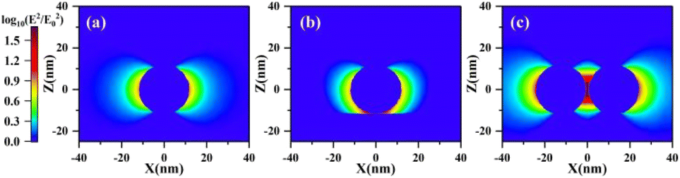 | ||
| Fig. 1 Calculated electromagnetic field enhancement for (a) single spherical NP with a diameter of 23 nm, (b) NP-substrate coupling system, and (c) NP–NP coupling system. | ||
In order to optimize the electromagnetic field enhancement, it is necessary to establish a plasmon coupling system with more “hot spots”. In the present work, we chose a coupling system consisting of 1D short nanochains to enhance LIBS signals. We first calculated the electric field distribution of Ag nanochain configuration as a function of nanochain lengths on the Si substrate (the interparticle distance was set to 2 nm) upon excitation at 532 nm, as shown in Fig. 2. The simulated results showed that for a single nanoparticle (Fig. 2a), the maximum electric field enhancement at the particle-substrate region was weaker than that in the regions between particle–particle junctions in Ag nanochains (Fig. 2b–h). The “hot spot” regions between the particle–particle connections were selected for near field enhancement analysis. Fig. 2i shows the maximum near field enhancement as a function of the number of particles in the silver nanochain. It can be clearly observed that the near-field enhancement effect of silver nanochains was significantly better than that of single nanoparticles, and it increases slowly with the increase of chain length. As the number of nanoparticles increases to four or more, the electric field reaches maximum enhancement. As a consequence, a short nanochain with 4 particles is sufficient for LIBS.
 | ||
| Fig. 2 (a–h) COMSOL simulation of the electric field distribution of Ag nanospheres (diameter ∼23 nm) on a Si substrate. The number of Ag NPs in the chain are (a) 1; (b) 2; (c) 3; (d) 4; (e) 5; (f) 6; (g) 7; (h) 8. (i) Maximum electric field enhancement as a function of the Ag NP number in the chain (diameter ∼23 nm), obtained from (a–h). | ||
Influence of CTAB concentration on formation of short silver nanochains
In order to examine the formation of a silver nanochain structure, we introduced varying concentrations of CTAB solution into Ag NPs. Fig. 3a shows a TEM image of Ag NPs, and well dispersed spherical Ag NPs with a diameter of approximately 23 nm can be clearly observed. In Fig. 3b–e, we observed that Ag NPs with a low concentration of CTAB formed chain-like configuration (interparticle distance was about 2 nm), and the chain length increased with an increasing concentration of CTAB. The further increasing concentration of CTAB to 0.1 mM led to the formation of clumps (Fig. 3f–h). Therefore, it is clear that the CTAB concentration plays an important role in the formation of Ag chain-like configuration.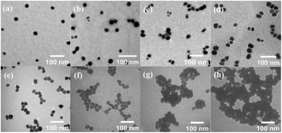 | ||
| Fig. 3 TEM images showing the morphologies of Ag NPs with different CTAB concentrations: (a) 0 mM; (b) 0.02 mM; (c) 0.04 mM; (d) 0.06 mM; (e) 0.08 mM; (f) 0.1 mM; (g) 0.5 mM; (h) 1 mM. | ||
Absorbance spectra
The surface plasmon resonance (SPR) of Ag NP colloids originates from extinction spectra depending on the size, shape and aggregation level of the NPs. The optical properties of the Ag NPs colloids were determined by UV-vis spectroscopy in the absorbance mode. Fig. 4 shows the absorption spectra of Ag NP colloids with varying concentrations of CTAB and MUA in the wavelength range of 300–780 nm. All the spectra exhibit strong absorption bands centered at ∼410 nm. As the concentration of CTAB increased, the absorbance peak intensity decreased, but no significant shift in the peak position was observed. This implies that the morphology of Ag NPs was unchanged and no aggregation occurs in solution, because the decrease of the main Ag NP absorption mode does not correspond to the appearance of a broad red-shifted shoulder. This observation is crucial because it demonstrates that the effect of CTAB is related to the deposition of NPs on the substrate during the drying process. In this view, the difference in SPR intensity in solution may be just related to the different extinction coefficient when CTAB is added to the solution.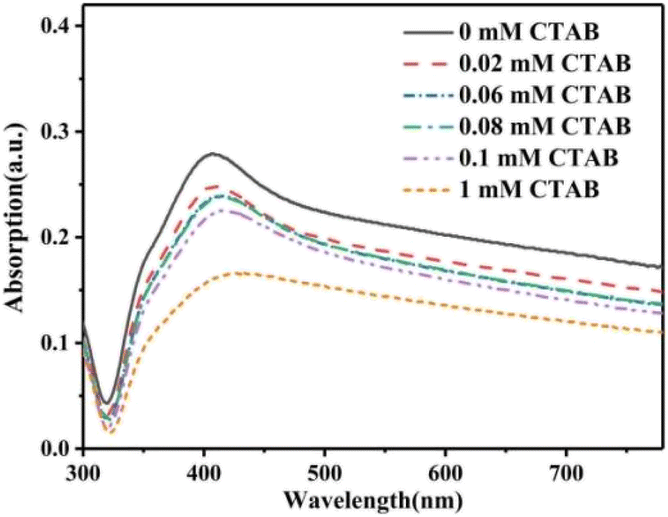 | ||
| Fig. 4 Absorption spectra of the Ag NPs with various concentrations of CTAB and MUA. | ||
Optimization of the analytical parameters
Both the energy of the incident laser and delay time of acquisition were two significant influencing parameters for LIBS quantitative analysis. The intensities of analytical lines (Ce II 418.66 nm, La II 394.91 nm and Pr II 422.29 nm) were measured as a function of laser pulse energies (gate delay and width were 0.5 and 5 μs, respectively) and gate delay times (laser energy was 60 mJ and gate width was 0.5 μs) and are shown in Fig. 5. As shown in Fig. 5a, the intensity of all analytical lines increased first and reached the maximum value at 60 mJ and then maintained a gentle trend with the increasing laser energy. Moreover, as shown in Fig. 5b, with a fixed laser energy of 60 mJ, the intensity of all analytical lines from 0.5 to 7 μs reduced with the increasing delay times, and the highest intensities of all analytical lines were obtained when the delay time was 0.5 μs. Therefore, the optimized parameters of nanochain enhanced LIBS for highly sensitive determination of lanthanides in an aqueous solution were 60 mJ for laser energy and 0.5 μs for delay time.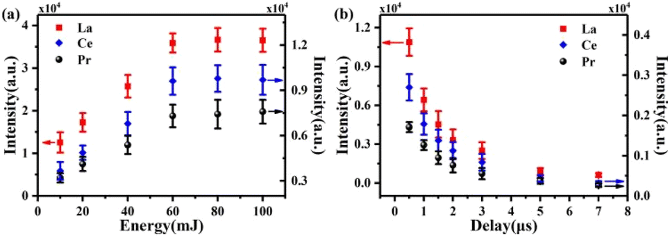 | ||
| Fig. 5 The intensities of analytical lines (Ce II 418.66 nm, La II 394.91 nm and Pr II 422.29 nm) measured as a function of laser pulse energies (a) and gate delay times (b). | ||
NELIBS signal enhancement
As described in the Experimental section, the sample was prepared by drying liquid solution on a Si substrate, which converted the analytical sample into a solid layer on the Si substrate surface. To verify the feasibility of short nanochain enhanced LIBS in quantitative determination of La, Ce and Pr elements, Fig. 6a–c present the comparison of a frame of NELIBS and LIBS spectra of Ce(NO3)3, La(NO3)3 and Pr(NO3)3 aqueous solutions. In detail, Fig. 6a shows a comparison of Ce II line spectra measured under different sample conditions with 5 mM Ce(NO3)3 solution. Spectrum 2 (dashed line) and spectrum 3 (solid line) were generated from mixed solutions of Ce(NO3)3 + Ag NPs and Ce(NO3)3 + Ag NPs + CTAB + MUA, respectively, deposited on a Si substrate. Spectrum 1 (dotted line) was acquired for Ce(NO3)3 solutions deposited on the Si substrate for comparison. The effective effect of nanochain configuration in LIBS signal enhancement is clearly demonstrated here. Several significant peaks at 395.25, 395.61, 396.09, 401.23, 429.67, 430.03 and 535.34 nm are characteristic bands of the Ce element; the intensities of these emission lines were strongly enhanced in the presence of CTAB and MUA. Similarly, we studied the results of La under the same experimental conditions, as shown in Fig. 6b, where a comparison of four La II line spectra measured under different sample conditions with 0.1 mM La(NO3)3 solution was shown. The effectiveness of nanochain configuration in enhancing LIBS signals has also been clearly demonstrated here. Four significant peaks at 394.91, 398.85, 399.58 and 429.61 nm are characteristic bands of the La element; the intensities of these emission lines were also strongly enhanced in the presence of CTAB and MUA. Moreover, we also studied the results of Pr under the same experimental conditions, as shown in Fig. 6c. Fig. 6c shows a comparison of four Pr II line (410.07, 417.94, 422.29 and 422.54 nm) spectra measured under different sample conditions with 4 mM Pr(NO3)3 solution. The intensities of these emission lines were also strongly enhanced in the presence of CTAB and MUA. | ||
| Fig. 6 (a) Comparison of NELIBS spectra of Ce II lines obtained for mixed solutions of Ce(NO3)3 (spectrum 1, dot line), Ce(NO3)3 + Ag NPs (spectrum 2, dashed line) and Ce(NO3)3 + Ag NPs + CTAB + MUA (spectrum 3, solid line) deposited on a Si substrate; (b) comparison of NELIBS spectra of La II lines obtained for mixed solutions of La(NO3)3 (spectrum 1, dotted line), La(NO3)3 + Ag NPs (spectrum 2, dashed line) and La(NO3)3 + Ag NPs + CTAB + MUA (spectrum 3, solid line) deposited on a Si substrate; (c) comparison of NELIBS spectra of Pr II lines obtained for mixed solutions of Pr(NO3)3 (spectrum 1, dot line), Pr(NO3)3 + Ag NPs (spectrum 2, dashed line) and Pr(NO3)3 + Ag NPs + CTAB + MUA (spectrum 3, solid line) deposited on a Si substrate; (d) NELIBS integral intensity of the Ce II line at 401.23 nm as a function of CTAB concentrations; (e) NELIBS integral intensity of the La II line at 394.91 nm as a function of CTAB concentrations; (f) NELIBS integral intensity of the Pr II line at 417.94 nm as a function of CTAB concentrations; (g) calibration curves of Ce; (h) calibration curves of La; (i) calibration curves of Pr. | ||
To describe the enhancement effect quantitatively, the intensities of the Ce II line at a wavelength of 401.23, La II line at a wavelength of 394.91 and Pr II line at a wavelength of 417.94 nm were measured as a function of the CTAB concentration as shown in Fig. 6d–f. For Ce, La and Pr, a critical CTAB concentration of 0.04–0.08 mM can be observed at which an evident increase in LIBS intensity occurred. On increasing the CTAB concentration to 0.04–0.08 mM as shown in Fig. 3c–e, the formation of a 1D short nanochain appeared and the NELIBS signal increased. When the chain becomes too large, it probably increases the probability of having large multilayer aggregation as shown in Fig. 3f and g with a consequent decrease in the NELIBS enhancement. As shown in Fig. 3h the NPs lose their individual structure and, consequently, appear fused with each other. This may be due to aging or to the surface melt of oxidized NPs; however, under this condition, the field enhancement is clearly hindered.
As shown in Fig. 6d–f, the highest NELIBS intensity was achieved with the sample containing 0.04–0.08 mM CTAB. Therefore, a concentration of 0.06 mM was selected to plot a calibration curve for determination of Ce, La and Pr, as shown in Fig. 6g–i. The results showed that the determination coefficients (R2) of Ce, La and Pr calibration curves were 0.989, 0.990 and 0.996, respectively. Using the calibration curve slope and background standard deviation multiplied by a factor of 3, we calculated the LOD of Ce, La and Pr which was 27.47, 85.71 and 65.11 ng mL−1, respectively. The LODs of the present method were lower than those reported previously.10–13 Therefore, nanochain enhanced LIBS shows a better analytical performance for determination of lanthanides in an aqueous solution.
Short nanochain enhanced LIBS on mixed solution of lanthanides
Finally, we report one last example on a mixed solution of lanthanides (Ce, La and Pr). The purpose of this example is to demonstrate the evident improvement on more complex matrices with nanochain enhanced LIBS compared to conventional NELIBS. Fig. 7 shows the comparison of nanochain and nanoparticle enhanced LIBS on the emission signal of the mixed solution of lanthanides (4 mM Ce(NO3)3, La(NO3)3 and Pr(NO3)3). It can be clearly seen that the intensities of all analytical lines (Ce II 401.23 and 429.67 nm; La II 394.91, 398.85 and 399.58 nm; Pr II 417.94, 422.29 and 422.54 nm) were strongly enhanced in the presence of CTAB and MUA, that is, the Ag chain-like configuration can effectively enhance LIBS signals. | ||
| Fig. 7 Comparison of LIBS spectra of analytical lines obtained for mixed solutions of lanthanides (Ce, La and Pr): Ce(NO3)3 + La(NO3)3 + Pr(NO3)3 (dotted line), Ce(NO3)3 + La(NO3)3 + Pr(NO3)3 + Ag NPs (dashed line) and Ce(NO3)3 + La(NO3)3 + Pr(NO3)3 + Ag NPs + CTAB + MUA (solid line). | ||
Conclusion
In summary, we successfully applied nanochain enhanced LIBS for the determination of Ce, La and Pr and LODs of 27.47, 85.71 and 65.11 ng mL−1 have been achieved in short nanochains with an appropriate concentration of CTAB serving as “glue”. The short silver nanochains exhibit strongly enhanced LIBS properties, due to the localized surface plasmon coupling at the interstitial sites of Ag nanochains. This method enables sensitive detection of trace analytes, showcasing practical potential in analytical sectors. For future work, assembling spherical Au/Ag NPs by physical methods could be introduced for nanochain formation to avoid toxicity in chemical synthesis for potential clinical utility.Data availability
Data will be made available on request.Author contributions
Zhifan Li: visualization, investigation, data curation, formal analysis, writing – original draft. Huiwei Wei: investigation, data curation. Gentao Gao: supervision, funding acquisition. Zhiyong Deng: funding acquisition, project administration. Shaohua Sun: supervision. Zuoye Liu: funding acquisition, supervision. Baowei Ding: supervision. Bitao Hu: resources, supervision. Jie Shen: conceptualization, methodology, writing – original draft, writing – review & editing.Conflicts of interest
The authors declare that they have no known competing financial interests or personal relationships that could have appeared to influence the work reported in this paper.Acknowledgements
This work was supported by the National Natural Science Foundation of China [grant number 12374266].Notes and references
- M. Z. Martin, S. Allman, D. J. Brice, R. C. Martin and N. O. Andre, Exploring laser-induced breakdown spectroscopy for nuclear materials analysis and in situ applications, Spectrochim. Acta, Part B, 2012, 74–75, 177–183 CrossRef CAS.
- A. P. Rao, P. R. Jenkins, J. D. Auxier II, M. B. Shattan and A. K. Patnaik, Analytical comparisons of handheld LIBS and XRF devices for rapid quantification of gallium in a plutonium surrogate matrix, J. Anal. At. Spectrom., 2022, 37, 1090–1098 RSC.
- Y. Lee, S. Yoon, N. Kim, D. Kang, H. Kim, W. Yang, M. Burger, I. Jovanovic and S. Choi, In situ measurement of Ce concentration in high-temperature molten salts using acoustic-assisted laser-induced breakdown spectroscopy with gas, Nucl. Eng. Technol., 2022, 54, 4431–4440 CrossRef CAS.
- Y. Gong, D. Choi, B.-Y. Han, J. Yoo, S.-H. Han and Y. Lee, Remote quantitative analysis of cerium through a shielding window by stand-off laser-induced breakdown spectroscopy, J. Nucl. Mater., 2014, 453, 8–15 CrossRef CAS.
- M. Rabiul Awual, M. Munjur Hasan, A. Shahat, M. Naushad, H. Shiwaku and T. Yaita, Investigation of ligand immobilized nano-composite adsorbent for efficient cerium(III) detection and recovery, Chem. Eng. J., 2015, 265, 210–218 CrossRef.
- R. Zare-Dorabei, K. Dashtian and V. Jalalat, Lanthanum (III) Ion Determination by a New Design Optical Sensor, IEEE Sens. J., 2015, 15, 6715–6721 CAS.
- J. Li, L. Guo, N. Zhao, X. Yang, R. Yi, K. Li, Q. Zeng, X. Li, X. Zeng and Y. Lu, Determination of cobalt in low-alloy steels using laser-induced breakdown spectroscopy combined with laser-induced fluorescence, Talanta, 2016, 151, 234–238 CrossRef CAS PubMed.
- D. Fernandes Andrade, E. Rodrigues Pereira-Filho and D. Amarasiriwardena, Current trends in laser-induced breakdown spectroscopy: a tutorial review, Appl. Spectrosc. Rev., 2020, 56, 98–114 CrossRef.
- N. Aizezi, Y. Ye, Z. Chen and Y. Liu, Impact of soldering temperatures on heavy metal and dust emissions: A LIBS-based environmental pollution analysis, Spectrochim. Acta, Part B, 2025, 225, 107124 CrossRef CAS.
- J.-I. Yun, T. Bundschuh, V. Neck and J.-I. Kim, Selective determination of europium (III) oxide and hydroxide colloids in aqueous solution by laser-induced breakdown spectroscopy, Appl. Spectrosc., 2001, 55, 273–278 CrossRef CAS.
- H. Zhang, F.-Y. Yueh and J. P. Singh, Laser-induced breakdown spectrometry as a multimetal continuous-emission monitor, Appl. Opt., 1999, 38, 1459–1466 CrossRef CAS PubMed.
- D. Alamelu, A. Sarkar and S. Aggarwal, Laser-induced breakdown spectroscopy for simultaneous determination of Sm, Eu and Gd in aqueous solution, Talanta, 2008, 77, 256–261 CrossRef CAS PubMed.
- X. Yang, Z. Hao, M. Shen, R. Yi, J. Li, H. Yu, L. Guo, X. Li, X. Zeng and Y. Lu, Simultaneous determination of La, Ce, Pr, and Nd elements in aqueous solution using surface-enhanced laser-induced breakdown spectroscopy, Talanta, 2017, 163, 127–131 CrossRef CAS PubMed.
- P. K. Diwakar, S. S. Harilal, J. R. Freeman and A. Hassanein, Role of laser pre-pulse wavelength and inter-pulse delay on signal enhancement in collinear double-pulse laser-induced breakdown spectroscopy, Spectrochim. Acta, Part B, 2013, 87, 65–73 CrossRef CAS.
- C. Goueguel, S. Laville, F. Vidal, M. Sabsabi and M. Chaker, Investigation of resonance-enhanced laser-induced breakdown spectroscopy for analysis of aluminium alloys, J. Anal. At. Spectrom., 2010, 25, 635–644 RSC.
- Z. Wang, Z. Hou, S. Lui, D. Jiang, J. Liu and Z. Li, Utilization of moderate cylindrical confinement for precision improvement of laser-induced breakdown spectroscopy signal, Opt. Express, 2012, 20, A1011–A1018 CrossRef PubMed.
- A. De Giacomo, R. Gaudiuso, C. Koral, M. Dell'Aglio and O. De Pascale, Nanoparticle Enhanced Laser-Induced Breakdown Spectroscopy of Metallic Samples, Anal. Chem., 2013, 85, 10180–10187 CrossRef CAS PubMed.
- A. De Giacomo, M. Dell'Aglio, R. Gaudiuso, C. Korala and G. Valenza, Perspective on the use of nanoparticles to improve LIBS analytical performance: Nanoparticle enhanced laser induced breakdown spectroscopy (NELIBS), J. Anal. At. Spectrom., 2016, 31, 1566–1573 RSC.
- C. Koral, M. Dell'Aglio, R. Gaudiusoa, R. Alrifai, M. Torelli and A. De Giacomo, Nanoparticle-Enhanced Laser Induced Breakdown Spectroscopy for the noninvasive analysis of transparent samples and gemstones, Talanta, 2018, 182, 253–258 CrossRef CAS PubMed.
- J. Shen, K. Wu, D. Cao, J. Wang and B. Hu, Effect of Ag nanoclusters deposited with magnetron sputtering on laser-induced breakdown spectroscopy enhancement, Spectrochim. Acta, Part B, 2019, 156, 59–65 CrossRef CAS.
- T. Ohta, M. Ito, T. Kotani and T. Hattori, Emission Enhancement of Laser-Induced Breakdown Spectroscopy by Localized Surface Plasmon Resonance for Analyzing Plant Nutrients, Appl. Spectrosc., 2009, 63, 555–558 CrossRef CAS PubMed.
- M. Abdelhamid, Y. A. Attia and M. Abdel-Harith, The significance of nano-shapes in nanoparticle-enhanced laser-induced breakdown spectroscopy, J. Anal. At. Spectrom., 2020, 35, 2982–2989 RSC.
- Z. Salajková, V. Gardette, J. Kaiser, M. Dell'Aglio and A. De Giacomo, Effect of spherical gold nanoparticles size on nanoparticle enhanced Laser Induced Breakdown Spectroscopy, Spectrochim. Acta, Part B, 2021, 179, 106105 CrossRef.
- Z. Li, K. Wu, J. Shen, W. Zhang, C. Zhou and B. Hu, Improved Sensitivity with Chloride Ions on Nanoparticle Enhanced Laser-Induced Breakdown Spectroscopy, J. Anal. At. Spectrom., 2021, 36, 2346–2352 RSC.
- J. Lim, M. Naveed, Y. Wang and R. Saraf, Kinetics of ion-mediated directed self-assembly of one-dimensional chains of metal nanoparticles in solution, Nanoscale, 2025, 17, 5012–5020 RSC.
- V. Nguyen, W. Qian, Y. Li, B. Liu, M. Aaberg, J. Henry, W. Zhang, X. Wang and Y. Paulus, Chain-like gold nanoparticle clusters for multimodal photoacoustic microscopy and optical coherence tomography enhanced molecular imaging, Nat. Commun., 2021, 12, 34 CrossRef CAS PubMed.
- H. Wang, H. Li, P. Gu, C. Huang, S. Chen, C. Hu, E. Lee, J. Xu and J. Zhu, Electric, magnetic, and shear field-directed assembly of inorganic nanoparticles, Nanoscale, 2023, 15, 2018–2035 RSC.
- A. Kuzyk, R. Schreiber, Z. Fan, G. Pardatscher, E. Roller, A. Högele, F. Simmel, A. Govorov and T. Liedl, DNA-based self-assembly of chiral plasmonic nanostructures with tailored optical response, Nature, 2012, 483, 311–314 CrossRef CAS PubMed.
- S. Mondal, P. Rehak, N. Ghosh, P. Kral and E. Gazit, Linear One-Dimensional Assembly of Metal Nanostructures onto an Asymmetric Peptide Nanofiber with High Persistence Length, ACS Nano, 2022, 16, 18307–18314 CrossRef CAS PubMed.
- Y. Yang, S. Matsubara, M. Nogami, J. Shi and W. Huang, One-dimensional self-assembly of gold nanoparticles for tunable surface plasmon resonance properties, Nanotechnology, 2006, 17, 2821–2827 CrossRef CAS.
- Y. Yang, J. Shi, T. Tanaka and M. Nogami, Self-assembled silver nanochains for surface-enhanced Raman scattering, Langmuir, 2007, 23, 12042–12047 CrossRef CAS PubMed.
- K. Wu, J. Shen, D. Cao, H. Cheng, S. Sun and B. Hu, Coulombic Effect of Amphiphiles with Metal Nanoparticles on Laser-Induced Breakdown Spectroscopy Enhancement, J. Phys. Chem. C, 2018, 122, 19133–19138 CrossRef CAS.
- S. Zhang, L. Zhang, K. Liu, M. Liu, Y. Yin and C. Gao, Digestive Ripening in the Formation of Monodisperse Silver Nanospheres, Mater. Chem. Front., 2018, 2, 1328–1333 RSC.
| This journal is © The Royal Society of Chemistry 2025 |