Vacuum degree detection performance improvement of vacuum switches based on NELIBS
Jiaqi
Liu
 ,
Xiaokang
Ding
,
Feilong
Zhang
,
Huan
Yuan
,
Xiaokang
Ding
,
Feilong
Zhang
,
Huan
Yuan
 *,
Xiaohua
Wang
*,
Aijun
Yang
,
Jifeng
Chu
*,
Xiaohua
Wang
*,
Aijun
Yang
,
Jifeng
Chu
 and
Mingzhe
Rong
School of Electrical Engineering, Xi'an Jiaotong University, Xi'an, 710049, PR China. E-mail: huanyuan@xjtu.edu.cn; yangaijun@mail.xjtu.edu.cn; 3121304047@stu.xjtu.edu.cn
First published on 30th November 2024
Abstract
The measurement of vacuum degree and electrification in vacuum switches has been a critical issue constraining the development of vacuum switches for over half a century, remaining unresolved. In recent years, our team has proposed the use of laser-induced breakdown spectroscopy (LIBS) for the non-contact measurement of vacuum switch pressure. However, its application is hindered by relatively high detection limits, low sensitivity, and susceptibility to background noise interference, which result in suboptimal precision. Extensive research has indicated that metal nanoparticles can effectively enhance signal detection capabilities, however, their enhancement effects under low-pressure conditions remain unclear. This study aimed to investigate the enhancement effects of silver nanoparticles (AgNPs) on signals under low-pressure conditions. By analyzing spectral data, plasma images, and radiation integral intensity, we seek to improve the accuracy of vacuum level measurements in vacuum switches. Results showed that in low-pressure environments, AgNPs enhanced spectral signals by up to 7.17-fold, increasing vacuum detection accuracy from 95.7% to 98.05% and extending the detection range by an order of magnitude to 10−3 Pa. This enhancement was regulated by particle size and concentration, with 10 nm AgNPs exhibiting better enhancement effects than 5 nm particles. The optimal concentration varied with both particle size and pressure. Nanoparticle-enhanced LIBS (NELIBS) increased the drop in plasma radiation integral intensity, prolonging the pre-expansion state of the plasma when excited and improving the accuracy of vacuum level measurements. This explores the potential of applying nanomaterials to conduct electrical detection in vacuum conditions and lays a theoretical and experimental foundation for optimizing spectroscopic analysis technologies.
1. Introduction
With the global low-carbon objectives, the green and low-carbon transformation of power systems is imperative. Vacuum switches are the preferred equipment for environmental upgrades of the power grid due to its zero greenhouse gas emissions feature. The internal vacuum level within the vacuum interrupter, which serves as the core component of the vacuum switch, plays a critical role in reliably interrupting the circuit. The degree of internal vacuum in the vacuum interrupter is the decisive factor for reliably breaking the circuit. To ensure the safety use of the device, the degree of vacuum detection for vacuum interrupter cannot be underestimated. The detection of online vacuum degree has been studied for over 70 years and is recognized as a difficult problem in the power equipment industry. CIGRE (Conseil International des Grands Réseaux Électriques) has evaluated it as a “bottleneck problem restricting the development of vacuum switches” and stated this “key problem urgently needs to be solved by users.” On-site, offline detection methods are commonly employed, necessitating the shutdown of vacuum switches. However, this approach severely constrains the development and application of vacuum switches, posing a threat to the safe and stable operation of the power grid. Therefore, vacuum degree online detection is the final barrier to overcome for the large-scale application of environmentally friendly vacuum switches of transmission level. Based on this international challenge, our team proposed a technology for vacuum degree online detection of vacuum switches based on LIBS (laser induced breakdown spectroscopy) in the preliminary stage. This technology utilized high-energy laser pulses focused on the surface of the target material to induce plasma generation, producing plasma signals. The vacuum level can be determined through the radiation integral intensity of the plasma signals,1 plasma contour images,2 and spectral information of specific elements.3 It can achieve detection capabilities down to 10−2 Pa while ensuring safe measurements.4–6 Cu, as a crucial component in the vacuum interrupter, has significant implications for online vacuum detection based on whether its signal is enhanced or not. According to industry regulations for electrical equipment, the internal pressure of a vacuum interrupter should not exceed 0.066 Pa during operation.7 Therefore, subsequent experiments mainly focus on pressure levels of 0.1 Pa and lower. However, plasma generated by LIBS technology dissipates rapidly under low pressure, resulting in weak and unstable spectral signals. A novel approach is required to overcome these challenges.Several technologies can enhance the signal intensity of LIBS, such as dual-pulse LIBS,8–12 ring magnet enhanced LIBS,13 and enhances the electric field effect.14,15 However, these methods rely on additional energy sources or tunable lasers, which are unsuitable for the closed environment of the vacuum interrupter in vacuum switches. In contrast, NELIBS (nanoparticle-enhanced kaser-induced breakdown spectroscopy) technology improves the laser ablation of samples to generate plasma using employing nanoparticles.16–18 Nanoparticles provide localized, highly enhanced near-field intensity, resulting in nanoscale ablation, which is the result of plasma enhancement by near-field electromagnetic waves incident on the particles. Upon laser irradiation, the nanoparticles are first affected, inducing localized surface plasmons (LSPs). Coupling between LSPs of adjacent nanoparticles generates a strong electric field and forms “hotspots”. “hotspots” refers to regions where localized surface plasmon resonance (LSPR) of neighbouring nanoparticles are coupled, generating intense electric fields. Within these regions, electromagnetic field energy is highly localized, resulting in elevated local surface temperatures. Under this strong electric field, field-induced electron emission from the sample leads to a stronger plasma, resulting in a significant enhancement of spectral signals by 1–2 orders of magnitude, with minimal impact on background radiation intensity. Consequently, the signal-to-noise ratio of plasma signals is enhanced, effectively improving the detection limit. Over the past decade, researchers worldwide have conducted extensive studies on NELIBS, and this technology has also been applied to non-metallic samples such as liquids and proteins,19,20 demonstrating significant enhancement effects on laser-induced plasma signals. The enhancement effect of NELIBS varies on different metal samples, after multiple laser pulses at the same point on the sample, the enhancement effect of nanoparticles on plasma signals gradually diminishes, which is related to the melting point of the metals.21 Experiments conducted by Abdelhamid et al. on nanoparticles of different shapes showed significant differences in enhancement effects.22 Sládková et al. studied the effect of nanoparticles on enhancing laser-induced plasma signals based on metallic lead in a low-pressure environment. They found little difference in signal enhancement between vacuum and low-pressure environments, which is one of the few experiments conducted under low-pressure conditions.23 Tang et al. proposed the use of a combination of magnetic fields and nanoparticles to enhance the LIBS signals. They found that the enhancement effect of magnetic confinement was weaker than that of nanoparticles and that the combined effect was stronger than either alone.24 Nanoparticles can also reduce plasma splashing and bubble formation, prevent plasma cooling, and improve the detection limits.25,26 For vacuum-switch equipment, reducing the detection limit and improving the signal intensity of laser-induced plasma can be achieved simply by uniformly coating the sample surface with metal nanoparticle reagents, achieving more precise detection results without additional equipment, thus meeting the requirements of its application scenarios.
Therefore, this paper proposes a method utilizing NELIBS to optimize vacuum degree online detection in vacuum interrupter. This method enhances spectral signal intensity to improve vacuum identification accuracy while also increasing the steepness of plasma radiation integral intensity decay and the resolution of plasma images to enhance the accuracy of vacuum level detection. We utilized silver nanoparticles due to their strong localized surface plasmon resonance effect. In this experiment, it aims to elucidate the effects of silver nanoparticle particle size, delay time, particle concentration on signal enhancement, as well as the integral intensity of plasma radiation under low pressure. To demonstrate the enhancement effect of AgNPs, the laser-induced plasma spectra of Cu were obtained, with 515.3 nm and 521.8 nm selected as characteristic spectral lines of copper.
2. Materials and methods
2.1 Experimental setup
The experiment employed a Q-switched Nd:YAG laser with a wavelength of 1064 nm, a pulse duration of 5 ns, and a repetition rate of 10 Hz. The pulse signal generated by the signal generator controls the laser pulse emission. The target material a T2 copper plate was placed in a vacuum chamber equipped with a quartz window. Laser focusing was achieved using a convex lens with a focal length of 150 mm, whereas a 90 mm convex lens was used to collect light. The laser and induced plasma were separated using a dichroic mirror, and the plasma spectral signals were analyzed using an ICCD camera and spectrometer. The exposure time of the ICCD was set to 30 ns. A three-dimensional stepper motor moved the copper plate inside the vacuum chamber. The experiment utilized water-soluble silver nanoparticle reagents with particle radius of 5 nm and 10 nm, with initial concentrations of 0.1 mg mL−1 and solutions modulated to concentrations of 0.05 mg mL−1 and 0.01 mg mL−1.The experimental setup is shown in Fig. 1, with the laser energy set at 30 mJ. Because of the relatively low laser energy, the influence of the gas generated by laser ablation and evaporation on the chamber's internal pressure can be disregarded during the experimental process.
During NELIBS low-pressure experiments, a vacuum chamber is utilized for evacuation. The volume of the vacuum chamber is 9 liters, employing a two-stage evacuation system comprising a mechanical pump and a turbomolecular pump. Initially, the mechanical pump is employed to reduce the pressure to approximately 10 pascals, followed by switching to the turbomolecular pump to achieve pressures ranging from 10−4 to 10−5 pascals. The pressure inside the vacuum chamber is continuously monitored in real-time using a wide-range combined vacuum gauge for thermal ionization. This gauge consists of a thermal cathode ionization measurement system and a Pirani measurement system, capable of measuring pressures down to 10−8 Pa.
Because the laser ablation of nanoparticles leading to their removal, only the results of the first ablation event at each point were recorded as the experimental data. To mitigate the influence of the rough ablation craters generated by laser irradiation on the experimental results, the step size of the motor was set to 1 mm. This distance was sufficient to ensure no mutual interference between the different irradiation points.
2.2 Experiment procedure
To comprehensively investigate the enhancement effect of silver nanoparticles on laser-induced plasma signals under low-pressure conditions, we systematically studied the combined effects of different pressures, silver nanoparticle particle sizes, concentrations, and delay times on the enhancement effect of NELIBS signals.Before the experiment, the copper plates were polished with sandpaper to remove surface oxidation and ensure the experiment's reliability. Subsequently, silver nanoparticle reagents with initial concentrations of 0.1 mg mL−1 were prepared for both 5 nm and 10 nm particle sizes, resulting in solutions of 0.05 mg mL−1 and 0.01 mg mL−1, respectively. The reagent was added to the copper plate sample to form a liquid droplet with a diameter of approximately 10 mm. The droplet was evenly spread into a rectangular shape using a glass rod and allowed to air dry before being placed in a vacuum chamber. During the experiment, the laser-irradiated the reagent-coated area at 1 mm intervals to ensure the independence of each experiment. After completing the NELIBS measurements, the area without the nanoparticle coating was irradiated again, and three sets of data were recorded as control groups.
3. Signals enhancement for improving detection accuracy
3.1 Effect of pressure
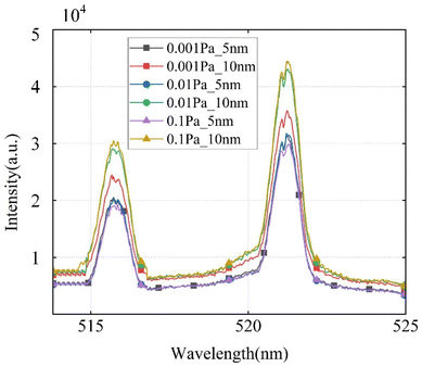
Fig. 2 illustrates the spectra obtained with and without applying 0.1 mg mL−1, 10 nm diameter silver nanoparticles on the copper target under a delay time of 100 ns, ranging from 10−3 Pa to 1 Pa. The spectral signals captured by a copper target with silver nanoparticles. (NELIBS) were generally stronger than those without silver nanoparticles (LIBS). However, owing to excessive intensity, self-absorption effects occur, leading to energy loss at the peak position.27 The average spectral intensity at 521.8 nm obtained from a blank copper plate was approximately 9.283 × 10³ a.u., whereas the minimum spectral intensity obtained from the copper plate coated with silver nanoparticles reached 1.9 × 10⁴ a.u., with the maximum intensity reaching 4.4 × 10⁴ a.u. at 0.1 Pa.Fig. 3 illustrates the enhancement factors at the Cu characteristic spectral lines as depicted in Fig. 2. It can be observed that under low pressure conditions, the enhancement of the Fig. 4 Performance comparison of different pre-treatment methods spectrum by silver nanoparticles ranges from 2 to 8-fold, with particularly pronounced enhancement observed at 515.3 nm, reaching a maximum of 7.17-fold. Furthermore, under 10−3 Pa, an enhancement factor of 4.97-fold is also demonstrated.
 | ||
| Fig. 3 Comparison of spectral enhancement factors under low pressure between 0.1 mg mL−1 silver nanoparticles (NELIBS) and uncoated silver nanoparticles (LIBS). | ||
At low pressure, the spectral signal from AgNPs was significantly enhanced. As seen in the spectral plot in Fig. 2, compared to the detection results at 10−2 Pa, NELIBS maintained strong detection intensity even at 10−3 Pa, while LIBS spectra were drowned out by noise under low pressure, leading to poorer resolution and stability. The characteristic spectral lines exhibited by the enhanced signals can be used for vacuum degree representation through the random forest algorithm. To this end, we built a vacuum model based on random forest, using the collected raw spectra to form a dataset, which was split into training and testing sets at a 3![[thin space (1/6-em)]](https://www.rsc.org/images/entities/char_2009.gif) :
:![[thin space (1/6-em)]](https://www.rsc.org/images/entities/char_2009.gif) 1 ratio. We used OOB error and accuracy as evaluation metrics and performed hyperparameter tuning for mtree and ntry to optimize the detection performance.3 In this context, the out-of-bag (OOB) error serves as an unbiased estimate of the generalization error of the random forest model, allowing for the identification of vacuum levels in different samples. Accuracy is used to assess the overall identification capability of the model, reflecting its reliability and precision in identifying vacuum levels.
1 ratio. We used OOB error and accuracy as evaluation metrics and performed hyperparameter tuning for mtree and ntry to optimize the detection performance.3 In this context, the out-of-bag (OOB) error serves as an unbiased estimate of the generalization error of the random forest model, allowing for the identification of vacuum levels in different samples. Accuracy is used to assess the overall identification capability of the model, reflecting its reliability and precision in identifying vacuum levels.
The spectral data collected from traditional LIBS and NELIBS experiments were input into the algorithm for detection, and the resulting OOB error, accuracy, R2, and RMSE are shown in Fig. 4. In this context, R2 represents the degree to which the spectral data explain the vacuum level in the model, while RMSE (root mean square error) indicates the deviation between the computed vacuum levels and the actual values. The results indicated that the accuracy of NELIBS reached 98.05%, surpassing traditional LIBS at 95.7%. This improvement was due to the AgNPs enhancing the peak intensity of element spectra and reducing the impact of background noise under high vacuum. The R2 value for NELIBS was 0.9432, while that for LIBS was 0.932. Additionally, the RMSE for NELIBS was lower than that for LIBS, indicating that NELIBS offered greater reliability in determining vacuum levels with a smaller deviation from the actual values. Therefore, NELIBS improves the precision of online vacuum detection and detection accuracy by enhancing the spectral signal strength.
3.2 Effects of particle size
Under atmospheric pressure, smaller-sized nanoparticles tend to generate stronger localized electric fields. This is attributed to their relatively higher curvature, which facilitates the excitation of free electrons, resulting in a higher surface plasmon resonance (SPR) frequency and positive effects on generating localized electric fields.14 However, previous studies exploring the influence of gold nanoparticles on the signal intensity used the intensity of Si I at 288.157 nm. It was found that AuNPs with a particle size of 7.5 nm exhibited an optimal Si I intensity compared to those with sizes of 5 and 10 nm.9 This discrepancy arises because various factors, including shape, surface modification, and the surrounding media, influence the SPR frequency of metal nanoparticles.In this study, the signal enhancement effects of different particle sizes under low pressure were investigated. The surface plasmon resonance frequency of nanoparticles may vary because of their lower dielectric constant in vacuum. Fig. 5 illustrates the spectra obtained under low pressure with a delay time of 100 ns and a concentration of 0.1 mg mL−1 for silver nanoparticles with particle sizes of 5 nm and 10 nm. Compared to the 5 nm particle size, the 10 nm silver nanoparticles exhibited better enhancement characteristics, with an average intensity exceeding 104 a.u. This is because under low pressure, in the absence of a higher dielectric constant atmosphere, the SPR frequency of 10 nm nanoparticles may be closer to the frequency of the laser source used, making it more conducive to generating a strong electric field and effectively enhancing the interaction signals with molecules near the surface.28 Meanwhile, smaller 5 nm nanoparticles may be constrained by shape and size, resulting in less significant local electric field enhancement effects than the 10 nm particle size. As the pressure decreases, the enhancement effect initially decreases and then increases, indicating that different particle sizes may have a certain critical pressure threshold at which they contribute significantly to signal enhancement.
 | ||
| Fig. 5 Comparison of signal enhancement of silver nanoparticles of different sizes under standard air pressure of vacuum switch operation. | ||
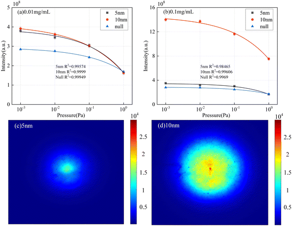
Fig. 6 illustrates the radiation integral intensity and plasma profile images of silver nanoparticles with particle sizes of 5 nm and 10 nm at concentrations of 0.01 mg mL−1 and 0.1 mg mL−1, respectively, with a delay time of 100 ns. Blank control were included in the experiments. Both Fig. 6(a) and (b) demonstrate similar characteristic patterns, with the radiation integral intensities of the nanoparticle-coated samples showing larger fluctuations than those of the blank control group. Additionally, the radiation integral intensity of the 10 nm particles was consistently higher than that of the 5 nm particles, consistent with the conclusion drawn from Fig. 5. At a concentration of 0.1 mg mL−1, the radiation intensity of 10 nm silver nanoparticles was approximately four times higher compared to the 5 nm particles and without nanoparticle coating. The 10 nm silver nanoparticles exhibited significantly higher absorption efficiency and a more pronounced SERS effect, thereby influencing the radiation intensity.
The relationship curve between pressure and radiation integral intensity can serve as a calibration curve for detecting vacuum levels. The degree of variation in this curve reflected the transformation of vacuum levels. The greater the difference in radiation integral intensity across different pressures, the more beneficial it is for improving the accuracy of vacuum detection. As observed in Fig. 6(a) and (b), the R2 values of the fitted curves are close to 1. As the pressure decreases, the rate of increase in plasma emission intensity slows, and based on this declining trend, the corresponding pressure can be determined.29 The steepness of the curve for the 10 nm particles exceeds that of the blank control group, indicating better characterization compared to the 5 nm silver nanoparticles, suggesting that nanoparticles can enhance the accuracy of vacuum level detection.
Furthermore, the plasma profile images in Fig. 6(c) and (d) also indicate that, at the same delay time, the plasma image of the 10 nm particles has a larger diffusion range and higher brightness.
3.3 Effect of concentrations
This section investigates the spectral images under different concentrations of AgNPs. Three concentrations of silver nanoparticles were used for comparison, and spectral images under a 10 nm particle size were observed, and results are shown in Fig. 7. It is evident that silver nanoparticles at any concentration significantly enhance the spectral signal. The increase in spectral intensity can improve vacuum identification accuracy, thereby enhancing the reliability of vacuum level detection. The signal enhancement was the greatest at 0.1 mg mL−1 under low pressure, followed by 0.05 mg mL−1. The signal enhancement was the smallest at 0.01 mg mL−1. We analysed this intriguing phenomenon.There are two choices for changing the concentration: one is to change the amount of solvent, and the other is to change the number of particles. In the enhancement of the NELIBS signals, the density of “hot spots” was closely related to the density of the nanoparticles. The strong electric field of the “hot spots” is the reason for field-induced electron emission. The higher the density of nanoparticles, the higher the density of “hot spots,” and the more frequent the field-induced electron emission, resulting in a stronger spectral signal. The solvent eventually evaporate from the sample, leaving only the particles. The only variable was the purity of water. Therefore, adjusting the concentration can achieve the optimal surface concentration of the sample during deposition,16 and the deposition level of the nanoparticles is the key to achieving good signal enhancement.30
Therefore, at a particle size of 10 nm, the higher the concentration, the better the enhancement effect, because at this time, the density of “hot spots” increases, and the critical distance between nanoparticles should be smaller than the diameter of the nanoparticle. However, at a particle size of 5 nm, the excessively high concentration of nanoparticles may prevent the laser from effectively reaching and ablating the sample's surface. Therefore, a lower concentration produces a better enhancement effect.31 This implies that there is an optimal concentration of AgNPs of different sizes.
Fig. 8(a) and (b) illustrate the integrated intensity of plasma radiation at a 100 ns delay for different concentrations of 5 nm and 10 nm particle sizes, respectively. Under a particle size of 5 nm, the intensity is maximum at a concentration of 0.05 mg mL−1, reaching 4.0 × 10⁸ a.u., while under a particle size of 10 nm, the intensity is maximum at a concentration of 0.1 mg mL−1, reaching 1.4 × 10⁹ a.u. However, a comparison with the spectral images reveals that the concentration resulting in maximum spectral intensity is not the same as the concentration resulting in maximum integrated plasma radiation intensity. This may involve nonlinear effects of nanoparticle concentration.
 | ||
| Fig. 8 Integrated plasma radiation intensity at different concentrations at low pressure at 100 ns delay: (a) 5 nm, (b) 10 nm. | ||
The spectral intensity is influenced by factors such as laser energy, the interaction between the laser and the sample, and the concentration of elements in the sample. Under conditions of high optical intensity, nanoparticles may exhibit saturation or other nonlinear effects, leading to an integrated radiation intensity that differs from a simple linear relationship.
The optimal concentration for 10 nm particles does not match that of 5 nm particles, indicating that the optimal solution for the concentration should be discussed separately for different particle sizes. This is also influenced by the distribution uniformity affected by the particle size. Nonetheless, as shown in Fig. 8, the steepness of the plasma radiation integral intensity curve increased with the addition of nanoparticles. Particularly, in Fig. 8(b), the curve at a particle size of 10 nm and a concentration of 0.1 mg mL−1 demonstrated a sharper gradient compared to the flatter blank control group, allowing for a more accurate assessment of the magnitude of the gas pressure.
3.4 Effect of time delays
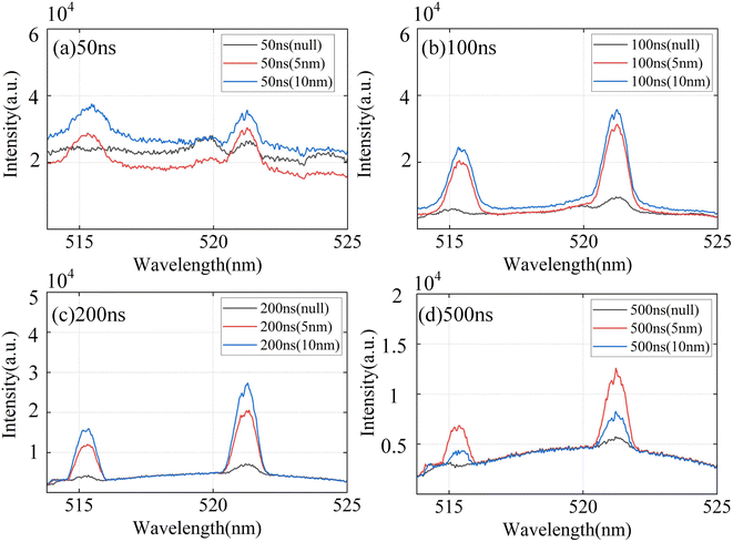
This section conducted experiments with fixed initial concentrations of 0.1 mg mL−1 and particle sizes of 10 nm and 5 nm silver nanoparticles, and spectra were measured at 10−3 Pa with delay times of 50 ns, 100 ns, 200 ns, and 500 ns. Fig. 9 illustrates that the signal intensity is significantly higher than that of no nanoparticle enhanced LIBS. The plasma signal undergoes an initial amplification process followed by attenuation owing to the concentration of laser energy within the pulse duration. This variation mirrors the results obtained under atmospheric pressure using NELIBS. The enhanced electromagnetic field generated by the nanoparticles persisted during laser irradiation, and upon the cessation of irradiation, the electrons within the nanoparticles gradually returned from the dipole state to a stable state. | ||
| Fig. 9 Spectra of 0.001 Pa, no nanoparticles and 0.1 mg mL−1 silver nanoparticles at different time delays: (a) 50 ns, (b) 100 ns, (c) 200 ns, (d) 500 ns. | ||
Because of plasma expansion and diffusion, by 500 ns, the no nanoparticle enhanced LIBS spectral signal intensity was significantly attenuated, being overwhelmed by noise. However, with the enhancement provided by the AgNPs, the duration of the plasma was prolonged, and the electron density within the plasma was high. This is attributed to the instantaneous emission of field-induced electrons, which results in higher ionization and electron emission rates. The NELIBS-generated plasma requires more time to cool completely.
Additionally, for silver nanoparticles (0.1 mg mL−1), the enhancement effect was superior for 10 nm particles compared to 5 nm particles.
Fig. 10(a) and (b) illustrate the variation of plasma radiation integral intensity over time with concentrations of 0.1 mg mL−1 and particle sizes of 5 nm and 10 nm, respectively. Fig. 10(b) shows that, upon laser irradiation of nanoparticles, an enhanced electromagnetic field was instantaneously applied to the sample, inducing field-induced emission initiation, consequently leading to an increase in the integrated intensity of plasma radiation. Before 200 ns, the plasma had not yet expanded significantly, and the plasma density generated by sample ionization continued accumulating, resulting in a higher radiation integral intensity.29 Additionally, ionization at high temperatures causes sample evaporation, producing gas molecules that rapidly accumulate near the plasma and constrain its expansion. During the electron field-induced emission period, the constraint exerted by the evaporated gas molecules further increases the plasma density. Because the short duration of the laser pulse, sample ionization primarily occurs during laser action, while NPs begin to play a role. As the plasma expanded, its density gradually decreased, the electric field effect weakened, and it tended to equilibrate with the environment over time.
 | ||
| Fig. 10 Integral diagram of plasma radiation intensity under low pressure with different particle sizes of 0.1 mg mL−1: (a) 5 nm, (b) 10 nm. | ||
Fig. 10(a) exhibits a trend similar to Fig. 10(b) at pressures of 10−3 Pa. However, as the pressure increases, the radiation integral intensity exhibits a direct attenuation trend. This is because, with increasing pressure, the collision frequency between particles in the plasma and surrounding gas molecules rises, leading to an increase in electron density. Consequently, more laser energy is absorbed, reducing the energy absorbed by the target material. This combined process reduces the plasma's radiation integral intensity. At low pressures below 10−2 Pa, the absorption and scattering of laser energy by gas molecules can be considered negligible.32,33
The enhanced temperature induced by the electromagnetic field of the silver nanoparticles led to the accumulation of a large amount of vapor near the plasma, causing the gas density and temperature to increase continuously. If the environmental pressure is low, the diffusion of the accumulated gas is hindered, thereby impeding plasma expansion. It took longer for the plasma enhancement effect to reach its maximum value and converge to equilibrium with the environment. Therefore, the critical expansion time of the plasma in NELIBS extended as the pressure decreased.
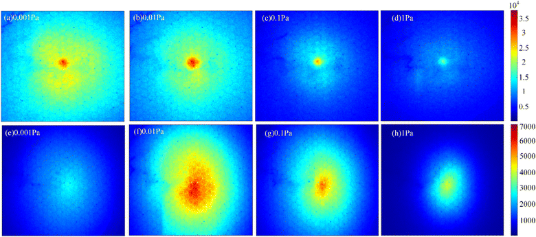
Fig. 11(a)–(d) presented the plasma profiles of 0.1 mg mL−1 silver nanoparticles with a particle size of 10 nm under various low pressures at a 500 ns delay, Fig. 11(e) and (f) represented the blank control group without the addition of silver nanoparticles. The ambient pressure can significantly affect the shape of the plasma. The distinct characteristics of plasma contours under different pressures can serve as a basis for determining the vacuum level. At pressures below 1 Pa, the length of the plasma plume can be used to quantify the pressure. As seen in Fig. 11(a)–(d), the overall plasma intensity gradually decreased with increasing pressure, which aligned with the radiation integral intensity trends shown in Fig. 10. The 200 ns delay, serving as the turning point for NELIBS plasma intensity, showed minimal variation along the vertical axis, whereas the 500 ns delay provided better differentiation. Comparing Fig. 11(a) and (d), it was evident that at 1 Pa and 500 ns, the plasma exhibited significant dissipation, nearing complete cooling. This was due to the presence of more gas molecules in the environment, which led to increased collisions. In contrast, lower pressures reduced the involvement of environmental gas molecules, allowing the plasma to remain at a higher density and temperature for a longer period. As shown in Fig. 11(e) and (h), conventional LIBS displays minimal differentiation in plasma images at 0.001 Pa and 1 Pa. This is because, at these pressures and times, the plasma has nearly dissipated, and without nanoparticles to sustain its expansion, the resulting signal is weak, making it difficult to distinguish vacuum levels. In contrast, the clear image differences shown in Fig. 11(a)–(d) allow for differentiation at 0.001 Pa.
4. Conclusions
Under low-pressure conditions, the NELIBS technology showed a promising potential of improving the accuracy of electrical detection in vacuum environments. The plasma generated by NELIBS differed from that generated by traditional LIBS, as nanoparticles provided locally and highly enhanced near-field intensities for generation of more “hot spots,” resulting in a larger area of sample ablation and increased emission of electrons. Under low-pressure conditions, NELIBS demonstrated superior signal enhancement, with an average spectral intensity reaching 3.5 × 10⁴ a.u., maintaining strong detection capability even at 10−3 Pa. In comparison, LIBS showed an average spectral intensity of around 9283 a.u. Additionally, the accuracy for NELIBS improved from 95.7% to 98.05%, indicating that NELIBS enhanced pressure detection accuracy by increasing the spectral peak intensity.Under low-pressure conditions, the enhancement effect of AgNPs is primarily influenced by the concentration of gas molecules. As pressure decreases, a more intense spectral signal can be observed. Experimental investigations on silver nanoparticles of different particle sizes under low-pressure conditions indicated that silver nanoparticles with a particle size of 10 nm exhibit a better enhancement effect compared to those with a particle size of 5 nm, with an average enhancement of 104 a.u. The integrated radiation intensity at 0.1 mg mL−1 exhibited a 3.13-fold enhancement compared to the 5 nm counterpart, while the intensity of the plasma profile chart was enhanced by 1.79 times compared to the 5 nm counterpart. This demonstrated a strong discriminative capability in LIBS, improving the detection accuracy of vacuum levels to 10−3 Pa.
This study also explored the effects of different concentrations and delay times on the spectra. Results indicate the existence of an optimal solution between the concentration of AgNPs and the enhancement effect. For silver nanoparticles with a particle size of 10 nm, a concentration of 0.1 mg mL−1 demonstrated a remarkable enhancement factor of up to 5.21 times, while for those with a particle size of 5 nm, a concentration of 0.01 mg mL−1 exhibited an enhancement factor of 5.17 times. The enhancement of the spectral signal improved the accuracy of vacuum identification and increased the precision of vacuum level detection. Moreover, under different delay times, shorter durations corresponded to greater spectral intensities. The integrated intensity of plasma radiation exhibited a trend of initially increasing and then decreasing, with the inflection point depending on the particle size. Smaller particle sizes led to earlier inflection times, while increasing the particle size can extend the expansion time of the plasma, resulting in a significantly increased enhancement factor. The variation in integrated radiation intensity can serve as real-time vacuum information, aiding in the optimization and adjustment of vacuum system operation. Increasing the steepness of the radiation integral intensity curve can provide a greater tolerance for identifying the magnitude of vacuum levels. Overall, silver nanoparticles with a particle size of 10 nm demonstrated outstanding performance in enhancing vacuum detection accuracy.
In short, this study provided an in-depth exploration of the factors that influence the spectral and radiation integral intensities of AgNPs under low-pressure conditions, laying a theoretical and experimental foundation for optimizing real-time vacuum monitoring using the NELIBS technology in vacuum environments. This application scenario includes but is not limited to, signal enhancement for vacuum switch vacuum detection. It can also be applied in environmental monitoring, medical devices, and aerospace, addressing the current lack of sensitivity in LIBS measurement signals.
Data availability
Data for this article, including observed wavelength of copper are available at NIST Atomic Spectra Database Lines Data at: https://physics.nist.gov/cgi-bin/ASD/lines1.pl?spectra=Cu%26output_type=0%26low_w=509%26upp_w=522%26unit=1%26submit=Retrieve+Data%26de=0%26plot_out=0%26I_scale_type=1%26format=0%26line_out=0%26en_unit=0%26output=0%26bibrefs=1%26page_size=15%26show_obs_wl=1%26show_calc_wl=1%26unc_out=1%26order_ou.Conflicts of interest
There are no conflicts to declare.Acknowledgements
We would like to thank the National Natural Science Foundation of China for funding this article with the project “Research on Theory and Method of LIBS-Raman Collaborative Detection of Multiple Impurity Particles in Transformer Oil”.References
- H. Yuan, W. Ke and X. Wang, et al., Research on vacuum degree detection of vacuum switch based on laser-induced plasma optical path multiplexing technology, High Voltage Eng., 2022, 48(3), 1160–1167 Search PubMed.
- H. Yuan, I. B. Gornushkin and A. B. Gojani, et al., Laser-induced plasma imaging for low-pressure detection, Opt. Express, 2018, 26(12), 15962–15971 CrossRef CAS PubMed.
- F. Zhang, H. Yuan and A. Yang, et al., On-line vacuum degree monitoring of vacuum circuit breakers based on laser-induced breakdown spectroscopy combined with random forest algorithm, J. Anal. At. Spectrom., 2024, 39(1), 281–292 RSC.
- H. Yuan, I. B. Gornushkin, A. B. Gojani, X. H. Wang and M. Z. Rong, Laser-induced plasma imaging for low-pressure detection, Opt. Express, 2018, 26(12), 15962–15971 CrossRef CAS PubMed.
- X. H. Wang, H. Yuan, D. X. Liu, A. J. Yang, P. Liu, L. Gao and M. Z. Rong, A pilot study on the vacuum degree online detection of vacuum interrupter using laser-induced breakdown spectroscopy, J. Phys. D: Appl. Phys., 2018, 49(44), 44LT01 CrossRef.
- H. Yuan, A. B. Gojani, I. B. Gornushkin and X. Wang, Investigation of laser-induced plasma at varying pressure and laser focusing, Spectrochim. Acta, Part B, 2018, 150, 33–37 CrossRef CAS.
- Waitangzhiku, DL/T 403-2017. DL/T 403-2017 High voltage AC Vacuum Circuit Breaker, 2022, https://www.waitang.com/report/236526.html Search PubMed.
- H. Heilbrunner, N. Huber, H. Wolfmeir, E. Arenholz, J. D. Pedarnig and J. Heitz, Double-pulse laser-induced breakdown spectroscopy for trace element analysis in sintered iron oxide ceramics, Appl. Phys. A, 2012, 106, 15–23 CrossRef CAS.
- F. Yang, L. Jiang, S. Wang, Z. Cao, L. Liu, M. Wang and Y. Lu, Emission enhancement of femtosecond laser-induced breakdown spectroscopy by combining nanoparticle and dual-pulse on crystal SiO2, Opt. Laser. Technol., 2017, 93, 194–200 CrossRef CAS.
- W. A. N. G. Songning, D. Zhang, C. H. E. N. Nan, H. E. Yaxiong, H. Zhang, K. E. Chuan and Z. H. A. O. Yong, Self-absorption effects of laser-induced breakdown spectroscopy under different gases and gas pressures, Plasma Sci. Technol., 2022, 25(2), 025501 Search PubMed.
- F. Poggialini, B. Campanella, S. Legnaioli, S. Pagnotta and V. Palleschi, Investigating double pulse nanoparticle enhanced laser induced breakdown spectroscopy, Spectrochim. Acta, Part B, 2020, 167, 105845 CrossRef CAS.
- E. Tognoni and G. Cristoforetti, Basic mechanisms of signal enhancement in ns double-pulse laser-induced breakdown spectroscopy in a gas environment, J. Anal. At. Spectrom., 2014, 29(8), 1318–1338 RSC.
- Z. Hao, L. Guo, C. Li, M. Shen, X. Zou, X. Li and X. Zeng, Sensitivity improvement in the detection of V and Mn elements in steel using laser-induced breakdown spectroscopy with ring-magnet confinement, J. Anal. At. Spectrom., 2014, 29(12), 2309–2314 RSC.
- D. A. Genov, A. K. Sarychev, V. M. Shalaev and A. Wei, Resonant field enhancements from metal nanoparticle arrays, Nano Lett., 2004, 4(1), 153–158 CrossRef CAS.
- G. N. Blackman III and D. A. Genov, Bounds on quantum confinement effects in metal nanoparticles, Phys. Rev. B, 2018, 97(11), 115440 CrossRef.
- M. Dell'Aglio, R. Alrifai and A. De Giacomo, Nanoparticle enhanced laser induced breakdown spectroscopy (NELIBS), a first review, Spectrochim. Acta, Part B, 2018, 148, 105–112 CrossRef.
- M. Dell'Aglio, Z. Salajková, A. Mallardi, M. C. Sportelli, J. Kaiser, N. Cioffi and A. De Giacomo, Sensing nanoparticle-protein corona using nanoparticle enhanced laser induced breakdown spectroscopy signal enhancement, Talanta, 2021, 235, 122741 CrossRef PubMed.
- C. Koral, A. De Giacomo, X. Mao, V. Zorba and R. E. Russo, Nanoparticle enhanced laser induced breakdown spectroscopy for improving the detection of molecular bands, Spectrochim. Acta, Part B, 2016, 125, 11–17 CrossRef CAS.
- A. De Giacomo, R. Gaudiuso, C. Koral, M. Dell'Aglio and O. De Pascale, Nanoparticle enhanced laser induced breakdown spectroscopy: effect of nanoparticles deposited on sample surface on laser ablation and plasma emission, Spectrochim. Acta, Part B, 2014, 98, 19–27 CrossRef CAS.
- M. Dell'Aglio, A. Mallardi, R. Gaudiuso and A. De Giacomo, Plasma parameters during nanoparticle-enhanced laser-induced breakdown spectroscopy (NELIBS) in the presence of nanoparticle–protein conjugates, Appl. Spectrosc., 2023, 77(11), 1253–1263 CrossRef PubMed.
- A. De Giacomo, R. Gaudiuso, C. Koral, M. Dell'Aglio and O. De Pascale, Nanoparticle-enhanced laser-induced breakdown spectroscopy of metallic samples, Anal. Chem., 2013, 85(21), 10180–10187 CrossRef CAS PubMed.
- M. Abdelhamid, Y. A. Attia and M. Abdel-Harith, The significance of nano-shapes in nanoparticle-enhanced laser-induced breakdown spectroscopy, J. Anal. At. Spectrom., 2020, 35(12), 2982–2989 RSC.
- L. Sládková, D. Prochazka, P. Pořízka, P. Škarková, M. Remešová, A. Hrdlička and J. Kaiser, Improvement of the laser-induced breakdown spectroscopy method sensitivity by the usage of combination of Ag-nanoparticles and vacuum conditions, Spectrochim. Acta, Part B, 2017, 127, 48–55 CrossRef.
- H. Tang, X. Hao and X. Hu, Research on spectral characteristics of laser-induced plasma by combining Au-nanoparticles and magnetic field confinement on Cu, Optik, 2018, 171, 625–631 CrossRef CAS.
- A. De Giacomo, R. Alrifai, V. Gardette, Z. Salajková and M. Dell'Aglio, Nanoparticle enhanced laser ablation and consequent effects on laser induced plasma optical emission, Spectrochim. Acta, Part B, 2020, 166, 105794 CrossRef CAS.
- A. Safi, J. E. Landis, H. G. Adler, H. Khadem, K. E. Eseller, Y. Markushin and N. Melikechi, Enhancing biomarker detection sensitivity through tag-laser induced breakdown spectroscopy with NELIBS, Talanta, 2024, 271, 125723 CrossRef CAS PubMed.
- W. Ke, H. Yuan, J. Q. Liu, X. H. Wang, A. J. Yang, J. F. Chu and M. Z. Rong, Effect of laser energy on temporal evolution of self-absorption at different air pressures, J. Phys. D: Appl. Phys., 2023, 57(9), 095204 CrossRef.
- Z. Salajková, V. Gardette, J. Kaiser, M. Dell'Aglio and A. De Giacomo, Effect of spherical gold nanoparticles size on nanoparticle enhanced laser induced breakdown spectroscopy, Spectrochim. Acta, Part B, 2021, 179, 106105 CrossRef.
- H. Yuan, W. Ke and X. Wang, et al., Research on vacuum degree detection of vacuum switch based on laser-induced plasma optical path multiplexing technology, High Voltage Eng., 2022, 48(3), 1160–1167 Search PubMed.
- F. Poggialini, B. Campanella, S. Giannarelli, E. Grifoni, S. Legnaioli, G. Lorenzetti and V. Palleschi, Green-synthetized silver nanoparticles for nanoparticle-enhanced laser induced breakdown spectroscopy (NELIBS) using a mobile instrument, Spectrochim. Acta, Part B, 2018, 141, 53–58 CrossRef CAS.
- A. De Giacomo, M. Dell'Aglio, R. Gaudiuso, C. Koral and G. Valenza, Perspective on the use of nanoparticles to improve LIBS analytical performance: nanoparticle enhanced laser induced breakdown spectroscopy (NELIBS), J. Anal. At. Spectrom., 2016, 31(8), 1566–1573 RSC.
- N. Farid, S. Bashir and K. Mahmood, Effect of ambient gas conditions on laser-induced copper plasma and surface morphology, Phys. Scr., 2011, 85(1), 015702 CrossRef.
- N. Farid, S. S. Harilal and H. Ding, et al., Emission features and expansion dynamics of nanosecond laser ablation plumes at different ambient pressures, J. Appl. Phys., 2014, 115(3), 033107 CrossRef.
| This journal is © The Royal Society of Chemistry 2025 |


