
Europium and calcium-co-doped TiO2 nanocrystals: tuning the biocompatibility and luminescence traceability of Drosophila melanogaster
Jerusa Maria
de Oliveira†
 ab,
Larissa Iolanda M.
de Almeida
ab,
Francisco
Rubens Alves dos Santos
ab,
João Paulo S. de
Carvalho†
ac,
Amanda I.
dos S. Barbosa†
a,
Marcus Andrei R. F. da
Costa
a,
Vanessa Tomaz
Maciel
ab,
Gabriela L. de
Souza
d,
Alysson N.
Magalhães
e,
Marcos V.
Vermelho
e,
Camilla Christian G.
Moura
d,
Felipe
Berti Valer
g,
Thiago Lopes
Rocha
f,
Sebastião William da
Silva
c,
Lucas
Anhezini†
b and
Anielle Christine A.
Silva
*ah
ab,
Larissa Iolanda M.
de Almeida
ab,
Francisco
Rubens Alves dos Santos
ab,
João Paulo S. de
Carvalho†
ac,
Amanda I.
dos S. Barbosa†
a,
Marcus Andrei R. F. da
Costa
a,
Vanessa Tomaz
Maciel
ab,
Gabriela L. de
Souza
d,
Alysson N.
Magalhães
e,
Marcos V.
Vermelho
e,
Camilla Christian G.
Moura
d,
Felipe
Berti Valer
g,
Thiago Lopes
Rocha
f,
Sebastião William da
Silva
c,
Lucas
Anhezini†
b and
Anielle Christine A.
Silva
*ah
aStrategic Materials Laboratory, Physics Institute, Federal University of Alagoas, Av. Lourival Melo Mota, S/N, Tabuleiro do Martins, Cep: 57072-970 Maceió, Alagoas, Brazil. E-mail: aniellechristineas@gmail.com
bLaboratory of In Vivo Toxicity Analysis and Neurodegenerative Diseases - Institute of Biological Sciences and Health, Federal University of Alagoas, Maceió, AL, Brazil
cOptical Spectroscopy Laboratory, Institute of Physics, University of Brasília, Brasília, Federal District, Brazil
dDepartment of Endodontics, School of Dentistry, Federal University of Uberlândia, Uberlândia, MG, Brazil
eApplied Microtechnologies Laboratories, Physics Institute, Federal University of Alagoas, Maceió, Alagoas, Brazil
fLaboratory of Environmental Biotechnology and Ecotoxicology, Institute of Tropical Pathology and Public Health, Federal University of Goiás, Goiânia, Goiás, Brazil
gDepartment of BioMolecular Sciences, School of Pharmaceutical Sciences of Ribeirão Preto, University of São Paulo, Ribeirão Preto 14040-900, São Paulo, Brazil
hNortheast Biotechnology Network, Federal University of Alagoas, Maceió, AL, Brazil
First published on 21st October 2024
Abstract
The incorporation of europium (Eu) ions improves the biocompatibility of TiO2 nanocrystals (NCs) and allows tracking by red luminescence. Calcium doping improves cellular compatibility while also facilitating better interaction with biological systems. Thus, in this work, were synthesized Eu and Ca co-doped TiO2 NCs and physical–chemical and biological properties were investigated. The physical–chemical properties were performed in order to analised the effects of the doping on the crystalline phase of TiO2 morphology, sized, zeta potential, hydrodynamic diameter, and photocatalytic properties. Biological assessments were conducted using in vitro assays with human osteosarcoma cells (SAOS-2) through cytotoxicity assays and in vivo assays with Drosophila melanogaster, where we evaluated the mortality rate during postembryonic development and the luminescence of nanomaterials in vivo. The physical–chemical properties confirmed with success the integration of Ca ions into the TiO2:Eu crystal (TiO2:Eu:xCa) NCs without additional phases. The co-doping of Ca led to a reduction of approximately 70% in photocatalytic activity. Moreover, co-doping with Ca was not cytotoxic to SAOS-2 cells. Our in vivo analysis showed no delays in postembryonic development and no larval or pupal lethality. The larval mortality rate and pupal formation rate were comparable to the control group when D. melanogaster was exposed to nanomaterials at concentrations of 1 mg mL−1 or lower. Luminescence of the NCs was detected in confocal microscopy images, indicating the presence of NCs in the larval brain and intestines. This luminescence was observed in TiO2:Eu:xCa NCs. These results showed that Ca doping improved the biocompatibility and enhanced the luminescence of these materials, making them traceable in biological tissues. Therefore, our research provides valuable insights into the tailored properties of TiO2 for potential applications in various fields of biomedicine.
Environmental significanceWe investigate the changes in the vibrational and photocatalytic properties of europium and calcium-co-doped TiO2 (TiO2:Eu:xCa) nanocrystals (NCs), and their toxicity in biological systems. Understanding the properties and toxicity of NCs is essential for their safe and efficient use in various applications, including biomedical ones. The results indicate that doping TiO2:Eu:xCa NCs modifies the structural characteristics and photocatalytic activity. Moreover, in vivo luminescence of the NCs exhibited increased biocompatibility and low toxicity, suggesting potential high biocompatibility. By evaluating their effects on biological systems, researchers can more effectively assess their potential environmental impacts if released into ecosystems. Moreover, the potential utilization of alternative NCs with high biocompatibility and intriguing physicochemical properties, such as luminescence, can be less likely to harm environmental systems. |
Introduction
In recent years, the integration of nanotechnology in biological research has gained significant attention due to its potential to revolutionize various aspects of experimental methodologies. Among the myriad of available nanomaterials, titanium dioxide nanoparticles (TiO2 NPs) have emerged as versatile candidates with unique physicochemical properties, making them particularly attractive for application in biological experiments.1–3 TiO2 NPs possess a high surface area and unique photocatalytic properties that can be harnessed for targeted applications in physical experiments. The large surface area allows for enhanced adsorption of biomolecules, facilitating interactions with cellular components.4 Additionally, the photocatalytic activity of TiO2 NPs under ultraviolet (UV) light exposure opens avenues for the controlled release of bioactive molecules or therapeutic agents, enabling precise modulation of biological responses.5,6The photocatalytic properties of TiO2 NPs also play a crucial role in photocatalysis-based experiments, where the nanomaterial can induce specific biological responses under controlled light exposure conditions. This capability provides researchers with a powerful tool to investigate light-triggered cellular processes or develop innovative therapeutic approaches that leverage the precise spatiotemporal control of light activation.7,8
Due to their pervasive application in consumer products and industrial processes, TiO2 NPs have garnered considerable interest across diverse scientific disciplines, focusing on nanotoxicology.7,9 This academic overview seeks to delve into the present understanding of the toxicity linked with TiO2 NPs, investigating pivotal factors that impact their potential adverse effects on biological systems.
Our research group has successfully established that the genotoxic effects of TiO2 NPs are contingent upon the specific crystalline phase in which they exist. Genotoxicity was evidenced by the induction of mutagenic activity by TiO2 in the rutile phase in Drosophila melanogaster. Various sizes of TiO2 NPs were assessed in the anatase phase (at 3.4 and 6.2 nm and in the rutile phase at 78.0 nm). Moreover, in both in vitro and in vivo tests, it was shown that the smaller anatase TiO2 NPs led to micronucleus formation. Notably, we observed that the rutile/brookite phase exhibited the highest degree of biocompatibility, whereas the anatase phase demonstrated the greatest level of toxicity.10,11 Another way to control biocompatibility is by doping elements into the crystal structure of the nanoparticle. Thus, depending on the doping ion, in addition to improving biocompatibility, it can provide innovative properties.12,13
Incorporating europium ions (Eu) into TiO2 NCs (TiO2:Eu) offers significant advantages, particularly photoluminescence enhancement. This augmentation makes it highly suitable for diverse optoelectronics and biomedical imaging applications. Our research group successfully demonstrated that incorporating Eu ions into TiO2 NCs enhances biocompatibility and enables the visualization of nanoparticles in the red region, which is characteristic of Eu3+ ions during luminescence biological assays.14
Incorporating calcium ions into materials in biological and biomedical fields is highly important. The presence of calcium ions is crucial for mimicking the physiological environment, as calcium is a fundamental component of the extracellular matrix and plays a vital role in various cellular processes. This is particularly relevant for applications in tissue engineering and regenerative medicine, where mimicking the natural cellular microenvironment is essential for successful integration with host tissues. Furthermore, calcium ions regulate cell signaling pathways, influencing cell adhesion, proliferation, and differentiation. As demonstrated by our group, materials doped with calcium ions can modulate these cellular responses, making them valuable for designing biomaterials that actively participate in cellular processes.15–17 However, there is significant concern about the potential adverse effects of any nanomaterial on living organisms and the environment. Therefore, studying the nanotoxicity of any nanomaterial, especially with possible medical and agricultural applications, is a big deal. The complex interaction of the physical and chemical properties of nanomaterials with biological systems necessitates thorough evaluations to ensure their safety and effectiveness.18,19 Therefore, adopting a cautious approach in the use and development of nanomaterials is crucial to minimize potential risks to human health and the environment. This involves comprehensive assessments of nanomaterial characteristics, including their biocompatibility, toxicity, and potential ecological impacts, to develop safe and beneficial applications in fields such as biomedicine, environmental remediation, and materials science. Continued research is essential to advance our understanding of these interactions and to establish guidelines that safeguard public health and environmental integrity while promoting innovation in nanotechnology.
Hence, this study harnessed the unique attributes of Eu doped TiO2 NCs by imparting red luminescence through europium ion doping while fine-tuning its biocompatibility through calcium (Ca) co-doping. Comprehensive physical characterization was conducted to assess the crystalline phase of TiO2:Eu and to elucidate how co-doping influences the phase, morphology, zeta potential, hydrodynamic diameter, and photocatalytic properties. Biological assessments were performed using in vitro (toxicity assays in human osteosarcoma cell line – SAOS-2) and in vivo tests (toxicity assay with D. melanogaster) to evaluate the toxicity of doping, with tracking facilitated through fluorescence imaging.
Results and discussion
Characterization of pure, Eu and Ca-co-doped TiO2 NCs
The vibrational and photocatalytic properties of the samples were investigated using Raman spectroscopy and methylene blue (MB) degradation under artificial light irradiation, respectively (Fig. 1). | ||
| Fig. 1 (A) Raman spectra and inset shows magnification around the band at 144 cm−1, (B) photocatalytic degradation, (C) photo discoloration percentage (%) of pure, Eu-doped, and Ca-co-doped TiO2 nanocrystals and inset shows in more detail in function of the dopings. | ||
The Raman spectra of the pure, Eu-doped, and Ca-co-doped TiO2:Eu NCs are shown in Fig. 1A. In all the spectra observed, Raman bands characteristic of the vibrational modes of anatase-phase TiO2 were localized at 144 (Eg), 197 (Eg), 399 (B1g), 513 (Ag), and 639 cm−1 (Eg). Raman bands corresponding to other phases or compounds formed are not observed, indicating that Ca ions efficiently integrate into the TiO2 crystal lattice. The inset shows magnification around the Raman band at 144 cm−1, with a red shift observed with increasing Ca content in the Eu-doped, 0.5Ca, and 1.0Ca samples, attributed to microscopic structural disorder. Thus, this result confirms the incorporation of Eu and Ca ions in the TiO2 crystal lattice. However, for samples 5.0Ca and 10.0Ca, a blue shift of the Raman band was observed, indicating that Ca ions no longer replace Ti ions in the structure but are adsorbed on the surface.
The photocatalytic activity of the NCs toward the degradation of MB at 25 °C was assayed. Fig. 1B shows the photocatalytic production of these NCs at different time points under UV-vis light irradiation. Fig. 1C shows the % photo discoloration of the NCs. The TiO2 NCs had the highest photocatalytic performance (73.73% photo discoloration) due to the low transfer barrier for electrons.20 Compared to the Eu-doped TiO2, a decrease in the degradation percentage was observed (Fig. 1B and C). This result is due to the rapid reduction of visible light absorbance and the increase in the defect concentration with Eu doping.
The introduction of Ca produced a flat degradation rate (Fig. 1B) and decreased photocatalytic activity (Fig. 1B and C), probably due to the rapid decrease in visible light absorbance and increased number of defects.21 The inset shows in more detail the photo discoloration % as a function of the doping. Excess defects can act as recombination centers for electron and hole pairs. Radical oxidative species, such as ˙OH and ˙O2−, generated on the surface of catalysts, are responsible for the degradation of MB. The decrease in MB degradation in the 0.5, 1.0, 5.0, and 10.0 Ca samples was 8.89%, 8.79%, 1.00%, and 0.7%, respectively. In general, the enhanced photocatalytic activity of TiO2 was mainly due to the accelerated generation of ˙O2− during the process.22,23 The rapid generation of ˙OH and ˙O2− species plays a significant role in the photocatalytic process.
This result contrasts with what Akpan and Hameed (2011) reported, who showed that Ca-doped TiO2 NCs exhibit complete dye degradation in a shorter UV irradiation time. High photocatalytic capability was also observed for TiO2 nanotubes (TNTs).24 Thus, these findings suggest that the morphological characteristics of TiO2 nanostructures play a fundamental role in ROS generation and photocatalytic activity.
Fig. 2 presents a panel with TEM images, followed by magnified views displaying the particle size (D), interplanar spacing (d), the SAED patterns and EDS results. TEM images of the pure and europium-doped TiO2 sample are found in ref. 15. It is observed that with Ca doping there was a decrease in the size of the TiO2:Eu nanocrystals from 2.48, 2.04, 1.96 to 1.6 nm with the doping of Ca (0.5, 1.0, 5.0, and 10.0). The small size of Ca ions can enhance doping density and stabilize the crystal structure, promoting competition for nucleation sites and inhibiting particle growth. The decrease in interplanar spacing (d) observed in Eu-doped TiO2 with increasing Ca concentration suggests a modification in the crystalline structure of the material. This effect can be attributed to the incorporation of Ca2+ ions into the TiO2:Eu lattice, substituting or positioning near Ti4+ ions. Given that the ionic radius of Ca2+ is larger than that of Ti4+, this substitution may induce lattice strain, resulting in a compression of interplanar distances as the structure adjusts to maintain stability, confirmed by SAED patterns. The EDS results revealed that Ca atoms increased with increasing Ca concentration in the Ca-co-doped TiO2:Eu and presence of Ti and Eu atoms (low intensity), and the Cu atoms is due to the sample holder support.
 | ||
| Fig. 2 TEM images (left panel), magnified views displaying the particle size, interplanar spacing (d) and the SAED patterns (central panel) and EDS results (right panel) for TiO2:Eu:xCa NCs. (A) x = 0.5, (B) x = 1.0, (C) x = 5.0 and (D) x = 10.0. | ||
In order to verify the interaction of these nanocrystals dispersed in water, zeta potential (ELS) and hydrodynamic diameter (Dh) DLS measurements were performed. Since the nanocrystals were not functionalized, the formation of aggregates could be observed which are strongly influenced by co-doping with Ca. ELS and DLS results showed that both TiO2:Eu NCs and Ca-co-doped TiO2:Eu NCs have negative surface charge in ultrapure water. However, the increase in Ca concentrations reduces zeta potential, especially for 5.0Ca and 10.0Ca. The zeta potential of TiO2:Eu was −37.16 ± 2.59 mV, while Ca-co-doped TiO2:Eu NCs had a zeta potential between −37.38 and 20.91 mV (0.5Ca = −37.38 ± 0.82 mV; 1.0Ca = −41.05 ± 1.01 mV; 5.0Ca = −22.32 ± 2.25 mV; 10.0Ca = −20.91 ± 1.32 mV). Furthermore, DLS analysis demonstrated that the Dh of TiO2:Eu was 887 ± 6.77 nm, while Ca-co-doped TiO2:Eu NCs had a Dh between 466 and 2073 nm (0.5Ca = 466.1 ± 73.28 nm; 1.0Ca = 1418 ± 92.17 nm; 5.0Ca = 034 ± 133.5 nm; 10.0Ca = 2073 ± 112.3 nm), indicating the formation of aggregates/agglomerates and a relatively stable state in ultrapure water. Thus, the current study showed that the co-doping of TiO2:Eu with Ca ions changes the Dh and zeta potential, and consequently its state of aggregation/agglomeration and toxicity. Particles possessing a negative charge tend to exhibit enhanced stability when immersed in an aqueous solution, facilitating their efficient removal from the body's system. Conversely, nanomaterials endowed with positive charges tend to engage in more immediate interactions with plasma membranes, consequently heightening the risk of accumulation within the biological tissues and potentially exacerbating toxic effects. This distinction in behavior underscores the nanotoxicology effects of particle charges on their behavior and potential consequences within biological systems. Therefore, as observed in this study, Ca co-doped nanomaterials showed smaller zeta potential they may present lower toxicity.
Nanocrystals do not induce cytotoxicity in vitro
The cytotoxicity of the pure, Eu-doped, and Ca-co-doped TiO2:Eu NCs to SAOS-2 cells based on the MTT formazan assay is shown in Fig. 2. The cells treated with 5Ca showed reduced cell viability at 50 and 100 μg mL−1 (p < 0.0001). The cells treated with the other nanomaterials did not show differences between the concentrations evaluated (p > 0.05). At 10 μg mL−1, 0.5Ca and 5Ca showed higher absorbance levels than 1Ca (p = 0.0003). At 50 μg mL−1, SAOS-2 cells treated with TiO2:Eu and 0.5Ca showed greater viability than those treated with TiO2 (p = 0.0003). At 100 μg mL−1, TiO2:Eu and 05Ca presented higher absorbance values than 1Ca (p = 0.0003).Compared with those of all the materials, the cells treated with 100 μg mL−1 5Ca showed lower viability (p = 0.0003), except for 1Ca, which presented similar viability (p > 0.05) to that of the control. Compared to the control group (SAOS-2 cells maintained in DMEM), the 1Ca group showed a reduction in viability at all tested concentrations (p < 0.0001). In contrast, the TiO2 and 5Ca groups showed reduced viability at 100 μg mL−1 (p < 0.0001). These Ca-co-doped TiO2 NCs induced very low toxicity in SAOS-2 cells compared to the positive control. Therefore, these NCs may have a reduced risk of toxicity; thus, these NCs can be considered biocompatible.
Calcium plays a pivotal role in cellular processes and signaling, and maintaining calcium homeostasis is essential for the normal functioning and survival of cells,25,26 suggesting that when SAOS-2 cells are exposed to hydroxyapatite nanoparticles (HA-NPs), they are internalized by cells in the phagolysosome and undergo calcium reprecipitation. This indicates that cells could effectively cope with the introduction of HA-NPs, emphasizing the resilience of the cellular system. Thus, in this study, the absence of toxic effects suggests that cells can also present the strength of the cellular system and control the concentrations of Ca ions. Studies on SAOS-2 and RAW 264.7 cells also did not indicate cytotoxicity when exposed to Ca-doped ZnO nanocrystals.15
Biocompatibility and in vivo luminescence of Eu and Ca co-doped TiO2 nanocrystals
Nanocrystals can cause toxic effects in Drosophila, inducing developmental delays and high lethality during postembryonic development.27,28 Therefore, evaluating the developmental process of fruit flies can predict the possible toxicity of NCs. In this study, we did not observe any delay in larval development when larvae were exposed to TiO2, Eu-doped TiO2, or Ca-co-doped TiO2 NCs at any doping percentage (Fig. 4). Only exposure of flies to the culture medium containing NCs with TiO2 co-doped with 0.5Ca showed a two-day developmental delay and only at high concentrations (2.0 and 4.0 mg mL−1) (Fig. 4C).The flies did show developmental delay of two days after exposure to media containing 0.5Ca-co-doped TiO2 NCs at high concentrations (2.0 and 4.0 mg mL−1) (Fig. 3C).
 | ||
| Fig. 3 MTT assay showing the cytotoxicity of pure, Eu-doped (TiO2:Eu), and Ca-co-doped TiO2 nanocrystals (TiO2:Eu:Ca) against SaOS cells at different concentrations. The values represent the means of two experiments. DMEM = Dulbecco's modified Eagle's medium. Different letters (A, B, and C) represent existing differences between concentrations and the control of a nanocrystal by the one-way ANOVA test followed by the Tukey mean test. | ||
 | ||
| Fig. 4 Daily pupation rate of Drosophila melanogaster following exposure to (A) pure, (B) Eu-doped, and Eu and Ca-co-doped TiO2 nanocrystals: (C) 0.5Ca; (D) 1.0Ca; (E) 5.0Ca; (F) 10.0Ca. The data are presented as the geometric means. The concentrations tested were 0.5, 1.0, 2.0, and 4.0 mg mL−1 for all NCs. | ||
One of the causes of delayed postembryonic development in Drosophila is a decrease in the protein glutathione (GSH), which can be caused by responses to oxidative stress caused by exposure to NCs.27,29 This occurs because GSH is a foundational protein to produce ecdysone. This principle hormone governs the progression of D. melanogaster throughout the larval phases and pupal transformation.27,29 This relationship is well documented in the scientific literature.27,29 Therefore, ingesting NCs does not induce toxicity, as it does not alter the time for larvae to reach the pupal stage. In contrast to our results, other authors have demonstrated the toxicity of TiO2 NCs27,30–32 or Eu-doped TiO2![[thin space (1/6-em)]](https://www.rsc.org/images/entities/char_2009.gif) 14 in fruit flies via developmental delay.
14 in fruit flies via developmental delay.
The larval lethality rate (Fig. 5A) and total pupa formation (Fig. 5B) were similar between treated and control animals. Only animals exposed to 4 mg mL−1 5.0Ca or 10.0Ca showed larval mortality 20% greater than the control (Fig. 5A). Our results show that 0.5–2.0 mg mL−1 pure TiO2, Eu-doped TiO2, and Ca-co-doped TiO2 NCs do not cause toxicity during larval development. Moreover, 0.05 Ca-co-doped TiO2:Eu was the most biocompatible sample at any concentration tested because the mortality rate was the same as that of the control (p = 0.004) (Fig. 5).
 | ||
| Fig. 5 Larval lethality (%) (A) and total pupation rates (%) (B) of Drosophila melanogaster exposed to pure, Eu-doped, and xCa-co-doped TiO2 nanocrystals (x = 0.5, 1.0, 5.0, 10.0). The data are presented as the means ± SEMs. The concentrations tested were 0.5, 1.0, 2.0, and 4.0 mg mL−1 for all NCs (n = 6 replicates comprising 30 larvae each, for each test concentration). | ||
TiO2 NCs32–34 and Eu-doped TiO2 in the anatase phase are considered toxic and pro-oxidants.14 The main toxicity pathway of these NCs is the induction of excessive formation of reactive oxygen species (ROS), mainly superoxide radical anions (˙O2−),35,36 and the reduction of antioxidant enzymes such as superoxide dismutase (SOD) and catalase (CAT),30 leading to oxidative stress and cell death. Thus, based on the low larval lethality rate, high pupal formation, and lack of delay in development, we suggest that in biological media, Ca-co-doped TiO2 NCs induce low formation of ROS. This reduction could prevent cellular oxidative stress and consequent cell death. However, further studies are still required to confirm the potential mechanism of NC toxicity. This is due to the physicochemical properties of TiO2 NCs, including size, crystallinity, shape, and the duration and dosage of exposure, which can impact cellular homeostasis.37 Once more, these findings highlight that the toxicity of nanomaterials is influenced by various physicochemical factors such as size, crystallinity, and shape, as well as the exposure period and concentration. These factors can affect cellular balance in diverse manners.38,39
The lifespan of adult animals exposed to Ca-co-doped TiO2 NCs was like that of the control group (Fig. 6). Other authors have reported similar results in fruit flies after exposure to TiO2.14,27 Previously, we also demonstrated that flies exposed to TiO2 and Eu-doped TiO2 in the anatase phase showed no difference in life expectancy.14 NCs in the standard culture medium may not interfere with the longevity of adult flies that emerge after exposure during postembryonic development. Additionally, we previously observed that NCs that may have accumulated during the pupal stage are excreted shortly after the emergence of adults.31,39 Therefore, evaluating life expectancy after postembryonic development in a standard culture medium containing NCs may not be the only parameter for measuring toxicity in this case.
 | ||
| Fig. 6 The survival rate (%) of adult Drosophila melanogaster after chronic exposure during the larval stage of pure, Eu-doped, and xCa-co-doped TiO2 nanocrystals (x = 0.5, 1.0, 5.0, 10.0). The lifespan in days of males (A, C, E and G) and females (B, D, F and H) for 0.5Ca, 1.0Ca, 5.0Ca, and 10.0Ca, respectively. The various doping treatments had no impact on the animals' lifespan. The concentrations tested were 0.5, 1.0, 2.0, and 4.0 mg mL−1 for all NCs. | ||
The crystallinity of NPs can be influenced by the type of synthesis,40 which in turn affects their biological interactions, potentially impacting their toxicity and behavior in biological systems.41–44 In this study, the high degree of crystallinity observed in the NCs can indicate that this characteristic can potentially enhance their biocompatibility. This is supported by the reduction in MB in the Ca-co-doped samples and the absence of cellular mortality in the fruit flies. Similar results were previously demonstrated by our group, which reported differences in the toxicity of ZnO in the amorphous and crystalline phases.45 The composition, size, shape, and structures of surface NCs influence their ability to cause toxic effects in a biological environment.46–48 The TiO2 samples synthesized in this study are of the anatase phase and are considered the most toxic.49 However, in this study, the NCs are highly crystalline and have a large surface area. This change in crystallinity intensity made the TiO2 NCs less toxic and did not cause larval or pupal lethality or delay postembryonic development in Drosophila. The crystalline structure of the NCs may promote better integration with biological tissues and better biomedical applications.50 Furthermore, crystallinity can also contribute to the physical structure of NCs, making them promising new candidates for biomedical implants.51,52
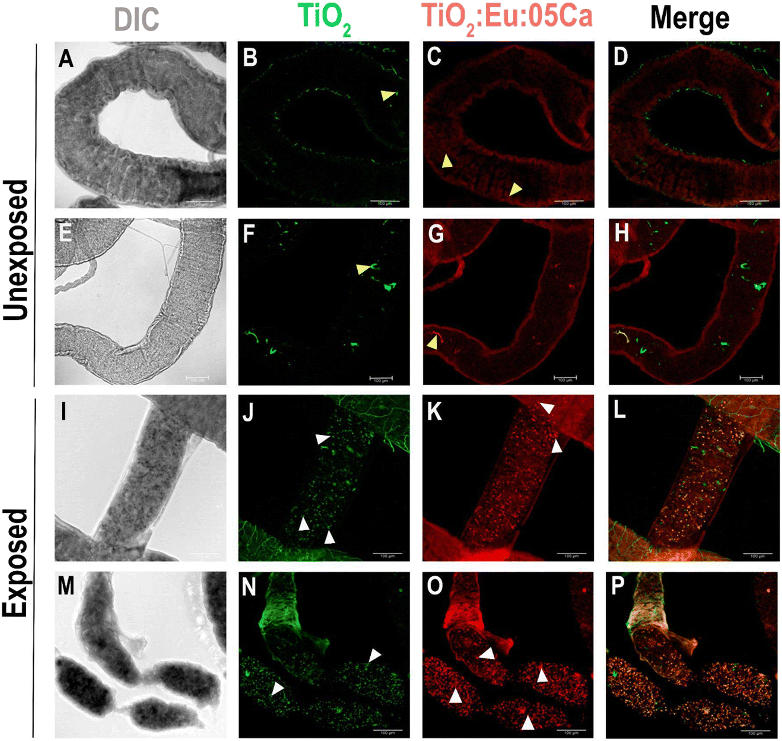
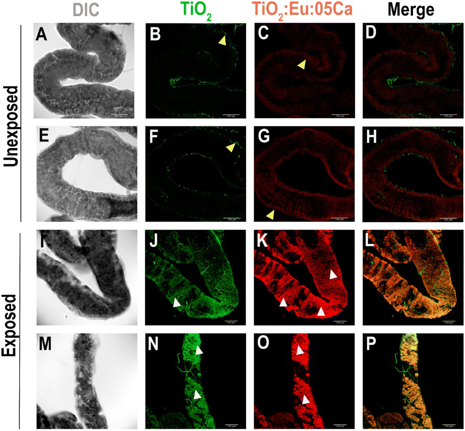
The next step in our analysis was to attempt to track NCs in the tissues of treated animals. Initially, we examined the larval brains of exposed and unexposed animals. As depicted in Fig. 8, control animals exhibited tracheal autofluorescence in the TiO2 NC excitation channel (405 nm), as indicated by the yellow arrowheads in Fig. 7B and F.
 | ||
| Fig. 7 Confocal images of Drosophila melanogaster larval brain showing the distribution of TiO2 and 0.5Ca NCs. (A–H) Larval brains dissected from unexposed larvae fed with a standard food medium showed autofluorescence of the central nervous system trachea (yellow arrowheads in B and F) and a poor signal of autofluorescence in the brain tissue (yellow arrowheads in C and G). (I–P) Larval brains dissected from animals that were treated with 4 mg mL−1 TiO2:Eu:0.5Ca NCs showed that these NCs were dispersed throughout the brain lobe (white arrowheads in J and K) and ventral nerve cord (white arrowheads in N and O). DIC: differential interference contrast; green: TiO2 NC fluorescence under 405 nm laser excitation; red: TiO2:Eu:05Ca NC fluorescence under 552 nm laser excitation. Scale bars = 100 μm. | ||
 | ||
| Fig. 8 Confocal images of the Drosophila melanogaster larval gut showing the distribution of TiO2 and 0.5Ca NCs. (A–H) Gut dissected from unexposed larvae fed with a standard medium showing the autofluorescence of the gut trachea (yellow arrowheads in B and F) and a low level of autofluorescence in gut cell walls (yellow arrowheads in C) as well as food autofluorescence in the gut lumen (yellow arrowhead in G). (I–P) Gut dissected from larvae that developed in 4 mg mL−1 TiO2 and TiO2:Eu:0.5Ca showed that these NCs dispersed along the lumen (white arrowheads in J, K, N, and O). DIC: differential interference contrast; green: TiO2 NC fluorescence under 405 nm laser excitation; red: Eu-doped TiO2 NC fluorescence under 552 nm laser excitation. Scale bars = 100 μm. | ||
Additionally, there was minimal autofluorescence in the TiO2:Eu:0.5Ca NC excitation channel (552 nm), as illustrated by the yellow arrowheads in panels C and G. Upon analyzing the larval brains of the exposed animals, we observed minor points of TiO2 accumulation, as indicated by the white arrowheads in panels J and N. This phenomenon became even more evident when examining the TiO2:Eu:0.5Ca NC excitation channel, where minor red points could be observed in the cerebral lobe of the central nervous system (white arrowhead in panel K) and the ventral nerve cord (white arrowheads in panel O). These data demonstrated that Ca co-doping enhanced in vivo biocompatibility and facilitated better NC dispersion throughout the nervous tissue.
In our subsequent analysis, we dissected larval intestines from animals exposed and not exposed to NCs. As exposure is conducted through a standard culture medium, a high concentration of NCs in the lumen of the larval intestine is expected. The intestines of control animals exhibited high tracheal autofluorescence in the TiO2 NC excitation channel (405 nm), as indicated by the yellow arrowheads in Fig. 8B and F. Additionally, autofluorescence is evident in the cells comprising the larval intestinal wall in the TiO2:Eu:0.5Ca NC excitation channel (552 nm), as indicated by the yellow arrowheads in Fig. 8C, along with autofluorescence from a few particles in the culture medium, as demonstrated in Fig. 8G. In the animals exposed to TiO2:Eu:0.5Ca NCs, a conspicuous abundance of luminescent particles within the larval intestinal lumen was observed, as indicated by the white arrowheads in Fig. 8J, K, N, and O. The notable concentration of particles at this site is anticipated, given that animal exposure to NCs occurs primarily through ingestion.
The presence of TiO2:Eu:0.5Ca NCs in larval gut cells becomes more evident in Fig. 9. The cells of the larval gut wall in control animals exhibited low autofluorescence, as shown in Fig. 9C and G (yellow arrowheads). In contrast, cells of the gut wall in animals exposed to TiO2:Eu:0.5Ca NCs showed noticeable luminescence (white arrowheads in Fig. 9J and N) in the 405 nm excitation channel when compared to control intestines, which displayed autofluorescence primarily in the trachea (yellow arrowheads in Fig. 9B and F). Surprisingly, luminescence was even more pronounced in the intestinal cells of animals exposed to TiO2:Eu:0.5Ca NCs when the excitation channel was exciting europium (552 nm), which became even more evident in the comparison between Fig. 9K and O (white arrowheads) and Fig. 9C and G (control, yellow arrowheads).
 | ||
| Fig. 9 Confocal images of the Drosophila melanogaster larval gut showing TiO2 and TiO2:Eu:0.5Ca NCs dispersed throughout gut cells. (A–H) Gut dissected from unexposed larvae fed with a standard medium showing the autofluorescence of the gut trachea (yellow arrowheads in B and F) and a low level of autofluorescence in the cells of the gut walls (yellow arrowheads in C and G). (I–P) Gut dissected from larvae that developed in 4 mg mL−1 TiO2 and TiO2:Eu:0.5Ca showed that these NCs were highly enriched in the cytoplasm of the cells that form the wall of the gut (white arrowheads in J, K, N, and O). DIC: differential interference contrast; green: TiO2 NC fluorescence under 405 nm laser excitation; red: Eu-doped TiO2 NC fluorescence under 552 nm laser excitation. The yellow arrowheads in (B, C, F, and G) represent autofluorescence in the Drosophila standard medium and tissues, while the white arrowheads in (J, K, N, and O) indicate the cells containing NC aggregates. Scale bars = 100 μm. | ||
Although not showing significant differences in toxicity between TiO2 and TiO2:Eu co-doped with Ca NCs, the presence of this chemical element in the NCs demonstrates some advantages. An improvement in luminescence and better particle dispersion in vivo can be observed. Additionally, we did not observe any changes in coloration in the intestinal cells of Drosophila larvae exposed to 0.5Ca-co-doped TiO2 NCs at a concentration of 4 mg mL−1 (Fig. 10). Cytotoxicity was only recorded in the intestines of larvae exposed to the positive control (62.5%) and only 20% of the animals exposed to TiO2:Eu showed blue coloring in the intestine (Fig. 10). This result differs from others that show high trypan blue staining in the intestine of larvae exposed to TiO2 NPs in the anatase phase, even at concentrations lower than those in this study.34,53 Trypan blue staining is based on the impermeability of the cell membrane to the blue dye, marking only cells in the process of death. Thus, we can infer that 0.5Ca-co-doped TiO2 NCs may not have caused cell death in the intestinal cells of Drosophila larvae under the conditions studied in this research. Therefore, the biocompatibility tests show no toxicity according to the parameters observed in this study, both in vitro and in vivo.
 | ||
| Fig. 10 Absence of cytotoxicity in intestinal cells of D. melanogaster after oral exposure to Eu-doped TiO2 NPs (TiO2:Eu), and to 0.5 calcium co-doped sample (TiO2:Eu:05Ca). (A) Representative larva from the control group exposed only to the standard culture medium. (B) Representative larva from the positive control exposed to the standard culture medium supplemented with 2,2′-azobis-2-amidinopropane (AAPH) (15 mM), where blue staining of trypan blue dye in the intestine is observed, indicating cytotoxicity. (C) and (D) Representative figures of larvae exposed to TiO2:Eu and TiO2:Eu:05Ca at a concentration of 4 mg ml−1, where no intestinal cytotoxicity was observed. (E) Percentage (%) of intestines stained per group (n = 8 per group). * means that there is a statistical difference between the treatments. | ||
Furthermore, the lower formation of ROS during the photocatalytic degradation process suggests that TiO2:Eu NCs possibly exhibit lower toxicity when co-doped with Ca. Regarding physical properties, Ca doping did not negatively affect the crystalline structure of TiO2 NCs, maintaining their high crystallinity, which can be advantageous for their integration with biological tissues and application in biomedical implants, for example.
Doping nanomaterials with Ca has numerous advantages that can contribute to biological and nanotechnological applications.54 Ca in NCs contributes to improved chemical stability and resistance and inhibits environmental degradation and corrosion, which are essential properties for applications in aggressive or long-term environments. It can also assist in energy conversion, as Ca can help modulate the electronic and optical properties of TiO2, favoring the generation and separation of electron–hole pairs, thus having potential photovoltaic and photoelectrochemical applications, for example. The increase in visible light absorption by TiO2 NCs is also enhanced with co-doping, extending their photocatalytic activity spectrum beyond the UV region, which is advantageous for applications requiring greater efficacy in solar light utilization. Due to its various bioactive properties like Ca also enhances the NC's ability to interact with biological systems (cells and tissues), promote osteoblast growth, secrete matrix, and accelerate osteogenesis.54–56 The TiO2:Eu doped with Ca, in addition to being more stable and having better luminescence, shows good biocompatibility, even at high concentrations (2 and 4 mg mL−1), making it a promising nanomaterial for biomedical applications, such as photodynamic therapies, gene therapy, radiotherapy, calcification therapy, controlled drug release, fluorescence imaging, and magnetic resonance imaging, for example. Therefore, TiO2:Eu: Ca NCs can be strong candidates for biomedical applications, treatments, and diagnosis, showing nanotheranostic properties.
Experimental
Synthesis and characterization of nanocrystals
The pure and Eu-doped TiO2 NCs were synthesized using the methodology described by Silva et al.11,14 Ca co-doping was incorporated during the synthesis using the same methods. All reagents were purchased from Sigma-Aldrich®.Transmission Electron Microscope (TEM), JEM-2100, Jeol, Tokyo, Japan, equipped with EDS, Thermo scientific and energy-dispersive X-ray spectroscopy (EDS) was obtained on a Zeiss EVO MA10 system. Raman spectra were obtained at room temperature using a LabRAM HR Evolution HORIBA spectrometer with a 633 nm excitation line.
The photocatalytic activities of the samples were tested for the degradation of methylene blue (MB) under artificial light irradiation, with a 300 W xenon lamp as the light source. In detail, in a cylindrical reactor, 8 mg of the photocatalyst was added to 50 ml of MB solution (0.02 mmol L−1). A magnetic stirrer was used to ensure the homogeneous dispersion of the photocatalyst during the reaction. The photocatalytic reactor was positioned 15 cm below the light source, and the photocatalytic reaction was initiated by adding the catalyst to the reactor and switching on the lamp. At controlled intervals, 3.0 mL of the suspension was collected for analysis. Adsorption experiments were conducted under the same conditions without xenon light irradiation. The MB concentration was measured using a UV-VIS-NIR spectrophotometer (Shimadzu) at a wavelength of 663 nm. The following equation was used to calculate the percentage of discoloration: % photo discoloration = (1 − (A/A0)) × 100, where A is the absorbance of the solution at time t > 0 and A0 is the initial absorbance.
The hydrodynamic diameter (Dh) of NCs was analyzed by dynamic light scattering (DLS), and the zeta potential (ζ potential) was determined by electrophoretic light scattering (ELS) using a ZetaSizer model Nano-ZS90 (Malvern®). For both analyses, a dispersion of NCs (100 μg mL−1) was prepared using ultrapure water (Milli-Q) at pH 7.0, and sonicated for 30 min (ultrasound bath, QUIMIS®), in triplicate.
In vitro assays in human osteosarcoma cell line (SAOS-2)
SAOS-2 cells were obtained from the Cell Bank of Rio de Janeiro (Rio de Janeiro, RJ, Brazil). The cells were cultured to confluence in Dulbecco's Modified Eagle's Medium (DMEM) (Vitrocell Embriolife, Campinas, SP, Brazil) supplemented with 10% heat-inactivated fetal bovine serum (FBS) (Gibco, Langley, OK, USA) and 1% penicillin–streptomycin (Sigma-Aldrich, St. Louis, MO, USA) in a humid atmosphere of 5% CO2 at 37 °C. SAOS-2 cells were plated in quintuplicate in 96-well plates (Costar Corp., Cambridge, MA, USA) (2 × 10⁴ cells per well) and allowed to adhere overnight. The cells were then stimulated with autoclaved solutions of pure, Eu-doped, and xCa-co-doped (x = 0.5, 1.0, 5.0, 10.0) TiO2 NCs at concentrations of 10, 50, and 100 μg mL−1 in a fresh medium for 24 hours. The control group contained cells maintained in DMEM. After the incubation period, the cells were prepared for cell viability analysis by the MTT (3-(4,5-dimethylthiazol-2yl)-2,5-diphenyl bromide tetrazoline]) formazan viability assay. This study was repeated twice using five samples for each group at every time point.The cell viability assay was evaluated 24 hours after treatment with the samples. MTT solution (Sigma-Aldrich) (5 mg mL−1) was added to each well, and the cells were incubated at 37 °C for 4 hours. The supernatant was removed, and 100 μL of dimethylsulfoxide (DMSO) (LGC Biotecnologia, Cotia, SP, Brazil) was added. The optical density at 570 nm was then measured using a microplate reader (Biochrom, Cambridge, UK). The results were expressed as absorbance levels, and differences between the mean values were statistically analyzed.
In vivo nanosafety assays
Nanocrystals (NCs) (pure, Eu-doped, and xCa-co-doped (x = 0.5, 1.0, 5.0, 10.0)) were diluted in ultrapure water to create a 10 mg mL−1 stock solution to prepare appropriate concentrations for all in vivo analyses. After mixing in water, the solution was dispersed in a 5 W probe sonicator (Elmasonic EASY 60 H, Misonix, USA) for 30 min before use.In vivo experiments focused on analyzing the total lifespan, mortality rates in each developmental stage, and potential aberrant phenotypes. For these investigations, we utilized fruit flies from the Canton S lineage of D. melanogaster (Diptera, Drosophilidae). The flies were raised in a standard cornmeal medium comprising cornmeal, agar, yeast extract, sugar, and propionic acid, maintained at 25 ± 1 °C with 60–70% humidity, and subjected to a 12-hour light/12-hour dark cycle.
Our initial assessment focused on the larval lethality rate, pupal formation rate, and daily pupal formation rate throughout the animals' development.14,57 Approximately 100 males and females were housed in a bottle with an agar cap containing grape juice at the bottom. The embryos were collected every 8 hours during cap replacement. After 24 hours of egg collection, the embryos developed into first-stage larvae, which were then introduced into experimental vials containing 4 mL of standard cornmeal medium with varying concentrations of Ca-co-doped samples (x = 0.5, 1.0, 5.0, 10.0) at concentrations of 0.50, 1.00, 2.00, and 4.00 mg mL−1. As a control, we employed a standard cornmeal medium. Six replicates comprising 30 larvae were prepared for every concentration tested with each NC doping. Subsequently, we closely monitored various stages of postembryonic development, documenting any observed changes. The delay in larval development was assessed by comparing the daily pupation frequency to that of the control group. Larval lethality was calculated as the disparity between the initial number of larvae transferred to the medium and the total number of pupae. The delay in larval development was assessed by comparing the daily incidence of pupa with that of the control group. Larval lethality was determined by subtracting the initial number of larvae transferred to the culture medium from the total number of pupae formed. Both the total pupal formation rate and the larval lethality rate were calculated only at the end of post-embryonic development. The daily pupal formation rate was measured daily, always taking into account the control treatment.
After the animals emerged, adulthoods were segregated into males and females and housed in vials with a standard control medium for the entirety of their lifespans. New vials were transferred every three days, with deceased animals meticulously recorded. This protocol was systematically executed in each experimental group until the death of all individuals. Consequently, we could ascertain the lifespan of individuals who developed in media containing NCs compared to control animals that evolved in standard media. The results were quantified and are expressed as a percentage of survival.
Statistical analysis
Statistical analyses were performed using GraphPad Prism 8 software (La Jolla, CA, USA). For in vitro assays, all the data were analyzed for normality using the Kolmogorov–Smirnov test. Two-way analysis of variance and the Tukey test were used to compare the nanoparticle and concentration parameters. The Dunnett test was used to compare the experimental groups with the control group.The in vivo nanosafety assays were also performed using GraphPad Prism 8 software. The pupation rate and larval and pupal lethality data were submitted for the analysis of homogeneity of variance with the Shapiro–Wilk test. Parametric data were subjected to analysis of variance (two-way ANOVA) followed by the Tukey test for mean comparisons. The log-rank test was used to test the survival curves (lifespan) for group comparisons. The chi-square test was used to analyze the presence and absence of the trypan blue dye and evaluate the level of cytotoxicity in the intestinal cells of the larvae exposed to the nanomaterials. A significance level of 5% was adopted for all analyses, and the data are presented as the mean ± standard error of the mean (SEM).
Conclusions
This study delves into the distinctive attributes of TiO2 NCs by introducing red luminescence through Eu-ion doping and optimizing biocompatibility via Ca codoping. Comprehensive physical characterization encompassing crystalline phase analysis, doping effects, morphology, and photocatalytic properties revealed the successful integration of Ca ions into the TiO2 crystal structure without the emergence of additional phases or compounds. Despite a reduction in photocatalytic activity due to codoping with Ca, the resulting NCs exhibit low toxicity to SAOS-2 cells, as evidenced by in vitro assays. Additionally, in vivo assessments using D. melanogaster demonstrated no adverse effects on postembryonic development or larval lethality. Confocal microscopy images of the larval brain and intestines revealed luminescence in the NCs, underscoring the successful enhancement of their biocompatibility and traceability in biological tissues through Ca doping. The optimized combination of luminescence and biocompatibility positions these NCs as promising candidates for advanced biomedical applications.Data availability
The authors confirm that the data supporting the findings of this study are available within the article in all figures shown.Author contributions
Jerusa Maria de Oliveira: conceptualization, experimental design, visualization, writing – original draft, review & editing, and supervision of assay in vivo. Amanda I. dos S. Barbosa: synthesis and characterization, photocatalytic activity measurements, data curation, writing – original draft, review & editing. Larissa Iolanda M. de Almeida: investigation of assay in vivo, data curation, analyses of results, writing – original draft preparation. Vanessa Tomaz Maciel: investigation of assay in vivo, data curation, analyses of results, writing – original draft preparation. João Paulo S. de Carvalho: characterization of samples and data curation, writing – original draft, review & editing. Marcus Andrei R. F. da Costa: photocatalytic activity measurements and data curation. Alysson N. Magalhães: EDS measurements and review & editing of the manuscript. Marcos V. Vermelho: EDS measurements and review & editing of the manuscript. Gabriela L. de Souza: investigation of cell assays, data curation, analyses of results, writing – original draft, review & editing. Camilla Christian G. Moura: supervision of cell assays, writing – original draft, review & editing, and funding acquisition. Sebastião William da Silva: characterization of samples, data curation, supervision of characterization, and funding acquisition. Thiago Lopes Rocha: experimental design in TEM images, zeta and DLS results, writing – original draft, and funding acquisition. Felipe B. Valer: experimental design in confocal images, supervision of confocal images, writing – original draft, and funding acquisition. Lucas Anhezini: conceptualization, assay in vivo, experimental design, visualization, writing – original draft, review & editing, and supervision. Anielle Christine A. Silva: conceptualization, experimental design, visualization, synthesis and characterization of samples, analyses of results, writing – original draft, review & editing, supervision, and funding acquisition.Conflicts of interest
There are no conflicts to declare.Acknowledgements
This study received financial support from Fundação de Amparo a Pesquisa de Alagoas (FAPEAL), Coordenação de Aperfeiçoamento de Pessoal de Nível Superior (CAPES), and Conselho Nacional do Desenvolvimento Científico e Tecnológico (CNPq). Rocha T. L. is granted with productivity scholarship from CNPq [no. 306329/2020-4] and Productivity the Nr. 309813/2023-9 (A. C. A. S.).References
- C. Rathore, V. K. Yadav, A. Gacem, S. K. AbdelRahim, R. K. Verma, R. S. Chundawat, G. Gnanamoorthy, K. K. Yadav, N. Choudhary, D. K. Sahoo and A. Patel, Microbial synthesis of titanium dioxide nanoparticles and their importance in wastewater treatment and antimicrobial activities: a review, Front. Microbiol., 2023, 14 DOI:10.3389/fmicb.2023.1270245.
- R. Mutsak Ahmed and I. Hasan, A review on properties and applications of TiO2 and associated nanocomposite materials., Mater. Today: Proc., 2021, 81, 1073–1078, DOI:10.1016/j.matpr.2021.04.381.
- P. C. Dathan, D. Nallaswamy, S. Rajeshkumar, S. Joseph, S. Ismail, L. Ajithan and L. Jose, A Review on Biomedical Applications of Titanium Dioxide, Trends Biomater. Artif. Organs, 2023, 37(1), 49–54 Search PubMed.
- C. A. Duarte, L. R. Goulart, L. de Souza Castro Filice, I. L. de Lima, E. Campos-Fernández, N. O. Dantas, A. C. A. Silva, M. B. P. Soares, R. R. dos Santos, C. M. A. Cardoso, L. S. de Aragão França, V. P. C. Rocha, A. R. L. P. Ribeiro, G. Perez, L. N. Carvalho and V. Alonso-Goulart, Characterization of Crystalline Phase of TiO2 Nanocrystals, Cytotoxicity and Cell Internalization Analysis on Human Adipose Tissue-Derived Mesenchymal Stem Cells, Materials, 2020, 13, 4071, DOI:10.3390/ma13184071.
- A. B. Younis, Y. Haddad, L. Kosaristanova and K. Smerkova, Titanium dioxide nanoparticles: Recent progress in antimicrobial applications, Wiley Interdiscip. Rev.: Nanomed. Nanobiotechnol., 2023, 15 DOI:10.1002/WNAN.1860.
- Y. Bai, I. Mora-Seró, F. De Angelis, J. Bisquert and P. Wang, Titanium Dioxide Nanomaterials for Photovoltaic Applications, Chem. Rev., 2014, 114, 10095–10130, DOI:10.1021/cr400606n.
- J. Wang, Z. Wang, W. Wang, Y. Wang, X. Hu, J. Liu, X. Gong, W. Miao, L. Ding, X. Li and J. Tang, Synthesis, modification and application of titanium dioxide nanoparticles: a review, Nanoscale, 2022, 14, 6709–6734, 10.1039/D1NR08349J.
- F. Hajareh Haghighi, M. Mercurio, S. Cerra, T. A. Salamone, R. Bianymotlagh, C. Palocci, V. Romano Spica and I. Fratoddi, Surface modification of TiO2 nanoparticles with organic molecules and their biological applications, J. Mater. Chem. B, 2023, 11, 2334–2366, 10.1039/d2tb02576k.
- T. Ayorinde and C. M. Sayes, An updated review of industrially relevant titanium dioxide and its environmental health effects, J. Hazard. Mater. Lett., 2023, 4, 100085, DOI:10.1016/j.hazl.2023.100085.
- M. P. Carvalho Naves, C. R. de Morais, A. C. A. Silva, N. O. Dantas, M. A. Spanó and A. A. A. de Rezende, Assessment of mutagenic, recombinogenic and carcinogenic potential of titanium dioxide nanocristals in somatic cells of Drosophila melanogaster, Food Chem. Toxicol., 2018, 112, 273–281, DOI:10.1016/J.FCT.2017.12.040.
- É. de M. Reis, A. A. A. de Rezende, P. F. de Oliveira, H. D. Nicolella, D. C. Tavares, A. C. A. Silva, N. O. Dantas and M. A. Spanó, Evaluation of titanium dioxide nanocrystal-induced genotoxicity by the cytokinesis-block micronucleus assay and the Drosophila wing spot test, Food Chem. Toxicol., 2016, 96, 309–319, DOI:10.1016/j.fct.2016.08.023.
- A. C. A. Silva, E. A. Alvin, F. R. A. dos Santos, S. L. M. de Matos, J. M. de Oliveira, A. S. Silva, É. V. Guimarães, M. S. Vieira, E. A. da S. Filho, R. S. Silva, L. Anhezini, N. D. Tebaldi, N. O. Dantas, A. C. A. Silva, E. A. Alvin, F. R. A. dos Santos, S. L. M. de Matos, J. M. de Oliveira, A. S. Silva, É. V. Guimarães, M. S. Vieira, E. A. da S. Filho, R. S. Silva, L. Anhezini, N. D. Tebaldi and N. O. Dantas, Doped Semiconductor Nanocrystals: Development and Applications, in Materials at the Nanoscale, 2021, DOI:10.5772/INTECHOPEN.96753.
- C. A. Silva, J. M. Oliveira, L. R. S. Floresta, M. V. da Silva, J. L. S. Duarte, K. B. da Silva, A. L. S. Borges, G. L. Fernandes, A. S. Silva, É. V. Guimarães, L. Anhezini and N. O. Dantas, Transition Metals Doped Nanocrystals: Synthesis, Characterization, and Applications, in Transition Metal Compounds - Synthesis, Properties, and Application, ed. S. Haider and A. Haider, 2021, pp. 1–16, DOI:10.5772/intechopen.97326.
- J. M. de Oliveira, K. T. R. da Silva, F. R. A. dos Santos, F. B. Valer, R. K. O. Takaki, J. P. S. de Carvalho, O. W. de Castro, T. L. Rocha, N. O. Dantas, A. C. A. Silva and L. Anhezini, Luminescence Tracking and In Vivo Toxicity Evaluation of TiO2 and Europium Doped TiO2 Nanocrystals during Drosophila Development, Chemosensors, 2023, 11, 55, DOI:10.3390/chemosensors11010055.
- G. L. de Souza, C. C. G. Moura, A. C. A. Silva, J. Z. Marinho, T. R. Silva, N. O. Dantas, J. F. S. Bonvicini and A. P. Turrioni, Effects of zinc oxide and calcium–doped zinc oxide nanocrystals on cytotoxicity and reactive oxygen species production in different cell culture models, Restor. Dent. Endod., 2020, 45, 1–1, DOI:10.5395/rde.2020.45.e54.
- G. L. Souza, A. C. A. Silva, N. O. Dantas, A. P. S. Turrioni and C. C. G. Moura, Cytotoxicity and Effects of a New Cacium Hydroxide Nanoparticle Material on Production of Reactive Oxygen Species by LPS-Stimulated Dental Pulp Cells, Iran. Endod. J., 2020, 15(4), 227–235, DOI:10.22037/iej.v15i4.28942.
- G. L. Souza, T. R. Silva, M. S. Vieira, N. O. Dantas, A. C. A. Silva and C. C. G. Moura, Development and study of cytotoxicity of calcium oxide nanocrystals, Dent. Mater., 2018, 34, e83, DOI:10.1016/j.dental.2018.08.172.
- M. Dong, D. Chen, L. Che, N. Gu, M. Yin, X. Du, J. Shen and S. Yan, Biotoxicity Evaluation of a Cationic Star Polymer on a Predatory Ladybird and Cooperative Pest Control by Polymer-Delivered Pesticides and Ladybird, ACS Appl. Mater. Interfaces, 2022, 14, 6083–6092, DOI:10.1021/acsami.1c24077.
- S. Yan, N. Li, Y. Guo, Y. Chen, C. Ji, M. Yin, J. Shen and J. Zhang, Chronic exposure to the star polycation (SPc) nanocarrier in the larval stage adversely impairs life history traits in Drosophila melanogaster, J. Nanobiotechnol., 2022, 20, 515, DOI:10.1186/s12951-022-01705-1.
- F. Meng, Y. Liu, J. Wang, X. Tan, H. Sun, S. Liu and S. Wang, Temperature dependent photocatalysis of g-C3N4, TiO2 and ZnO: Differences in photoactive mechanism, J. Colloid Interface Sci., 2018, 532, 321–330, DOI:10.1016/j.jcis.2018.07.131.
- S. Wei, G. Zhang and X. Xu, Activating BaTaO2N by Ca modifications and cobalt oxide for visible light photocatalytic water oxidation reactions, Appl. Catal., B, 2018, 237, 373–381 CrossRef CAS.
- J. Liu, G. Dong, J. Jing, S. Zhang, Y. Huang and W. Ho, Photocatalytic reactive oxygen species generation activity of TiO2 improved by the modification of persistent free radicals, Environ. Sci.: Nano, 2021, 12 10.1039/D1EN00832C.
- S. Y. Lee and S. J. Park, TiO2 photocatalyst for water treatment applications, J. Ind. Eng. Chem., 2013, 19, 1761–1769, DOI:10.1016/j.jiec.2013.07.012.
- M. Rosales, T. Zoltan, C. Yadarola, E. Mosquera, F. Gracia and A. García, The influence of the morphology of 1D TiO 2 nanostructures on photogeneration of reactive oxygen species and enhanced photocatalytic activity, J. Mol. Liq., 2019, 281, 59–69, DOI:10.1016/j.molliq.2019.02.070.
- Y. Wu, M. Cheng, Y. Jiang, X. Zhang, J. Li, Y. Zhu and Q. Yao, Calcium-based biomaterials: Unveiling features and expanding applications in osteosarcoma treatment, Bioact. Mater., 2024, 32, 385–399, DOI:10.1016/J.BIOACTMAT.2023.10.008.
- A. L. Rossi, M. M. Longuinho, M. N. Tanaka, M. Farina, R. Borojevic and A. M. Rossi, Intracellular pathway and subsequent transformation of hydroxyapatite nanoparticles in the SAOS-2 osteoblast cell line, J. Biomed. Mater. Res., Part A, 2018, 106, 428–439, DOI:10.1002/jbm.a.36256.
- R. Posgai, C. B. Cipolla-McCulloch, K. R. Murphy, S. M. Hussain, J. J. Rowe and M. G. Nielsen, Differential toxicity of silver and titanium dioxide nanoparticles on Drosophila melanogaster development, reproductive effort, and viability: Size, coatings and antioxidants matter, Chemosphere, 2011, 85, 34–42, DOI:10.1016/J.CHEMOSPHERE.2011.06.040.
- M. Alaraby, B. Annangi, R. Marcos and A. Hernández, Drosophila melanogaster as a suitable in vivo model to determine potential side effects of nanomaterials: A review, J. Toxicol. Environ. Health, Part B, 2016, 19, 65–104, DOI:10.1080/10937404.2016.1166466.
- X. Pan, R. P. Connacher and M. B. O'Connor, Control of the insect metamorphic transition by ecdysteroid production and secretion, Curr. Opin. Insect Sci., 2021, 43, 11, DOI:10.1016/J.COIS.2020.09.004.
- B. Jovanović, Review of titanium dioxide nanoparticle phototoxicity: Developing a phototoxicity ratio to correct the endpoint values of toxicity tests, Environ. Toxicol. Chem., 2015, 34, 1070–1077, DOI:10.1002/ETC.2891.
- B. Jovanovi, V. J. Cvetkovi and T. L. Mitrovi, Effects of human food grade titanium dioxide nanoparticle dietary exposure on Drosophila melanogaster survival, fecundity, pupation and expression of antioxidant genes, Chemosphere, 2016, 144, 43–49, DOI:10.1016/j.chemosphere.2015.08.054.
- B. Jovanović, N. Jovanović, V. J. Cvetković, S. Matić, S. Stanić, E. M. Whitley and T. L. Mitrović, The effects of a human food additive, titanium dioxide nanoparticles E171, on Drosophila melanogaster - a 20 generation dietary exposure experiment, Sci. Rep., 2018, 8, 1–12, DOI:10.1038/s41598-018-36174-w.
- V. J. Cvetković, B. Jovanović, M. Lazarević, N. Jovanović, D. Savić-Zdravković, T. Mitrović and V. Žikić, Changes in the wing shape and size in Drosophila melanogaster treated with food grade titanium dioxide nanoparticles (E171) – A multigenerational study, Chemosphere, 2020, 261, 127787, DOI:10.1016/j.chemosphere.2020.127787.
- E. R. Carmona, B. Escobar, G. Vales and R. Marcos, Genotoxic testing of titanium dioxide anatase nanoparticles using the wing-spot test and the comet assay in Drosophila, Mutat. Res., Genet. Toxicol. Environ. Mutagen., 2015, 778, 12–21, DOI:10.1016/j.mrgentox.2014.12.004.
- G. I. Dar, M. Saeed and A. Wu, Toxicity of TiO2 Nanoparticles, in TiO2 Nanoparticles: Applications in Nanobiotechnology and Nanomedicine, 2020, pp. 67–103, DOI:10.1002/9783527825431.ch2.
- D. T. Jayaram and C. K. Payne, Bioconjugate Chem., 2020, 31, 1354–1361 CrossRef CAS PubMed.
- T. Ayorinde and C. M. Sayes, An updated review of industrially relevant titanium dioxide and its environmental health effects, J. Hazard. Mater. Lett., 2023, 249–259, DOI:10.1016/j.hazl.2023.100085.
- J. Du, S. Xu, Q. Zhou, H. Li, L. Fu and Jun-Hong Tangand, The ecotoxicology of titanium dioxide nanoparticles, an important engineering nanomaterial, Toxicol. Environ. Chem., 2019, 101, 165–189, DOI:10.1080/02772248.2019.1693572.
- H. Chang, Q. Wang, X. Meng, X. Chen, Y. Deng, L. Li, Y. Yang, G. Song and H. Jia, Effect of Titanium Dioxide Nanoparticles on Mammalian Cell Cycle In Vitro: A Systematic Review and Meta-Analysis, Chem. Res. Toxicol., 2022, 35(9), 1435–1456, DOI:10.1021/acs.chemrestox.1c00402.
- S. S. Watson, D. Beydoun, J. A. Scott and R. Amal, The effect of preparation method on the photoactivity of crystalline titanium dioxide particles, Chem. Eng. J., 2003, 95, 213–220, DOI:10.1016/S1385-8947(03)00107-4.
- A. Nel, T. Xia, L. Mädler and N. Li, Toxic potential of materials at the nanolevel, Science, 2006, 311, 622–627, DOI:10.1126/SCIENCE.1114397.
- C. Egbuna, V. K. Parmar, J. Jeevanandam, S. M. Ezzat, K. C. Patrick-Iwuanyanwu, C. O. Adetunji, J. Khan, E. N. Onyeike, C. Z. Uche, M. Akram, M. S. Ibrahim, N. M. El Mahdy, C. G. Awuchi, K. Saravanan, H. Tijjani, U. E. Odoh, M. Messaoudi, J. C. Ifemeje, M. C. Olisah, N. J. Ezeofor, C. J. Chikwendu and C. G. Ibeabuchi, Toxicity of Nanoparticles in Biomedical Application: Nanotoxicology, J. Toxicol., 2021,(1), 1–21, DOI:10.1155/2021/9954443.
- A. Sukhanova, S. Bozrova, P. Sokolov, M. Berestovoy, A. Karaulov and I. Nabiev, Dependence of Nanoparticle Toxicity on Their Physical and Chemical Properties, Nanoscale Res. Lett., 2018, 13, 1–21, DOI:10.1186/S11671-018-2457-X.
- A. Manuja, B. Kumar, R. Kumar, D. Chhabra, M. Ghosh, M. Manuja, B. Brar, Y. Pal, B. N. Tripathi and M. Prasad, Metal/metal oxide nanoparticles: Toxicity concerns associated with their physical state and remediation for biomedical applications, Toxicol. Rep., 2021, 8, 1970–1978, DOI:10.1016/J.TOXREP.2021.11.020.
- É. de M. Reis, A. A. A. de Rezende, D. V. Santos, P. F. de Oliveria, H. D. Nicolella, D. C. Tavares, A. C. A. Silva, N. O. Dantas and M. A. Spanó, Assessment of the genotoxic potential of two zinc oxide sources (amorphous and nanoparticles) using the in vitro micronucleus test and the in vivo wing somatic mutation and recombination test, Food Chem. Toxicol., 2015, 84, 55–63 CrossRef.
- C. Domingues, A. Santos, C. Alvarez-Lorenzo, A. Concheiro, I. Jarak, F. Veiga, I. Barbosa, M. Dourado and A. Figueiras, Where Is Nano Today and Where Is It Headed? A Review of Nanomedicine and the Dilemma of Nanotoxicology, ACS Nano, 2022, 16, 9994–10041, DOI:10.1021/acsnano.2c00128.
- O. Kose, M. Tomatis, L. Leclerc, N.-B. Belblidia, J.-F. Hochepied, F. Turci, J. Pourchez and V. Forest, Impact of the Physicochemical Features of TiO 2 Nanoparticles on Their In Vitro Toxicity, Chem. Res. Toxicol., 2020, 33, 2324–2337, DOI:10.1021/acs.chemrestox.0c00106.
- Y. Xuan, W. Zhang, X. Zhu and S. Zhang, An updated overview of some factors that influence the biological effects of nanoparticles, Front. Bioeng. Biotechnol., 2023, 11, 1254861, DOI:10.3389/FBIOE.2023.1254861/BIBTEX.
- K. Gerloff, I. Fenoglio, E. Carella, J. Kolling, C. Albrecht, A. W. Boots, I. Förster and R. P. F. Schins, Distinctive toxicity of TiO 2 rutile/anatase mixed phase nanoparticles on Caco-2 cells., Chem. Res. Toxicol., 2012, 25, 646–655, DOI:10.1021/tx200334k.
- M. Benčina, A. Iglič, M. Mozetič and I. Junkar, Crystallized TiO2 Nanosurfaces in Biomedical Applications, Nanomaterials, 2020, 10, 1121, DOI:10.3390/NANO10061121.
- P. J. Ter Brugge, J. G. C. Wolke and J. A. Jansen, Effect of calcium phosphate coating crystallinity and implant surface roughness on differentiation of rat bone marrow cells, J. Biomed. Mater. Res., 2002, 60, 70–78, DOI:10.1002/jbm.10031.
- A. Bandyopadhyay, I. Mitra, S. B. Goodman, M. Kumar and S. Bose, Improving biocompatibility for next generation of metallic implants, Prog. Mater. Sci., 2023, 133, 101053, DOI:10.1016/J.PMATSCI.2022.101053.
- D. Sabat, A. Patnaik, B. Ekka, P. Dash and M. Mishra, Investigation of titania nanoparticles on behaviour and mechanosensory organ of Drosophila melanogaster, Physiol. Behav., 2016, 167, 76–85, DOI:10.1016/j.physbeh.2016.08.032.
- C. Qi, J. Lin, L. H. Fu and P. Huang, Calcium-based biomaterials for diagnosis, treatment, and theranostics, Chem. Soc. Rev., 2018, 47, 357–403, 10.1039/c6cs00746e.
- L. Bai, P. Song and J. Su, Biomater Transl., 2023, 4(4), 248–269, DOI:10.12336/biomatertransl.2023.04.005.
- H. Cao, H. Qin, Y. Zhao, G. Jin, T. Lu, F. Meng, X. Zhang and X. Liu, Nano-thick calcium oxide armed titanium: Boosts bone cells against methicillin-resistant Staphylococcus aureus, Sci. Rep., 2016, 6, 1–10, DOI:10.1038/srep21761.
- J. F. Silva, J. M. de Oliveira, W. F. Silva, A. C. Costa Soares, U. Rocha, N. Oliveira Dantas, E. A. da Silva Filho, M. Duzzioni, A. Helmut Rulf Cofré, O. W. de Castro, L. Anhezini, A. C. Almeida Silva and C. Jacinto, Supersensitive nanothermometer based on CdSe/CdSxSe1-x magic-sized quantum dots with in vivo low toxicity, Chem. Eng. Sci., 2022, 264, 118153, DOI:10.1016/j.ces.2022.118153.
- F. S. E. Rener Mateus Francisco Duarte, S. M. Malta, F. N. A. do Prado Mascarenhas, V. Prado Bittar, A. L. Borges, R. R. Teixeira, R. G. Zanon and C. U. Vieira, Environ. Toxicol. Pharmacol., 2024, 106 DOI:10.1016/j.etap.2024.104388.
- Y. Zhang, M. B. Wolosker, Y. Zhao, H. Ren and B. Lemos, Exposure to microplastics cause gut damage, locomotor dysfunction, epigenetic silencing, and aggravate cadmium (Cd) toxicity in Drosophila, Sci. Total Environ., 2020, 744, 140979, DOI:10.1016/j.scitotenv.2020.140979.
Footnote |
| † These authors contributed equally to this work. |
| This journal is © The Royal Society of Chemistry 2025 |