
 Open Access Article
 This Open Access Article is licensed under a
 Creative Commons Attribution 3.0 Unported Licence
Translational nanorobotics breaking through biological membranes
Alzbeta
Ressnerova
 ab,
Zbynek
Heger
bc and
Martin
Pumera
*adef
ab,
Zbynek
Heger
bc and
Martin
Pumera
*adef
aCentral European Institute of Technology, Brno University of Technology, Purkynova 123, CZ-612 00, Brno, Czech Republic. E-mail: pumera.research@gmail.com
bResearch Group for Molecular Biology and Nanomedicine, Department of Chemistry and Biochemistry, Mendel University in Brno, Zemedelska 1, CZ-613 00, Brno, Czech Republic
cCenter of Advanced Innovation Technologies, Faculty of Materials Science and Technology, VSB – Technical University of Ostrava, 17. Listopadu 2172/15, 70800 Ostrava, Czech Republic
dAdvanced Nanorobots & Multiscale Robotics Laboratory, Faculty of Electrical Engineering and Computer Science, VSB – Technical University of Ostrava, 17. listopadu 2172/15, 70800 Ostrava, Czech Republic
eDepartment of Chemical and Biomolecular Engineering, Yonsei University, 50 Yonsei-ro, Seodaemun-gu, Seoul 03722, South Korea
fDepartment of Medical Research, China Medical University Hospital, China Medical University, No. 91 Hsueh-Shih Road, Taichung, Taiwan
First published on 14th January 2025
Abstract
In the dynamic realm of translational nanorobotics, the endeavor to develop nanorobots carrying therapeutics in rational in vivo applications necessitates a profound understanding of the biological landscape of the human body and its complexity. Within this landscape, biological membranes stand as critical barriers to the successful delivery of therapeutic cargo to the target site. Their crossing is not only a challenge for nanorobotics but also a pivotal criterion for the clinical success of therapeutic-carrying nanorobots. Nevertheless, despite their urgency, strategies for membrane crossing in translational nanorobotics remain relatively underrepresented in the scientific literature, signaling an opportunity for further research and innovation. This review focuses on nanorobots with various propulsion mechanisms from chemical and physical to hybrid mechanisms, and it identifies and describes four essential biological membranes that represent the barriers needed to be crossed in the therapeutic journey of nanorobots in in vivo applications. First is the entry point into the blood stream, which is the skin or mucosa or intravenous injection; next is the exit from the bloodstream across the endothelium to the target site; further is the entry to the cell through the plasma membrane and, finally, the escape from the lysosome, which otherwise destroys the cargo. The review also discusses design challenges inherent in translating nanorobot technologies to real-world applications and provides a critical overview of documented membrane crossings. The aim is to underscore the need for further interdisciplinary collaborations between chemists, materials scientists and chemical biologists in this vital domain of translational nanorobotics that has the potential to revolutionize the field of precision medicine.
 Alzbeta Ressnerova | Alžběta is a postdoctoral researcher at the Innovative Genomics Institute, University of California, Berkeley, USA, as of late 2024. She earned her MSc in Molecular Medicine from Humboldt University in Berlin, Germany, in 2018. She completed her PhD in Advanced Nanotechnologies and Microtechnologies at the Central European Institute of Technology in Brno, Czech Republic, in 2024. During her doctoral studies, she was a researcher at the Mendel University in Brno in the Department of Chemistry and Biochemistry. In 2022, Alžběta was awarded Fulbright-Masaryk Scholarship. Her current research focuses on advancing delivery systems for CRISPR-based gene therapy and other therapeutic agents through innovative nanotechnology approaches. |
 Zbynek Heger | Zbynek received his PhD from the University of Veterinary and Pharmaceutical Sciences in Brno, CZ, in 2016. He became associate professor of Biochemistry at Charles University (2021). He is currently group leader of Research Group for Molecular Biology and Nanomedicine and head of the Department of Chemistry and Biochemistry (Mendel University in Brno, Czech Republic). He had received several awards such as the Award of League against Cancer Prague, Award of Purkynje Foundation, Award of the Czech Urological Society or Award of the Rector of Mendel University in Brno. He has interests in nanoscaled devices for drug delivery and theranostics. |
 Martin Pumera | Professor Martin Pumera is Chief Investigator of Future Energy & Innovation Lab at CEITEC, Brno, and the Head of the Advanced Nanorobots and Multiscale Robotics Laboratory at Technical University Ostrava, Czech Republic. He founded the Center for the Advanced Functional Nanorobots at UCT Prague, where he served as a director (2017–2023). He was a tenured group leader at the National Institute for Materials Science, Japan, in 2006. In 2010, Martin joined Nanyang Technological University, Singapore, where he worked as a tenured associate professor for almost a decade. Prof. Pumera has diverse research interests in nanomaterials and microsystems, in the specific areas of micro and nanomachines, quantum materials, machine intelligence and 3D printing. |
Nanorobots and membranes: interdisciplinary dialogues for biological breakthroughs
Emerging about 20 years ago, nanorobotics is the youngest offspring of the nanotechnology family (Box 1).1–11 Over this short period, nanorobots have seen remarkable advancements in their materials,12–14 power sources,15 modifications,16 and applications.17 A multitude of sophisticated medical applications, particularly in drug delivery, have emerged, holding the potential to revolutionize precision medicine.18–21 While developing nano- and microdevices for cargo delivery within biological systems, substantial focus must be placed on their ability to effectively cross biological membranes (Box 1). This becomes especially essential while envisioning their clinical application for delivering therapeutics of any type in vivo, requiring these nanorobots to effectively cross several biological membranes on their journey to deliver a payload into the target cell. Crossing biological membranes is among the most crucial aspects in the successful delivery of all therapeutics intended for use in patients.22–25
Box 1Membrane: a selective barrier around cells or certain organs, dividing and protecting the inside from the outside and orchestrating the intricate interplay necessary for sustaining life's diverse functions.Robot: the word “robot” was coined in 1920 by Karel Čapek, a Czech writer who for the first time described robots in his thought-provoking science fiction play R.U.R.1 R.U.R. stands for Rossum's Universal Robots where “rossum” means “intellect” in the Czech language. Since then, smart robots have deserted the realm of sci-fi and become part of our society as helpers to mankind.2–6 Nanorobot: a nanorobot is a tiny autonomous machine capable of active propulsion. It blends physical, biological, and computational sciences with the potential to revolutionize healthcare, industry, and everyday life. The ultimate vision is advanced intelligence of these minuscule marvels, allowing them to tackle complex problems within the human body or in any other environment they operate. |
There are four essential biological membranes that nanorobots need to pass through to fulfill their mission (Fig. 1). These intricate barriers exist to shelter the “inside” from the “outside” and do just that when they are faced with foreign entities such as nanoconstructs. To enable the clinical implementation of nanorobots, they need to be able to fearlessly conquer them. Surprisingly, the exploration of strategies for crossing biological membranes within the realm of nanorobotics has remained quite underrepresented, despite its profound importance and potential to advance the field significantly. The state-of-the-art therapies often come with unwelcome side effects. The domain of nanoparticle-based nanomedicine has its undisputable limitations regarding on-target delivery26,27 and efficient barrier crossing. These challenges might be elegantly addressed by motorization.28–31
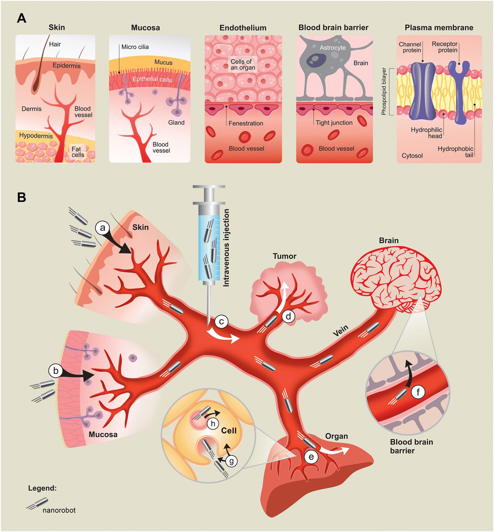 | ||
| Fig. 1 Nanorobots crossing biological membranes. (A) Anatomical details of types of barriers that must be crossed. From left to right: the skin, mucosa, endothelium, blood–brain barrier and plasma membrane. (B) Graphical representation of membrane crossings by nanorobots. Nanorobots are entering the body either through the skin (a), mucosa (b) or bypassing the first barrier by being injected intravenously (c). Nanorobots navigating through blood circulation cross the endothelial membrane to their target site, which is either a tumor (d) or organ (e) or they cross the blood–brain barrier (f). Nanorobots entering the cell cross the plasma membrane (g). By escaping the endosome into the cytoplasm (h), nanorobots reach their destination. (a)–(c) Represent the first barrier. (d)–(f) Represent the second barrier. (g) Is the third barrier and (h) is the fourth barrier. | ||
For that to happen, it is necessary that materials scientists and biologists align their efforts. Today, more than ever are these two disciplines collaborating in the pursuit of identifying gaps in the treatment of various diseases that can potentially be addressed through nanomedicines. The significance of this interdisciplinary partnership is by all means obvious, yet effective communication and mutual understanding of scientists representing these two distinct fields can be troublesome.
Materials scientists working in isolation often lack the necessary biological or medical expertise needed to evaluate the potential application of their nanoconstructs. Frequently they are not fully aware of the biological barriers that need to be conquered when using these nanoconstructs in biological contexts. Conversely, biologists when working in isolation may overlook the vast potential of the latest developments in nanoscience, potentially missing opportunities to successfully tackle biological hurdles through the application of nanotechnology. However, it is evident that harnessing the prospects of nanomaterials for useful biological applications could truly enhance the well-being of humans.
We should strive to bring researchers from various fields together transcending the borders of their primary disciplines and merging the best aspects of their fields in the quest for scientific insights. Numerous research advancements have blossomed from collaborations between scientists in biology and material sciences. Notable examples include scaffolds for tissue regeneration,32 biosensors for cancer diagnostics,33 nanoparticles for drug delivery34 and the mRNA COVID-19 vaccine.35 It has been shown that an interdisciplinary approach combining two different fields in the highly cited paper cluster significantly boosted research impact by 20% as assessed by the citation-based method.36 Hence, it is vital to cultivate interdisciplinary collaborations. This involves understandably communicating the perspectives of our own fields while also listening closely to those from different disciplines. Interdisciplinary research broadens the horizons of researchers and fosters innovative visions on the intersection when two or more fields merge. This has a notable potential to be a scientific hotspot from which unique and groundbreaking ideas arise.37 Nanorobotics is a shining example of this interplay, a field increasingly intertwined with biology and medicine. This collaborative approach creates a dynamic research area that bridges disciplinary boundaries, resulting in so-called translational solutions to clinically relevant challenges.
While we set off on this journey together, it is of utmost importance to clearly communicate biological concepts and struggles to scientists outside the realm of biology, so we could come together and work on tackling them. This review aims to serve exactly as that bridge, offering perspectives important to biologists clearly communicated to materials scientists. In this review, we will introduce the topic of biological membranes and elucidate why it is essential to cross them in the pursuit of rational in vivo applications. Design challenges in in vivo membrane crossing will be discussed as well. We will also provide a comprehensive summary of up-to-date crossings of these barriers by variously powered nano- and microrobots. In the quest for nanorobotic excellence, the challenges illuminated in our exploration serve as signposts guiding us toward a future where unprecedented precision in medical intervention is the norm.
Membrane as the stumbling block in nanorobot's journey
The capacity to cross biological membranes is undeniably pivotal for the effectiveness of any therapeutic drug carried by nanorobots, particularly in the context of treating tumors or diseases located in organs that are hard to access through non-invasive methods. In such scenarios, the bloodstream emerges as an attractive route as it provides access to virtually all organs and cells within the human body. This is among the most complex applications to research and master, yet the most appealing one.The ideal smart nanorobot would navigate in the bloodstream, delivering therapy to the designated organ, right to the cells that need it, all while safeguarding the cargo from loss and minimizing accumulation in non-target tissues. While this goal has not been fully achieved yet, it is a vision that materials scientists collaborating closely with biologists are working towards fulfilling in the near future.
The human body has several biological barriers, safeguarding it from outside elements and agents (Fig. 1). These very barriers are what stand in the way of nanorobots delivering a therapeutic payload of any type. In this chapter, we will delve into a discussion of the biological barriers nanorobots encounter and ideally overcome during their journey throughout the body into target cells.
First barrier: the body's initial gateway
There are several ways of possible administration of nanorobots as pharmaceutical delivery systems. Some of them act as more difficult barriers to cross than others. The human body is equipped with two primary biological barriers shielding it against external elements: the skin and the mucous membrane often called mucosa. The first barrier of the human body can be bypassed by intravenous injection. There has been only limited research focused on nanorobots crossing the first biological barrier.Skin with its impressive 1.5–2 square meter surface area is one of the largest organs in the human body. It consists of multiple layers (Fig. 1A) and serves a myriad of functions from providing protection to facilitating the synthesis of vitamin D. While technically not classified as a membrane, we include the skin here because it might serve as an entry point of nanorobots into the body. Crossing this biological barrier to get into the bloodstream becomes necessary if this form of administration is chosen (Fig. 1B(a)). Transdermal drug delivery is viewed as patient-friendly and non-invasive, making it an ideal method for patients to self-administer without the need for a healthcare professional. Notably, dozens of approved drugs are available on the market utilizing this delivery method.38 The rate-limiting step in transdermal delivery is the crossing of the outer layer known as stratum corneum, which is part of epidermis (Fig. 1A), a hydrophobic barrier rich in lipids that poses significant resistance to penetration.39
The mucosa is further safeguarded by a layer of mucus (Fig. 1A) that makes it hard for external agents to penetrate. Mucosa is present at the body's “openings” such as the mouth, nose, ears, and vagina, and it lines the respiratory, digestive, and reproductive tract. It serves as a critical protective barrier against invading pathogens. Additionally, the mucous membrane houses an integral part of the immune system, contributing significantly to the body's immune response to pathogens.40 Transmucosal drug delivery has been in the view finder due to its non-invasive nature being pain-free compared to needle-based injections.41,42 Furthermore, the proximity of mucosal surfaces to the bloodstream makes this administration route attractive (Fig. 1B(b)). Even though transmucosal and transdermal deliveries involve the challenge of crossing these barriers, down the line motorization and thus nanorobots hold the potential to outperform passive nanoparticles due to their active movement, and therefore, active transport of therapeutic agents.
Intravenous injection is the standard administration route of the majority of therapeutics intended to reach the circulatory system. Despite being associated with some degree of pain or discomfort, intravenous delivery circumvents the initial and significant biological barrier that must be crossed represented by the skin or mucosa (Fig. 1B(c)). However, this method also has its disadvantages as it calls for trained hospital staff, sterile needles, and disinfectant, rendering such administration less convenient, particularly in developing countries or regions affected by war conflict, or natural disasters.
There are other possible ways of administration. In many studies exploring the use of nanorobots for the treatment of solid tumors, researchers opt for intratumor injection of nanorobots.20,43 This approach allows them to circumvent not just one but two barriers simultaneously, as nanorobots are not required to exit the bloodstream to reach the tumor. This method prevents systemic exposure to the treatment and potentially reduces side effects while using a lower amount of the drug.44 However, this approach can be burdensome for the patient as it requires a deep tissue injection, and trained staff and a clinical setting advanced enough for this approach. In addition, the versatility of this approach is low and can be exploited only for limited types of solid tumors.
The administration of nanorobotic therapy through the skin, mucosa, or even intravenously (Fig. 1B(a–c)) offers distinct advantages. Such an approach would lessen the burden on patients. It might not require anesthesia or hospitalization and potentially expand the pool of healthcare providers who can administer the treatment, which would make such treatment more accessible. We should endeavor to investigate less invasive approaches in the treatment of various diseases. Smart nanorobots could potentially serve as the cornerstone in achieving this goal. In this review, we will focus on transdermal, transmucosal, and intravenous delivery by nanorobots.
Second barrier: voyage through the veins
After entering the bloodstream, the next crossing that nanorobot undertakes is the transit from the circulation to the site of its intended therapeutic action, which can be an organ or a tumor (Fig. 1B(d and e)). To reach this destination, a nanorobot must traverse the endothelium, a single layer of cells that lines the interior of blood vessels (Fig. 1A). Physiologically, the endothelium mediates the shuttling of elements into and out of the tissues served by the blood vessel. These elements can be white blood cells, macromolecules present in the blood such as antibodies, nutrients, and fluids. The same route awaits any nanomachine trying to access the target tissue from the bloodstream. The extent to which the mentioned elements permeate the endothelium varies, depending on factors such as the presence and size of gaps between the endothelial cells or lack of them. Fenestrations are pores in the endothelium through which larger molecules can penetrate into organs. Conversely, tight junctions bind cells together without any space in between them, which makes the crossing much harder. Consequently, the permeability of the endothelium varies across different organs. It is notably higher in the liver, intestine, or kidneys, whereas it is significantly decreased in skeletal muscles, lungs, heart, or blood–brain barrier (Fig. 1A).45,46Endothelial permeability is also highly influenced by various disease conditions. The “enhanced permeation and retention effect” initially described in 198647 highlights the increased permeability of solid tumor vessels. It has been observed that gaps between endothelial cells in solid tumors can reach sizes up to 2000 nm.48 As a result, the field of cancer nanomedicine began engineering nanoparticles of specific sizes to pass to a tumor based on the size of those gaps. However, recent findings challenge this notion. It has been reported that these gaps are not primarily responsible for the entry of nanoparticles into tumors. Instead up to 97% of nanoparticles actively traverse the endothelial cells.49 Nevertheless, it remains clear that tumor vasculature is highly permeable and leaky compared to healthy organ tissues. Inflammation is another factor known to increase endothelial permeability. This can manifest in the context of infections, burns, and in chronic inflammation such as asthma or chronic bronchitis.50,51
Despite the inherent leakiness of tumors, nanoparticles often struggle with their delivery and effective penetration into tumor tissues.52 This is due to the increase in collagen production, leading to tumor stiffness.53 Numerous studies have pointed to this phenomenon as a contributing factor to the poor penetration of therapeutic molecules and nanoparticles in tumors.52,54,55 A similar barrier for delivery is the elevated interstitial tumor pressure caused by abnormal growth of blood vessels and the absence of proper lymphatic drainage, leading to the accumulation of fluid.56 The lack of pressure gradient hinders the movement of therapeutic molecules and nanoparticles across the endothelial barrier into the tumor.57,58 Hypoxia, characterized by insufficient oxygen supply is yet another common feature of solid tumors, which is associated with an elevated risk of metastasis, aggressiveness, and ultimately poorer prognosis. It acts as an additional hurdle for therapeutics.59 Nanoparticles struggle to reach these sites and to penetrate deep enough into hypoxic regions.60 In this context, nanorobots hold a distinct advantage over nanoparticles due to their motorization, enabling active movement that does not rely on passive transport through pressure gradients and diffusion. This active movement also facilitates improved penetration of stiff tumor tissues, ensuring the effective delivery of therapeutic agents throughout the entire tumor mass.61 Studies of nanorobots successfully crossing the endothelial membrane will be covered in a later chapter.
It is important to emphasize that not all types of nanorobots are required to cross the endothelial membrane. Some nanorobots are specifically engineered to target entities within the bloodstream rather than aiming for organs or tumors. Such an example is a nanorobot designed for the purpose of destructing blood clots in veins,62 concluding its mission within the confines of the bloodstream.
The blood brain barrier (BBB), an incredibly selective blood–brain interface is often viewed as the most formidable barrier in the human body, seemingly almost impenetrable. Rich in tight junctions and lacking fenestrations (Fig. 1A) this barrier establishes a meticulously regulated microenvironment that is vital for the proper functioning of the brain. It acts as a barrier, blocking pathogens and immune cells from entering the brain, and even makes it challenging for drugs to enter the organ.63 The quest to cross the BBB has been the focal point of a vast amount of drug research, given its pivotal role in the treatment of central nervous system diseases (Fig. 1B(f)).
Numerous approaches have been explored in the quest to facilitate the passage of therapeutic agents through the BBB for the treatment of brain diseases, yet a definitive solution remains elusive as there has been no silver bullet. Current strategies for BBB crossing can be broadly categorized into two main groups: invasive and non-invasive.
Within the non-invasive realm, several noteworthy approaches have been investigated. Focused ultrasound (FUS) creates microbubbles that transiently disrupt the BBB and make it more penetrable, allowing for enhanced delivery of systemically administered drugs.64 However, the FUS transducer can be bulky, costly, and not always readily available. Additionally, magnetic resonance imaging is often used in conjunction with FUS to improve the guidance and evaluation of the BBB opening and closure,65 which can make this procedure even more costly. The safety of FUS-mediated BBB opening has been the subject of debate,66 as FUS has been reported to induce vessel damage.66,67 Furthermore, an open BBB may lead to the leakage of immunoglobulins into the brain, which could contribute to inflammation.68
Intranasal delivery through nasal mucosa however avoids the systemic administration of drugs and completely bypasses the BBB by utilizing the olfactory nerve for direct drug delivery.69 The limitations of this way of BBB crossing are mainly the need for low volume of the administered drug, which means that only a small amount of a drug can be administered.70 Such drug must also be hydrophobic for it to be successfully delivered through the nasal mucosa into the brain; moreover, the safety of such drug must be thoroughly evaluated not to disturb the physiological environment of the brain.69
In addition, drugs can be transported across the BBB using viral vectors or by targeting specific receptors expressed on the BBB, or combination of both.71,72 Constructing delivery vehicles with the affinity to receptors expressed on the BBB such as Transferrin receptor (TfR) or Insulin receptor (IR) has proved to be a promising strategy for BBB crossing.23,73,74 However, in the case of using viruses as delivery vehicles regardless of the targeting strategy, they are known to pose the risk of immunogenicity, which can sometimes have fatal consequences, making them a less ideal delivery system.75,76
Recently, cell-membrane-coated delivery systems for BBB crossing started to emerge using cell membranes from various cells such as red blood cells,77 macrophages78 or other cells of the immune system.79 They generally possess low immunogenicity and recognition by the immune system by masking the delivery vehicle.23
The exploration of various types of nanoparticles for BBB penetration has been ongoing, although none have received approval for the treatment of brain diseases thus far. Despite extensive research, overcoming the challenge of BBB penetration remains a significant hurdle, with only a limited number of strategies progressing to clinical trials in humans.80 The quest for an effective and safe method to breach the BBB for therapeutic purposes continues to be a pressing concern in the field of medical research. Nanorobots might therefore offer some innovative solutions in this ongoing endeavor, which will be discussed later in the text.
When crossing the endothelial membrane from the bloodstream to the target site, nanorobots must possess the capability to efficiently deliver their payload to the desired organ while not distressing the endothelium, thereby avoiding the complications such as wounding, bleeding, and scarring. The ideal nanorobot should not only navigate this task of crossing the endothelial membrane at a location leading to the target organ, but also avoid entering into other tissues where it serves no purpose. It is necessary to think of endothelium permeability when designing nanorobots for in vivo applications, as it provides insights into where the nanomachines will most probably accumulate. Further insights into the various design variables related to this aspect will be provided later on.
Third barrier: intracellular access and path inward
In the next part of their mission, nanorobots should be capable of gently entering the cells of the target organ through the plasma membrane without causing any damage (Fig. 1B(g)). This ensures that all components of the cells remain intact and fully functional while safeguarding the integrity of the precious cargo. Gentle entry might not be needed as much in the case of treating solid cancer. Membranes in cells are formed around the cell itself (i.e. plasma membrane), but also around certain cellular compartments such as the mitochondria or nuclei. However, the role of the plasma membrane extends beyond merely being a barrier, a wall between the cell interior and the external environment. It also serves as a vital communication interface, facilitating essential interactions between the cell and the outside world.The plasma membranes of eukaryotic cells, which are the building blocks of the human body, consist of two molecules thick phospholipid bilayer possessing numerous functions (Fig. 1A).81,82 Phospholipids forming the plasma membrane are mostly glycerophospholipids: phosphatidylcholine, phosphatidylethanolamine and phosphatidylserine. These consist of a glycerol backbone, a phosphate group esterified to choline, serine or ethanolamine and two fatty acids.83,84 Phospholipids form a bilayer by grouping hydrophobic tails in the center and hydrophilic heads on the outer side, effectively separating two aqueous environments from one another. These environments are cytosol, which is the liquid inside of a cell where a nanorobot is aiming to end up, and an extracellular space from which the nanorobot is coming. The plasma membrane also accommodates a diverse array of other molecules with designated functions, making it a highly heterogeneous and dynamic layer. Proteins can find their docking platforms in the plasma membrane, which facilitates important cell signaling (Fig. 1A). This is a key element of cell communication with other cells and their surroundings.85,86 It is worth noting that the plasma membrane of a resting cell tends to have a negative charge, a factor that holds significance in the design of nanorobots that will be further discussed later.
Biological membranes exhibit selective permeability, allowing only specific molecules that meet the cell's requirements to pass through. While small molecules can traverse the plasma membrane without the need for a delivery vehicle, this process, facilitated by diffusion through channel proteins (Fig. 1A)87, lacks cell specificity. Doxorubicin (DOX) (see structure in Fig. 6D) is a chemotherapeutic agent, a member of the anthracycline drug class. It is a 14-hydroxylated derivative of daunorubicin, the precursor of DOX in its biosynthesis, produced by Streptomyces bacterium. DOX is also known to enter healthy cells, leading to the notorious side effects associated with systemic administration. Additional targeting mechanisms of these small molecules are therefore critical. Numerous targeted small molecules, designed to spare healthy cells, are being developed for the treatment of various cancers; however, they still face substantial challenges.88 Despite small molecule's ability to penetrate the plasma membrane independently, enhancing their delivery to the target site could significantly improve efficiency and mitigate side effects. This is where the field of nanorobotics comes into play, offering a promising avenue for the development of sophisticated delivery vehicles.
Conversely to small molecules, larger molecules can be shuttled into the cell through specialized transport proteins (Fig. 1A) or via a process referred to as endocytosis. Antibodies are known to promote receptor-mediated endocytosis, which ensures their efficient transport across the plasma membrane. Leveraging this mechanism, antibodies have been successfully conjugated with anti-cancer drugs, effectively serving as delivery vehicles. This conjugation facilitates the endocytosis of the antibody–drug complex by cancer cells, leading to subsequent drug release within the cell.89,90
However, many large molecules are unable to effectively cross the plasma membrane without the aid of a delivery or targeting system. Certain proteins or advanced tools like CRISPR genome-editing system composed of a protein and a nucleic acid vitally depend on a delivery vehicle to gain entry into the target cell and function effectively. In these instances, the delivery system plays a critical role in crossing the plasma membrane and releasing its cargo once inside the target cell. Recent reports suggest that the active motion of nanorobots significantly aids in the internalization of large molecules into cells compared to passive delivery systems.28,29,31,91 This underscores the potential of nanorobotics to enhance the delivery of complex therapeutic agents, ensuring their effective entry into target cells. However, nanorobots still encounter challenges similar to those faced by large molecules such as the risk of being trapped and digested by the cell during endocytosis. The successful plasma membrane crossings will be explored further in a subsequent section.
Fourth barrier: the art of escaping cell's booby trap
After the nanorobot successfully conquers the endothelium and the plasma membrane, it might seem that the hard part is done. However, what awaits now is the cell's booby trap: the endolysosomal compartment. Before releasing the cargo into the cytosol where it should do its therapeutic magic, the nanorobot must perform an endosomal escape to evade being digested and thus destroyed (Fig. 1B(h)). Endocytosis is the process that unfolds when a large molecule or entity tries to pass through the cell's plasma membrane. A portion of the membrane forms a vesicle that encapsulates the transported entity, pulling it off the surface and internalizing it into a so-called endosome. That is the place where a nanorobot might get trapped for good if it fails to escape. The composition of the endosomal membrane mirrors that of the plasma membrane in terms of the types of phospholipid molecules present; however, it differs in the proportions of these phospholipids.92 The whole process of endocytosis is complex and not yet fully comprehended, with several possible pathways and mechanisms at play.93,94 Nevertheless, once an endosome is formed, it progresses through various stages. During this process, the number of protein degradative enzymes increases, and the pH gradually decreases.95 Subsequently, about just 30 min after the initial formation of the endosome, it reaches its final destination – another vesicle called lysosome. These two structures fuse, forming what is known as endolysosome.96The lysosome, characterized by its low pH of 4.5–5, serves several functions including the degradation, digestion, and break down of whatever is inside of endosome.97 In the context of nanorobots, this implies the degradation of both the nanorobot and its cargo. The only way the nanorobot can avert this fate is through a process known as endosomal or endolysosomal escape (Fig. 1B(h)). Endosomal escape has been the subject of extensive research, particularly in the context of nanoparticles. It is often described as the rate-limiting step for nanoparticle-mediated delivery.25,98,99 Understanding the capability of nanorobots to achieve endosomal escape, as well as the potential role of active movement in this process, is of utmost importance. Such knowledge would highly contribute to the rational design of therapeutically successful nanorobots. Successful endosomal escapes of nanorobots will be covered extensively in a later part of this review.
From lab to life: design challenges
The success of nanorobots conquering the mentioned biological barriers hinges on their design (Fig. 2). At the beginning of every nanomaterial used for biological purposes is its testing on in vitro models such as cell lines. However, in vitro does not show us all the pitfalls of applications in living organisms and gives us a slack when the design would actually never be viable in vivo. There are several questions materials scientists could ponder when developing nanorobots for future use in vivo that might help with creating a nanorobot that has a rational application: is the nanorobot manufactured to be invisible for the immune system and will it be able to circulate long enough to find its target? Is the target in blood or in an organ or solid tumor and how many biological barriers the delivery system has to cross? Is the cargo a small molecule or macromolecular therapeutic agent and does it therefore need to be transported across the plasma membrane with the nanorobot? Is the fuel biocompatible and is it available at the site of the nanorobot's action in the concentration that is needed? How does this nanorobot or its descendant outperform the state-of-the-art treatment? The design determines whether a nanorobot will be the appealing and marketable solution for therapeutics delivery. Terms such as “biocompatibility”, “biodegradability” and “prolonged circulation” are recurrent in nanomedicine discussions and must be considered when evaluating nanorobotics clinical prospects (Fig. 2). It is valuable to envision even if only faintly, the future patient that will benefit from this advancement or the descendant of such advancement and to simply bear this in mind when designing sensible translational nanosystems. This chapter will explore design challenges that could hinder the ability of nanorobots to effectively traverse membranes, thereby affecting their clinical translation. We will discuss the means of movement, and the influence of size, shape, surface coating, and other properties in the context of potential biological applications involving membrane crossing (Fig. 2). | ||
| Fig. 2 Design challenges of nanorobots that need to be addressed for successful in vivo membrane crossing. (A) Fuel availability at the site of nanorobot's action, (B) ability of movement in biological fluids, (C) degree of non-target organ accumulation, (D) sufficient biocompatibility and biodegradability, (E) surface coating and charge, (F) material and its properties, (G) loss of nanorobots due to macrophage ingestion, (H) adhesion of plasma proteins on nanorobot (formation of protein corona) and (I) size requirements for specific in vivo applications. | ||
The future is in motion
A vigorous debate exists among scientists developing static nanosystems such as nanoparticles and those dedicated to dynamic nano and microrobots. The fundamental question being contemplated is: does the capability of motion, and thus the active transport of cargo enhance the effectiveness of a nanosystem within biological environments? Nanorobots are not always necessary as nanoparticles can provide exquisite solutions in certain applications, particularly those involving short delivery routes such as topical applications or vaccinations. Nanoparticles are shown to be effective in areas such as transcutaneous100,101 or mucosal102 needle-free vaccination and in treating antibiotic-resistant bacterial infections in wounds103 or fungal skin infections.104 In these areas of research, there is no pressing need to advance nanoparticles towards motorization or smartness.Nonetheless, a drawback that persists is that nanoparticles, despite their long-standing presence, appear to be unable to jump over their own shadow, struggling to break through their limitations. A typically cited example is the efficiency of nanoparticle delivery to tumor tissues. The number 0.7% which represents the portion of injected nanoparticles that reach the tumor26 has sparked discussions about the effectiveness of this approach. Even with cell-specific targeting involving functionalization of a nanoparticle surface for improved tumor targeting, efficiency increased only marginally to 0.9%.26 This has led to questions about the future direction and potential of nanoparticle-based therapies. A recent study has investigated the threshold for nanoparticle quantities required for improved tumor delivery and reported that administering 1 trillion of nanoparticles (larger than 10 nm) to a mouse increased the delivery efficiency to 12%.27 This implies, however, that a staggering 88% of the injected nanoparticles get lost somewhere during their venture throughout the body. Nanorobotics, despite being a relatively new addition to the nanotechnology family, is bringing forward the ideas of how to overcome the longstanding challenges of nanomedicine. The active transportation of molecules facilitated by movement, whether directional or chaotic, may address the issue of low nanoconstruct accumulation in the target tissue.
Nanorobots can be driven by various energy sources. These locomotion methods are becoming increasingly sophisticated creating a space for many exciting applications (Fig. 3). While crossing the biological membranes is a crucial challenge, it is not the only one. In biological contexts, such as the in vivo membrane crossing, even the best membrane crossers cannot be used in future applications if they disrupt the body's delicate homeostasis with their presence or their fuel or are not efficiently eliminated from the body once their task is over (Fig. 2). Many excellent in-depth reviews have been written on this topic,15,105 hence we will offer only a brief summary, aiming to highlight issues or achievements relevant to the full understanding of our topic, focusing on fuel types and their concentration for rational in vivo applications, nanorobot's ability to propel in body fluids and biocompatibility or lack thereof.
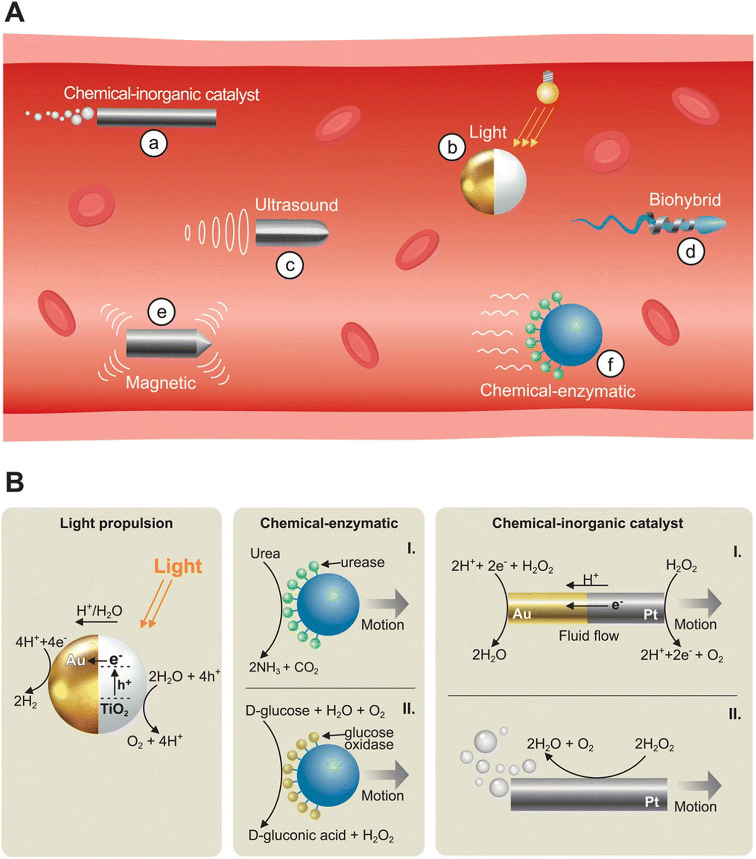 | ||
| Fig. 3 Types of propulsion mechanisms of nanorobots. (A) Nanorobots journeying through a blood vessel. Externally powered nanorobots utilize ultrasound propulsion (c), magnetic propulsion (e), and light propulsion (b). Chemically powered nanorobots can use inorganic catalysts such as hydrogen peroxide (a) or enzymatic propulsion (f). Biohybrid nanorobots utilize cells or microorganisms together with other types of propulsion such as magnetic propulsion (d). Red elements depict red blood cells. (B) Details of selected propulsion mechanisms. From left to right: light propulsion of Au Janus-type nanorobots, enzymatic propulsion based on urease (I) and glucose oxidase (II), chemical propulsion with inorganic catalysts based on self-electrophoresis (I) and bubble propulsion (II). | ||
Externally powered
Nanorobots powered by external physical forces have an undisputable advantage over those propelled chemically. That is mainly due to their fuel-free locomotion that does not utilize any toxic fuel that would have to be added. Nevertheless, these nanorobots face their own set of biocompatibility challenges.Magnetic guidance using static or dynamic magnetic fields stands as a prominent and widely researched type of propulsion in nanorobotics in vivo for a good reason (Fig. 3A(e)). Magnetic actuation offers many exciting advantages by providing highly controllable directional motion, which has been used in many scenarios involving miniaturized robots.19,20,30,106–112 The speed of magnetically actuated nanorobots depends on the source of the magnetic field and the robot's size and shape, typically decreasing as the robot size increases. In vitro and ex vivo research proved that magnetically guided micro and nanorobots are capable of propulsion in the fast-paced blood flow.113–115 This has been validated by several in vivo uses with intravenous administration of magnetically guided nanorobots.19,20,30,111 Nonetheless, integrating magnetic materials for guidance poses biocompatibility challenges as these materials must be present either on the nanorobot's surface or in its core. Most commonly used materials such as superparamagnetic iron oxide,116 NdFeB,117 nickel118 or cobalt119 are not inherently biocompatible and biodegradable. Overcoming these issues during the manufacturing process is vital to ensure that the presence of nanorobots in complex biological systems will not result in cytotoxicity or immune response. A review focusing on magnetically actuated nanorobots can be found elsewhere.112
Light-driven nanorobots are on the contrary much easier to be made biocompatible because their propulsion mechanisms allow for using soft light-responsive polymers either alone or in combination with biological materials120 (Fig. 3A(b)). The advantage of polymers is their better biodegradability than metal materials.121 Other materials such as gold or inorganic salts can also be used.122 Propulsion is usually achieved through an asymmetric morphology or shape, such as Janus type of nanorobots, which respond to light stimuli.123,124 Three main propulsion mechanisms have been described in light-driven nanomotors that operate without the need for external fuels: self-diffusiophoresis, self-electrophoresis, and self-thermophoresis. Self-electrophoresis (Fig. 3B) for example, involves the nanorobot generating its own electric field, which drives its motion. When a photocatalytically active nanorobot, such as an Au/TiO2 Janus nanorobot, gets irradiated by a suitable light source, it leads to electron–hole pair generation. The holes remain in the semiconductor, where water is oxidized to oxygen and H+, while the metal component reduces H+ to H2. This results in a proton gradient and the generation of an electric field, resulting in self-propulsion,122,123 while magnetically actuated nanorobots typically achieve a higher speed than that of light-driven ones.120 The limitations of the light source as optical propulsion are the penetration depth of the light and the speed of the nanorobot. Near-infra-red light (NIR) can safely penetrate tissues but its penetration depth is limited to several millimeters,125–127 restricting the use of nanorobots powered by this light spectrum to near-surface areas. However, NIR-II provides reduced photon scattering and therefore deeper tissue penetration.128–131 NIR is oftentimes successfully used for triggered drug release.20,132–134 The sole use of NIR for propulsion is rare.61 More in vivo research is therefore needed, as current studies are largely limited to in vitro models or often rely on alternative propulsion methods. This limitation hinders the complete assessment of the functionality of solely light propelled nanorobots in complex biological systems. A comprehensive review on light-driven nanorobots has been previously published elsewhere.135
Ultrasound, routinely used for imaging for decades and overall considered to be safe,136 can generate large propulsive force by creating a pressure gradient along the nanorobot (Fig. 3A(c)). A remarkable speed of 6.3 m s−1 with deep tissue penetration of ultrasound propelled microbullets has been reported.137 However, it has been shown that the ultrasound propulsion velocity decreases with the increasing shear viscosity of the fluid.138 This factor needs to be taken into consideration when propelling nanorobots in the bloodstream. Numerous exciting in vitro applications of nanorobots utilizing ultrasonic propulsion have been documented.29,31,139 However, there is a pressing need for studies aimed at applying these forces in translational in vivo approaches. This is because encouraging in vitro results do not automatically translate to successful in vivo applications. Ultrasound-driven nanorobots have been summarized elsewhere.140
Chemically powered
Chemical propulsion, also known as self-propulsion, relies on the conversion of a chemical or biological fuel source to generate motion. Understanding the environment where the nanorobot will operate is critical, to know if it naturally contains the fuel source, to avoid introducing foreign substrates into unrelated biological environments (Fig. 2). Additionally, it is essential for the nanorobot to have adequate fuel supply to complete its task without running out of fuel prematurely.The earliest form of self-propulsion involved the catalytic decomposition of hydrogen peroxide using noble metals such as platinum141,142 or silver143 (Fig. 3A(a)). The decomposition of hydrogen peroxide can drive bubble propulsion as the catalytic layer of the nanorobot converts hydrogen peroxide into water and oxygen. This process produces bubbles that propel the nanorobot forward14 (Fig. 3B). However, in the case of bimetallic nanorobots, such as those made from gold and platinum, motion is achieved through self-electrophoresis rather than bubble generation. In these bimetallic nanorobots, the gold and platinum ends function as an anode and cathode, respectively, with redox reactions occurring at both ends. At the platinum end, hydrogen peroxide is oxidized, producing protons, which are then consumed at the gold end. This process creates an electric field that propels the nanorobot forward (Fig. 3B).144 The disadvantage of hydrogen peroxide as fuel is its obvious toxicity to biological systems and its negligible natural presence in the human body, making it unsuitable as a fuel source. Although some studies demonstrated propulsion in very low peroxide concentrations in vitro,145,146 applying this method in in vivo scenarios is practically impossible. Nonetheless, hydrogen peroxide propulsion still has a place in in vitro environments where it can be valuable, for instance, in cell transfection.28
Enzymatic propulsion has recently been in the spotlight due to its superior biocompatibility, though it also requires a constant fuel source (Fig. 3A(f)). Unlike other methods, its substrates are commonly found in the human body eliminating the need for their artificial addition. Several reviews have been published on this topic147 and we will therefore focus only on two most extensively studied enzymatic substrates.
Glucose (C6H12O6), used as a fuel to propel nanomachines, seems an ideal solution for blood-propelled nanorobots. Glucose oxidase can be utilized to decorate the surface of a nanorobot, where it catalyzes β-D-glucose to D-glucono-δ-lactone (gluconic acid) and hydrogen peroxide in the presence of molecular oxygen, which creates propulsion (Fig. 3B).148
However, its blood concentration, approximately 5 mM, is often too low for the function of the majority of glucose-operated nanorobots, which require several times higher glucose concentrations.149–152 A study using polymeric stomatocyte nanomotors containing glucose oxidase and catalase in their cavity achieved a speed of 11 μm s−1 and 6 μm s−1 in glucose concentrations of 10 and 5 mM, respectively, marking the first successful use at biologically relevant glucose levels.153 Glucose conversion is not limited to glucose oxidase. Alternative methods of glucose conversion can overcome the weak propulsion of glucose oxidase and help nanorobots reach a much higher speed; however, they often need several times higher glucose concentration than the physiological levels.151,152
Urea (CO(NH2)2) serves as the substrate for urease powered nanorobots. Urea is being converted by enzyme urease to ammonia and carbon dioxide, creating propulsion (Fig. 3B).148 However, urease propelled nanorobots face similar struggles to those using glucose oxidase, primarily needing sufficient fuel concentration to start actively moving. Studies indicated that 50 mM of urea is required to mobilize urease-powered robots. This concentration does not occur in any tissue except for the bladder.147 Demonstrating this, urease-powered nanorobots have shown locomotion in a living mouse's bladder154,155 and hold significant potential for bladder cancer treatment.154–156 Such applications highlight the rational use of a nanorobot in biologically relevant environments, paving the way for translational research and clinical application.
Biohybrid nanorobots
Biohybrid nanorobots represent the endeavor towards better biocompatibility and biodegradability, blending the natural characteristics of cells or microorganisms with advancements in modern technology (Fig. 3A(d)). This approach aims to harness the best from both biological and technological realms. Detailed reviews on this subject are available elsewhere.16,17Biohybrid nanorobots have been constructed using various body cells including red blood cells,157,158 platelets,157,159,160 cancer cells,161,162 white blood cells such as neutrophils or macrophages,18,19 or even their membranes. Many such endeavors led to exciting in vivo applications with the potential for clinical translation.18,19,160,161 Neutrophils and macrophages particularly possess innate chemotactic abilities, which allow them to migrate in concentration gradients of chemical stimuli such as pro-inflammatory cytokines towards the source which can be harnessed for tumor or tissue inflammation targeting.18,19,133 Such sensing capabilities that would utilize biocompatible strategies are predominantly found in hybrid nanorobots. However, enzymatically powered nanorobots that migrate toward substrate gradients, such as glucose or urea, also exhibit sensing capabilities. An in-depth review of various sensing mechanisms in micro- and nanorobots in various applications not solely focused on in vivo scenarios is available elsewhere.163 Due to the small size of nanorobots, integrating manufactured sensors can be technologically challenging. Therefore, it has not been performed to date. For these reasons, biohybrid nanorobots often excel in sensing capabilities due to their inherent nature of chemotaxis. Spermbots, created using sperm cells, leverage their motility for propulsion and chemotactic skills for navigation.21,164 Their natural capability of chemotaxis in the female reproductive system suggests a potential in treating ovarian cancer.164 Bacteriobots similarly to spermbots use the cell's ability of movement for actuation.110,111,165 Their ability to adhere to epithelial cells in the urinary and gastrointestinal tract offers prospects for targeted drug delivery in these systems.166 Magnetically guided bacteriabots were also utilized for in vivo cancer treatment in a mouse model with no obvious toxicity.111 The improved biocompatibility and natural ability to move in biological fluids make biohybrid nanorobots an attractive option for targeted drug delivery within the human body.
In summary, the quest for an ideal propulsion mechanism in nanorobotics often feels like trying to eat our cake and have it too. The trade-off between biocompatibility/biodegradability and speed is a common consideration; similarly, speed must be balanced against size. This raises the question: have we reached the zenith of our creativity or are there still untapped propulsion mechanisms awaiting discovery? Is a groundbreaking actuation mechanism necessary to open doors toward translational research and human application or can a “mix and match” strategy suffice? Thus far, diversification in propulsion approaches seems to yield better performance, biocompatibility, and ultimately greater translational potential for real-world applications. Nature has engineered plenty of nanorobots that are living within us or around us. These are immune cells, capable of sensing, independent assessment of the situation, collaboration with other cells and thread elimination, sperm cells excelling in chemotaxis and fast locomotion towards the target, bacterial cells skilled in immune evasion, and many more. Drawing inspiration from these natural systems seems to right now guide the development of nanorobotics toward practical and realistic uses.
Design beyond propulsion
The performance and interaction characteristics of nanorobots with biological systems are not solely determined by their propulsion mechanism and material composition. Additional factors such as size, shape, charge, and surface coating play crucial roles in shaping their behavior (Fig. 2). The following chapter delves into design considerations beyond propulsion that significantly influence membrane crossing. Due to membrane crossing being a largely unexplored territory of nanorobotics, there are only handful of studies explaining how design of a nanorobot beyond propulsion influences its ability to interact with and cross biological membranes in in vivo applications. Research on nanorobots that demonstrates membrane crossing often focuses on a binary assessment of whether nanorobots can cross membranes, rather than investigating the underlying mechanisms. Given the nascent nature of nanorobotics, drawing insights from nanoparticle research is advantageous, as their interactions with biological systems have undergone extensive scrutiny for over a decade.167–170 Several in-depth reviews on this topic have been written.171,172 While nanorobots may exhibit distinct behaviors compared to nanoparticles, there are certain aspects where it is reasonable to anticipate similar interactions with the biological system. Specific examples of nanorobots conquering biological membranes will be discussed in detail in the following chapter.Design for prolonged circulation
Both nanorobots and nanoparticles have been struggling with off-target accumulation since their first use in in vivo applications (Fig. 2). Only recently have nanorobots been introduced in in vivo scenarios through intravenous administration. Although the initial results are promising, the understanding of design aspects remains limited due to the novelty of this approach. Almost all studies employing intravenous administration of nanorobots encounter some level of accumulation in non-target tissues. Notably, in some cases, the accumulation in organs such as the liver and spleen surpasses that in the intended target, such as tumor tissue.20,133,173 A thorough understanding of the mechanisms behind off-target accumulation is crucial for developing nanorobots with better targeting abilities.Nanoparticles with a size of 5 nm or less are typically eliminated through renal clearance.174,175 Conversely, particles exceeding 150 nm tend to accumulate in the liver and spleen. The liver, characterized by fenestrations ranging from 50 to 300 nm, has the highest endothelial permeability.176 A similar pattern is observed in the spleen where interendothelial slits, 200–500 nm in size, create spaces in between endothelial cells, which make them easy to cross.177,178 As the nanoparticle size increases to several micrometers, the accumulation in lung capillaries also occurs.171
Prolonged circulation appears to be associated with nanoparticles of approximately 100 nm size.171 Nevertheless, adjusting the nanoparticle size might be a potential avenue for targeting specific organs such as the liver, spleen, or lungs. Importantly, larger nanoparticles do not solely accumulate in these organs due to large fenestrations; macrophages also play a significant role in this process (Fig. 2).
Tissue macrophages, primarily located in the liver, spleen, and lungs, play a pivotal role in clearing foreign objects from the bloodstream through ingestion. Particularly, nanoparticles of sizes 2–3 μm are recognized by macrophages the easiest, possibly due to their similarity in size to bacteria.179 It was also found that macrophages exhibit shape preference, favoring spherical nanoparticles over ellipsoids180,181 or worm-shaped structures.182 This preference has been corroborated in vivo in pigs.183 Nevertheless, this effect might be dependent on the material and surface coating of nanoparticles and not just on their shape because contradicting results exist as cetyltrimethylammonium bromide (CTAB)-coated gold nanorods were reported to be favored by macrophages 230-times more than CTAB-coated gold spheres.184
In terms of nanoparticle charge, both strongly positive and strongly negative charges have been associated with increased uptake in the liver, potentially stemming from recognition by macrophages185 (Fig. 2). Importantly, neutral nanoparticles demonstrated lower clearance from the blood compared to charged nanoparticles, with positively charged nanoparticles exhibiting the fastest clearance.186 These studies point out that a neutral or slightly negative charge is connected with the optimal prolonged circulation and half-life in vivo.
Macrophage recognition is also closely related to the opsonization of a nanoparticle, a process involving the adhesion of plasma proteins onto the nanoparticle surface, forming a protein corona187,188 (Fig. 2). This protein corona is subsequently identified by macrophages. Polymer coatings such as polyethylene glycol (PEG) have shown the ability to reduce the amount of plasma proteins adsorbed onto the nanoparticle's surface189 as well as the ingestion by macrophages.190 However, it is essential to acknowledge that such coatings also diminish the nanoparticle's ability to interact with target cells. However, research has demonstrated that in certain cases, the protein corona can have various advantageous effects. These include acting as a cell protectant, shielding cells from possible damage induced by cationic191 and anionic192 nanoparticles. It has also been shown that protein corona can help with cell internalization,193,194in vivo biodistribution,195 and tissue-specific delivery.196
It is worth noting that the interaction between nanorobots and macrophages may differ due to the movement properties of nanorobots; however, this has not been studied yet. The opsonization of nanorobots could potentially have adverse effects on their response to stimuli, whether chemical or external, leading to the inhibition of their movement. While this aspect has not been thoroughly investigated, understanding these dynamics is crucial for the successful design of nanorobots for in vivo applications.
The in vivo studies discussed in the upcoming chapter predominantly utilize magnetic field or NIR light for actuation following intravenous injection.19,20,30,61,111 The prevalence of these propulsion mechanisms in documented in vivo applications suggests that they may not be significantly inhibited by opsonization. Additionally, many in vivo studies use biohybrid nanorobots which in the case of the body's own cells should theoretically avoid opsonization and clearance as invading agents.18–20 However, even biohybrid nanorobots remain susceptible to off-target accumulation, probably influenced by factors other than opsonization.
Design for cell-specific delivery
The ultimate goal in intravenous administration for all delivery vehicles targeting a particular tissue is to achieve cell-specific targeting. If delivery platforms could precisely navigate to the designated organ and release their cargo directly to target cells, it would significantly reduce side effects and enhance efficiency, minimizing cargo loss in off-target tissues. Such a scenario would also offer economic advantages, as a lower quantity of delivery vehicles could be utilized to achieve the desired therapeutic effect. Cell-specific delivery stands as the future paradigm for intravenously administered therapies across various diseases.For example, the demand for cell-specific technology is urgent in the realm of CRISPR-based genetic therapy. Current methods rely on delivery via viruses, which raises safety concerns,197–200 or on ex vivo administration that involves removing patient cells, editing them with CRISPR, and then reintroducing them back to patient's body—a costly and hospitalization-dependent procedure that is not universally accessible. Intravenously administered CRISPR, delivered in a cell-specific manner could democratize CRISPR-based genetic therapy, making CRISPR-based treatments accessible to a broader range of patients afflicted by genetic diseases.
In oncological treatment with chemotherapeutic agents, cell-specific delivery holds promise for significantly reducing the well-known side effects associated with this form of treatment. As highlighted earlier, conventional nanoparticle delivery methods have not provided substantial gains in efficiency for targeting tumor tissues, with reported increases as modest as 0.2%.26 However, the emergence of nanorobotics opens up possibilities for revolutionary strategies that could transform cell-specific delivery of therapeutics.
Investigations into targeting strategies for the cell-specific delivery of drugs extend to various organs, such as the brain, where receptors like TfR or IR are commonly expressed on the BBB. The binding of these receptors facilitated by targeting antibodies initiates receptor-mediated transcytosis, triggering the transport of cargo linked to the antibody across the BBB.71,201 Transferrin which is also known to bind to TfR has been used for nanorobot functionalization for BBB crossing.202 LRP-1 targeting peptides have also been utilized for the crossing of nanorobots across the BBB leveraging the abundant expression of LRP-1 receptor on the BBB.203 These cases will be fully discussed in the following chapter.
Antibody-based targeting has found particular application in oncological treatments, with numerous antibodies, basically FDA-approved, as delivery vehicles for a wide array of anti-cancer drugs.204 Small molecules, including folic acid, have also been explored for targeted cargo delivery to cancer and immune cells by binding to folate receptors and facilitating receptor-mediated endocytosis.205
Additionally, targeting peptides and aptamers, which are nucleic acid fragments, have been studied for receptor targeting in various diseases.206 Although many of these strategies have advanced to clinical trials and gained FDA approval, they primarily involve targeting moiety–drug conjugation scenarios, not using nanocarriers. Nanomaterials with their advanced properties could enhance the characteristics of such cell-targeted therapies. The fusion of the worlds of cell-specific targeting and nanorobotics has the potential to introduce cutting-edge strategies for treating various diseases. Despite these prospects, to our knowledge, there are limited reports of in vivo scenarios demonstrating cell-specific targeting of nanorobots.203 Nanorobot could be also constructed in a way that targeting of certain cells is ensured without relying on antibodies or targeting peptides. For instance, the CD44 receptor is expressed on various cell types including stem cells, hematopoietic cells, and many types of cancer cells.207 Hyaluronic acid, a linear anionic polymer naturally present in the extracellular matrix, is the major binder of CD44.208 Therefore, surface coating of nanorobots with hyaluronic acid might not only enhance their surface and stability, but also serve as a targeting moiety while offering opportunities for additional surface modification through conjugation.
Design for cellular internalization
Targeting moieties discussed in the section about cell-specific delivery also generally aid in cellular internalization oftentimes by receptor-mediated endocytosis.205 Cell penetrating peptides (CPPs) such as the transactivator of transcription (TAT) derived from the HIV virus are also known to help with cellular internalization despite them not being cell-specific moieties. CPP contains 5 to 30 amino acids and a relatively high number of positively charged amino acids arginine and lysine, which makes most CPPs being positively charged.209 CPPs have been used with nanorobots for the purpose of cellular internalization for in vivo delivery.30 However, studies on the specific mechanisms of cellular internalization of nanorobots are largely missing.The majority of cells maintain a negative plasma membrane potential, resulting in a faster internalization of positively charged nanoparticles compared to their neutral or negatively charged counterparts.185,210–212 However, the paradoxical outcome arises wherein positively charged nanoparticles are typically cleared from the bloodstream the fastest of all variants of charge and also possess higher inherent cytotoxicity (Fig. 2). In response to this contradiction, there has been an effort to engineer zwitterionic nanoparticles characterized by a negative charge profile, thereby promoting prolonged circulation. Upon reaching the acidic microenvironment of a tumor, these nanoparticles undergo a charge-switch transitioning to a positive charge state, which facilitates cell uptake.213,214
Cellular uptake and endocytosis of nanoparticles are not solely dependent on charge but are also significantly influenced by shape and size (Fig. 2). Citrate-stabilized gold nanoparticles functionalized with DNA through 5′-thiol moiety demonstrated reduced cellular internalization as the hydrodynamic diameter increased, with 10 nm nanoparticles exhibiting the most efficient internalization.170 Paradoxically, it was observed that endocytosis of gold nanoparticles increased with size progression from 14 nm, 30 nm to 50 nm, reaching its peak, and subsequently declined for sizes of 74 nm and 100 nm, which exhibited the lowest internalization rate. Moreover, the aspect ratio proved to be also important with spherical nanoparticles outperforming their rod-shaped counterparts in cellular internalization.215 Notably, the former study used nanoparticles functionalized with thiolated DNA that had a negative charge, while the latter study employed nanoparticles stabilized with citric acid ligands giving the nanoparticles also negative charge.
This observation underscores the pivotal role played by the manufacturing process. A comprehensive examination of the interplay between size, shape, and charge in nanorobots traversing plasma membranes is essential.
Nevertheless, several studies featured in the subsequent chapter demonstrate that nanorobots show much faster and more efficient internalization into cells compared to static nanoparticles.28,29,31,91 The diversity in their actuation mechanisms suggests that the mere presence of motion can significantly enhance their ability to cross the plasma membrane.
Design for endosomal escape
Endosomal escape, long recognized as the rate-limiting step in intracellular cargo delivery within the field of nanomedicine, has prompted numerous nanoparticle designs that could inspire advancements in nanorobotics. Various strategies have been explored to overcome the acidic environment of lysosomes, and in-depth reviews have delved into these approaches.25,216,217 Various polymers have been utilized for the purposes of endosomal escape, such as pH-sensitive PEG that cleaves at low pH218 or polycationic polymers such as polyethyleneimine (PEI) that protonate in the endosome, inducing a proton sponge effect leading to high osmotic pressure, endosome lysis and release of the cargo.219 Similarly, endosomal escape can be facilitated by ionizable lipids destabilizing the endosomal membrane in acidic pH.220 CPPs are also known to contribute to the endosomal escape.221,222 Numerous other polymers, lipids, and peptides have been designed to equip the delivery vehicle with the ability of endosomal escape.The nanorobots that were able to escape the endosome and release their cargo to the intracellular space will be discussed in the upcoming chapter along with the mechanisms they employed to possess this ability. Encouragingly it appears that the ability of movement is often efficient enough to facilitate endosomal escape,223,224 offering promising prospects for the field of nanorobotic drug delivery. It is crucial to note that achieving a faster release from endo-lysosomal compartments is more desirable than release from late lysosomes that reach a highly acidic pH of 4.5–5. Some studies detailing nanorobots capable of escaping endo-lysosomes employ designs facilitating escape in the mildly acidic pH range of 6–6.5,134,224 while others use strategies for escape later at a pH of 5.202 However, the design of nanorobots should carefully consider that many therapeutic payloads are sensitive to highly acidic conditions, and prolonged exposure to such environments can cause prototropic and pleiotropic structural alterations225 compromising the functionality of these molecules,226 including DOX that is probably the most used payload in nanorobotic studies.227 Therefore, an escape from the endosome is greatly preferred to preserve the functionality of the cargo.
The exploration of nanoparticle behaviors highlights the intricate nature of biological systems and the myriad variables that must be considered when crafting nanorobots (Fig. 2). Assumptions regarding the equivalence of nanorobot behaviors, particularly in terms of endocytosis or macrophage phagocytosis, cannot be made based solely on the behaviors exhibited by nanoparticles. Despite this, an unchanging factor for any agents introduced into the bloodstream remains the necessity to adhere to specific size requirements. It is highly likely that nanorobots with weaker propelling strength may be carried along by the bloodstream and deposited in the same organs as nanoparticles if they do not have the required dimensions and characteristics.
The heart is a powerful organ pumping about 5 liters of blood per minute, which is a significant force, nanorobot development should prioritize achieving prolonged circulation. Elastic materials that offer flexibility may partially overcome the size requirements. Moreover, the size of a nanorobot influences their speed with a general rule, indicating a larger size correlated with a greater speed. However, this must be delicately balanced against biocompatibility and size requirements for the bloodstream (Fig. 2). Understandably, the use of microrobots in direct bloodstream injection scenarios may pose challenges, especially given capillaries’ narrow diameter of 5–10 μm. Red blood cells have to pass one by one in these capillaries and a swarm of microrobots might cause serious clotting. However, microrobots still have their place in in vivo fields such as microsurgery228.
While the notion of a single nanorobot performing a multitude of tasks across diverse biological environments may currently seem ambitious, it represents the ultimate trajectory of the field. To unlock the full potential of miniaturized machines for translational applications—ones that are readily marketable and surpass current state-of-the-art therapies—confronting these challenges is imperative. Only through this coordinated effort can nanorobots be deemed prepared for the final phase of their journey: clinical trials and integration into treatment protocols for various diseases.
Nanorobotic applications
Entering the body through the first barrier
The first biological barrier that nanorobots encounter and have to cross is dependent on the administration route. This chapter delves into nanorobotic types that successfully conquered the first barrier of the human body, which is the skin or mucosa, or bypassed it by intravenous injection. The effect on the disease model will be discussed as well as the biocompatibility of these nanomachines.The penetration of stiff spinal metastasis is one of the examples demonstrating the power of nanorobots in a scenario with translational potential. Asymmetric nanorobots with urchin-like head and hollow tail were used for the penetration of stiff spinal metastasis of difficult-to-treat breast cancer in a mouse model.61
These nanorobots with a spikey head were crafted by partially coating Au nanostars with silica. The Janus-like segregation of the Au nanostar surface was achieved by binding 4-mercaptophenylacetic acid and poly acrylic acid asymmetrically on the surface of the Au nanostar. Added silica reacted with 4-mercaptophenylacetic acid and formed an asymmetric SiO2 coating on the head of the Au nanostar. Polyacrylic acid was allowed to react with CTAB, which promoted further deposition of silica that created a hollow tail. The tail was then loaded with fatty acids and DOX. It was revealed that upon NIR irradiation, the nanorobots were propelled by means of thermophoresis due to Au nanostars converting the adsorbed photons to heat, which created a local thermal gradient throughout the nanorobot. Mice intravenously injected with these DOX-loaded nanorobots subjected to NIR irradiation showed an 80% improvement in survival rate and a substantial decrease in tumor volume compared to controls. Group with DOX only demonstrated a 0% survival rate. These nanorobots were also capable of near eradication of other types of stiff tumors after intravenous injection, showing that this approach is feasible for other types of solid tumors with dense stroma. Hematological analysis of main organs revealed no damage and pointed to good biocompatibility of nanorobots.61
Enhanced tumor penetration and DOX delivery was also achieved using deformable polymeric nanocarriers with a flowable polyphosphoester core.30 A TAT-functionalized diblock copolymer TAT-PeG-b-PHEP where PHEP stands for poly(2-hexoxy-2-oxo-1,3,2-dioxaphospholane) was synthesized and used for the manufacturing of the nanocarriers. The used TAT CPPs were designed to activate in the acidic environment typical of solid tumors, aiding cell uptake. The formed nanocarriers were loaded with DOX and ferromagnetic nanocubes (Fig. 4A). When injected intravenously into mice with tumors, and with a magnet placed over the tumor, the magnetic field facilitated the nanocarrier's penetration into the tumor. This triggered the CPP's pH-sensitive activation, leading to cell internalization, DOX release, and approximately 86% tumor growth inhibition. The highest DOX tumor accumulation was observed group of mice that received magnetically actuated deformable nanocarriers containing pH-responsive CPPs. This was in comparison to groups receiving deformable nanocarriers with non-pH-responsive CPPs and to nanocarriers with rigid core either with or without magnetic actuation. A remarkable tenfold enhancement in the accumulation of DOX within tumor tissue was noted when employing deformable nanocarriers featuring pH-responsive CPPs navigated by a magnetic field, as opposed to their counterparts lacking magnetic guidance. Although the biocompatibility of the nanocarriers was declared to be sufficient, the assessment relied solely on the stable weight of the experimental animals.30
 | ||
| Fig. 4 Various nanorobots for membrane crossing. (A) Nanocarriers with a flowable core are deformable under a magnetic field and possess pH-responsive transactivator of transcription (TAT) cell penetrating peptides, which aid in tumor penetration and DOX drug delivery after intravenous injection. Reproduced with permission.30 (B) Macrophage-based cellular microrobots with chemotactic properties and magnetic guidance for NIR-triggered DOX drug release and photothermal therapy of solid tumors following intravenous administration. Reproduced and modified with permission.20 (C) Nanorobots created from neutrophils (neutrobots) loaded with magnetic nanogels containing the PTX drug sense inflammatory factors released from malignant glioma, cross the blood brain barrier (BBB) and guided by the magnetic field deliver PTX drugs after intravenous injection. Reproduced and modified with permission.19 (D) Orally administered enzymatically powered microcapsules cross the mucosa and switch to macrophage bioengine nanorobots in Peyer's patch, continuing to inflammation sites, delivering therapy. Reproduced with permission.18 | ||
Switching to the realm of biohybrid micro and nanorobots in translationally relevant applications, tumor growth inhibition was observed when using bacteria-hybrid microrobots. The non-pathogenic bacterium Escherichia coli Nissle1917 (EcN) with inherent hypoxia sensing abilities served as the foundation for this type of microrobot. The microrobot was constructed by loading EcN with citric acid-coated Zn-doped Fe3O4 magnetic nanoparticles by forming amide bonds between the nanoparticles and the surface of EcN. The created microrobots were subsequently utilized for targeted cancer therapy.111 EcN was further modified to incorporate a genetically encoded magnetothermal bioswitch. This bioswitch activated by an alternating magnetic field, facilitated the thermally triggered expression of fluorescent protein mCherry, enabling the visualization of the microrobot. Additionally, the bioswitch triggered the expression of NDH-2 enzyme capable of generating hydrogen peroxide for enhanced anticancer treatment. Microrobots were injected intravenously into a tumor-bearing mouse. While being guided by the rotating magnetic field, the microrobots were visualized through thermal imaging. The temperature in the tumor tissue increased to 45 °C within 10 min. Microrobots without magnetic guidance were able to elevate the tumor temperature albeit to a lesser extent. After a 24-hour period, the mCherry fluorescence in the tumor tissue was detected to be 1.15-fold higher under conditions involving magnetic guidance than microrobots without magnetic guidance. Furthermore, the microrobots manifested an enhanced ability to penetrate tumor stroma through a combination of hypoxia sensing and magnetic propulsion. In contrast to control groups, the magnetically guided microrobots facilitated the most substantial inhibition of tumor growth, attributed to the synergistic impact of the magnetothermal effect and the bioswitch-triggered generation of hydrogen peroxide. Immunohistochemical analysis of main organs and analysis of blood biomarkers revealed no signs of toxicity associated with the bacteria-hybrid microrobots.111
Another example is the controlled DOX release in a tumor tissue by cellular microrobots, which further highlights the potential of biohybrid micro- and nanorobots in cancer treatment. Macrophages were used as the building blocks for cellular microrobots engineered to execute photothermal and chemotherapeutic interventions for solid tumors.133 A specific subset of macrophages known for their tumor-homing capabilities was selected for this purpose. Gold nanorods were prepared by an in situ reaction with simultaneous growth and nucleation processes and they were loaded into liposomes together with DOX. Further, liposomes were ingested by these macrophages, and subsequently, intravenously administered into a tumor-bearing mouse. Demonstrating an autonomous migratory capacity guided by chemotaxis, these cellular microrobots precisely homed in on the tumor site. Upon NIR irradiation, the gold nanorods induced a photothermal effect, converting absorbed light to heat and triggering the controlled release of DOX, which resulted in significant necrosis of cancer cells. Remarkably, this approach shielded other organs from the well-documented toxic effects associated with DOX.133 In a subsequent investigation, the same research group used the same type of macrophages.20 This time, macrophages engulfed citric acid-coated superparamagnetic nanoparticles and thermosensitive liposomes carrying DOX (Fig. 4B). Upon intravenous injection into tumor bearing mice, these microrobots not only employed chemotaxis for navigation but their movement was further enhanced by magnetic field through the attachment of a magnet to the tumor. Subsequent NIR irradiation induced a photothermic effect, prompting the release of DOX and resulting in a substantial inhibition of tumor growth. Groups lacking magnetic guidance or NIR irradiation exhibited less significant tumor growth inhibition. Importantly, the presented cellular microrobots possessed exceptional biocompatibility in vivo.20
Similarly to macrophages, neutrophils, as phagocytic cells of the immune system, have also been harnessed for innovative therapeutic applications, in this case the treatment of brain tumor. The intrinsic ability of neutrophils to cross the BBB was leveraged by transforming them into “neutrobots” for this cause19 (Fig. 4C). Hydrophobic Fe3O4 nanoparticles were encapsulated into gelatin nanogels together with chemotherapeutic drug paclitaxel (PTX), which is a tetracyclic diterpenoid (see structure in Fig. 6D), using an emulsion/solvent evaporation technique. Further, these magnetic nanogels were enveloped with an E. coli membrane promoting phagocytosis by neutrophils and preventing drug leakage into the carrier cell. The in vivo study used a post-operative glioma model, selected for the common presence of residual tumor cells and inflammatory factors that would attract neutrobots to the tumor site. Upon intravenous administration, a rotating magnetic field guided the movement of neutrobots. Their innate chemotaxis then enabled them to pass through the BBB homing in on the tumor site where they released PTX. Notably, in this study, mice treated with neutrobots guided by a magnetic field demonstrated the highest survival rates when compared to other treatment groups.19 In a follow-up study, the research focus shifted to a comprehensive assessment of the pharmacology and toxicity associated with intravenously administered neutrobots. The results were highly encouraging, revealing negligible immunotoxicity and minimal impact on liver and kidney functions. The reported minimal toxicity towards healthy tissues underscores the potential of neutrobots in further biomedical applications.229
Exploring the transdermal and transmucosal applications of nanorobots unveils futuristic possibilities that carry significant promise, especially for treatments in developing countries or challenging conditions where maintaining sufficient sterility may be a challenge. Despite these highly favorable prospects, research in the field of transdermal delivery remains limited. Nanoparticles have been reported to enhance transdermal delivery and facilitate the penetration of both hydrophilic and hydrophobic drugs.230,231 The introduction of nanorobots into this arena could further augment the transdermal penetration of these therapeutic molecules, potentially reducing the required drug dosage driven by increased transport efficiency.
Contrary to the transdermal delivery, transmucosal delivery utilizing nanorobots has already seen its first translationally relevant application. Twin-bioengine yeast micro/nanorobot displaying impressive abilities was able to cross multiple membranes including the mucous membrane18 (Fig. 4D). The nanorobots were created in an asymmetric Janus manner. Anti-inflammatory nanoparticles were self-deposited in a hollow yeast microcapsule which was subsequently coated with an asymmetric film, and through imidazole–carbamate linkers, –NH2-containing glucose oxidase and catalase were immobilized onto its surface. In an in vivo setup, mice were orally administered this microcapsule. Once ingested, the nanorobots demonstrated active directional propulsion responding to the glucose concentration gradient in the small intestine, converting D-glucose to D-gluconic acid and hydrogen peroxide with glucose oxidase and further decomposing hydrogen peroxide to water and oxygen by the action of catalase. This propulsion was guiding the microcapsules toward the mucosa where the nanorobots successfully traversed the mucosa barrier and reached Peyer's patches. Payer's patches are follicular structures with immune function housing macrophages ready to engulf invading pathogens. Exploiting this natural mechanism, the nanorobots were ingested by macrophages, initiating a transformative journey as macrophage bioengines. These bioengines harnessed the innate chemotaxis ability of macrophages, allowing them to travel through the bloodstream over long distances. Subsequently, they exited the bloodstream, reaching deep-seated sites of gastrointestinal inflammation, where they successfully delivered anti-inflammatory treatment, which significantly reduced the inflammation. Further testing proved good biocompatibility of these nanorobots.18
Investigating biohybrid nanorobots as promising candidates for membrane crossing in transmucosal transport is a compelling avenue for exploration. The utilization of macrophages to create bioengines or cellular microrobots carries distinct advantages, not only due to their innate ability to migrate towards sites of inflammation or solid tumors through chemotaxis but also because of their autonomous and self-sustained nature, eliminating the necessity for external fuel sources. This approach ultimately offers a biocompatible way of addressing the treatment of deep-seated inflammation and solid cancers.
Exiting the bloodstream by crossing the endothelium
In their quest to fulfill designated roles, nanorobots face a critical hurdle—departing the circulation by crossing the endothelial membrane to access the intended target, whether it be a tissue, organ, or tumor. This chapter will focus on nanorobots that have been documented to cross the endothelium or BBB. Additionally, we will discuss instances of nanorobots crossing the endothelium to off-target organs.Urchin-like nanorobots mentioned in the previous chapter were used for both photothermal therapy and the delivery of DOX to tumors characterized by rigid stroma. This study also described the ability of these nanorobots to exit the bloodstream, an essential step in targeted drug delivery.61 Initially, in vitro tests on experimental models were performed to validate the hypothesis that nanorobots could cross the endothelial membrane into the tumor. Subsequent in vivo compared the performance of nanorobots to passive nanoparticles possessing similar properties but lacking active movement. When injected intravenously, nanorobots successfully extravasated from the bloodstream and evenly penetrated the entire tumor within 120 min. In contrast, the passive nanoparticles remained trapped in blood vessels at the 120 min mark and showed only poor diffusion into the tumor tissue (Fig. 5A). Notably, despite the absence of an active tumor targeting mechanism, the tumor had the highest concentration of DOX among all tested tissues.61 These results highlight that nanorobots might be key to successful drug delivery into solid tumors with stiff stroma, which often present obstacles to conventional therapies and even nanoparticle-based approaches.
 | ||
| Fig. 5 Designs of nanorobots for exiting the blood stream. (A) Nanorobots cross the endothelium and penetrate the stiff stroma of a tumor in 120 min after NIR irradiation compared to passive morphologically similar nanoparticles that are trapped in blood vessels. Reproduced and modified with permission.61 (B) Twin-bioengine nanorobots (TBY-robot) travel with the blood flow and exit the bloodstream at the site of inflammation. Reproduced and modified with permission.18 (C) Nanorobots (AIEdots) coated with natural-killer (NK) cell membrane cross the blood–brain barrier (BBB) upon NIR irradiation inducing photothermal effects in glioblastoma tumors. Nanorobots without NK cell membrane fail to cross the BBB.79 | ||
The ability to cross the endothelial membrane and penetrate tumor tissue was also described in the study exploring deformable polymeric nanocarriers with flowable core for magnetically guided delivery of DOX deep into the tumor tissue.30 An in vitro study was first conducted, centering on a comparative analysis between nanocarriers possessing a flowable core and their counterparts characterized by a rigid core, each exhibiting a diameter of 180 nm. Deformable nanocarriers showed the capability to traverse a polycarbonate membrane with 80–100 nm pores, contrary to rigid nanocarriers that were not able to accomplish that. The knowledge gained from in vitro experiments of nanocarrier's deformability was instrumental in an experiment focusing on crossing the endothelial membrane. Mouse models bearing tumors received intravenous injections of either deformable or control nanocarriers, and after two hours, the mice were sacrificed for tissue analysis. Deformable nanocarriers that were subjected to a magnetic field were successfully able to cross the endothelial membrane and penetrate the tumor tissue. In the absence of a magnet, nanocarriers, both deformable and rigid, remained trapped in blood vessels. Importantly, even with a magnetic field, rigid nanocarriers accumulated in vessels unable to cross to the tumor site. All nanocarriers tended to accumulate in non-target tissues, especially the spleen. However, combining deformable nanocarriers with a magnetic field significantly reduced spleen accumulation, directing the majority of nanocarriers to the tumor.30 These findings highlight the advantage of actively propelled nanocarriers over passive ones in tumor targeting.
Another successful endothelial crossing was achieved by previously discussed macrophage-based cellular microrobots. The efficacy in traversing the endothelial membrane was due to the utilization of the chemotactic properties inherent to macrophages. This ability was leveraged for photothermal therapy and the delivery of DOX into solid tumors.20,133 A subsequent study underscored the pivotal contribution of supplementary magnetic guidance, which significantly augmented the efficiency of this infiltration process.20 Despite this tumor targeted approach, both studies showed that cellular microrobots exhibited infiltration into non-target tissues, including the lungs, spleen, and liver, with the lungs receiving a disproportionately higher dosage of microrobots than the intended tumor site.20,133 The incorporation of magnetic guidance, however, decreased this off-target penetration, concurrently amplifying the tumor targeting.20 Nevertheless, even with the implementation of magnetic propulsion, the microrobots remained more predominant in the lungs than in the targeted tumor site, highlighting a persistent challenge in achieving optimal targeting specificity.20,133
As previously highlighted, twin bioengine nanorobots were constructed using yeast microcapsule with an anti-inflammatory drug that was propelled in the glucose gradient of the small intestine. Once ingested by macrophages in Peyer's patches, the bioengines had the capability to navigate the bloodstream and traverse the endothelium, reaching the site of inflammation within 24 h (Fig. 5B). This ability was attributed to the innate ability of macrophages in this regard.18
Expanding upon this theme, a biohybrid nanorobot was used in a study that utilized Magnetococcus marinus bacterium for bloodstream extravasation and subsequent deep tumor penetration. M. marinus is a magnetotactic bacterium renowned for using geomagnetic field lines as a navigational reference during its swimming endeavors. Notably, this bacterium also exhibits a natural inclination to migrate toward regions with low oxygen levels. The study capitalized on this intrinsic capability by loading M. marinus with nanoliposomes carrying a therapeutic payload.110 The –COOH functionalized liposomes were conjugated to M. marinus surface by a covalent bond to –NH2 groups naturally present on the bacterial cell membrane. These nanorobots were then injected in proximity to a tumor and guided towards the tumor mass through the application of a directional magnetic field. Upon sensing the oxygen gradient, the nanorobots autonomously initiated their journey toward the core of the tumor, where hypoxia is most prevalent. For that to happen, they must have exited the bloodstream before permeating the tumor, even though the study does not explicitly detail this step. This approach notably outperformed the passive diffusion of dead bacteria and nanoparticles used as controls. Importantly, the employment of a magnetic field proved instrumental in facilitating profound tumor penetration, as the bacteria failed to achieve such efficacy without magnetic guidance. The study did not delve into investigating off-target effects, potentially due to the localized administration rather than systemic.110
BBB safeguards the brain, and it is no surprise that it is the toughest barrier in the body to cross. Despite this, some nanorobots have successfully crossed the BBB, showcasing the potential of active propulsion over passive nanoconstructs in this context.
The BBB has been crossed by neutrobots that were utilized for the targeted delivery of PTX to post-resection gliomas.19 Neutrophils were selected for their ability to migrate towards the concentration gradient of inflammatory factors, often released from post-resection gliomas. Initial in vitro experiments utilized a BBB model with a chemokine gradient and glioma cancer cells on the other side of the barrier. Neutrobots demonstrated the capability to migrate through the model BBB, reach the cancer cells, and release PTX. This prompted an in vivo experiment on the mouse model. Upon the intravenous administration of neutrobots, mice were subjected to imaging at 1- and 3-hour intervals. Remarkably, the group treated with neutrobots under magnetic guidance exhibited a significantly higher accumulation of these nanorobots in the brain compared to the control groups. Further histological analysis provided evidence of the neutrobots successfully migrating through the BBB and accumulating in the tumor tissue in mice receiving neutrobots steered by magnetic guidance. These findings underscore the potential of neutrophil-based nanorobots in the efficient transportation of drugs across the BBB.19 However, the study does not provide insights into the potential accumulation of these neutrobots in other tissues.
Polymersome swimmers were also reported to successfully conquer the BBB. These swimmers were assembled by mixing amphiphilic copolymer poly(ethylene oxide) poly(butylene oxide) with poly[oligo(ethylene glycol) methyl methacrylate] and encapsulated glucose oxidase and catalase for self-propulsion in a glucose concentration gradient converting glucose with glucose oxidase and the resulting hydrogen peroxide with catalase.203 Notably, the high expression of glucose transporters on endothelial cells in the brain suggested the presence of a glucose gradient towards the blood vessel walls, forming the foundation for polymersome chemotaxis. The polymersome swimmers were further conjugated with a peptide targeting LRP-1 receptor abundantly expressed on the BBB that facilitates the BBB crossing. This BBB crossing was proved through an in vitro BBB model and validated in an in vivo experiments on rats. The in situ brain perfusion technique achieved 20% uptake of swimmers into the brain tissue in contrast to the 5% uptake observed with passive polymersomes.203In situ brain perfusion is a technique used to measure whole brain uptake of molecules, in this context, polymersomes. It involves perfusion of the brain from the carotid artery with a buffer containing the tested agents. One hour after the start of the perfusion, the brain is removed and examined.232 This method however cuts out systemic circulation, cannot be used to evaluate systemic biocompatibility, and does not provide insights into the behavior of these agents when administered intravenously, including the success of brain targeting, BBB crossing, and potential accumulation in organs other than the brain.
Nanorobots were also able to target glioblastoma tumors by crossing the BBB. The capability to transverse BBB was due to the design of nanorobots that emulated natural-killer (NK) cells79. Glioblastoma, the most prevalent adult brain tumor, renowned for its aggressiveness and difficulty of surgical resection leading to poor prognoses, presents significant clinical challenges.233 The nanorobots were constructed from aggregation-induced emission-active polymeric nanoendoskeletons (AIEdots) with NIR II fluorescence. The NIR II emission-active polymer PBPTV was created by utilizing regioregular bispyridal [2,1,3] thiadiazole and alkyl-substituted (E)-2-(2-(thiophen-2-yl)vinyl)-thiophene units where the first was the acceptor and the latter one the donor. AIE dots were then coated with the plasma membrane of NK cells using biomimicking virus-budding process. In vitro assessments on BBB model revealed that the membrane proteins inherent to the NK cell plasma membrane bind to endothelial cells. This interaction resulted in the disruption of tight junctions that make BBB so impermeable, creating openings for the nanorobots to pass through. This finding was substantiated by in vivo experiments in mouse models. Following intravenous administration, the NIR II fluorescence tracking confirmed nanorobot's ability to cross BBB and accumulate in the tumor. In contrast, AIEdots lacking the NK cell membrane failed to accumulate in the tumor, indicating the membrane's key role in BBB crossing and tumor targeting (Fig. 5C). Importantly, upon NIR laser irradiation, the nanorobots induced a photothermal effect that significantly inhibited tumor growth of the tumor. It is noteworthy, however, that both nanorobots and control AIEdots exhibited high accumulation in non-target tissues, specifically the liver and spleen.79
Crossing the plasma membrane
The efficacy of a drug hinges on its ability to permeate the cell's plasma membrane. While numerous examples exist of nanorobots successfully crossing the plasma membrane and gaining entry into cells in vitro, the translational potential of these findings requires consideration. In many cases, these studies, though intriguing, do not use biologically relevant fuel sources, optimal fuel concentrations, or biocompatible materials, or simply in vivo study still needs to be performed to confirm the functionality in complex biological systems. Nevertheless, the promising aspect of in vitro studies lies in the comparison between the performance of propelled nanorobots and static nanorobots or nanoparticles in delivering cargo into cells. The dynamic movement of nanorobots often translates into a substantially faster and more effective cargo delivery process compared to their static counterparts, highlighting the potential advantages of propelled nanorobots in therapeutic applications.All variations of gene therapy need a solid delivery vehicle to get into the target cell. Nanorobots have surfaced as a promising platform for this purpose. Gene therapy has many flavors, and small interfering RNA (siRNA) capable of silencing genes is one of them. siRNA is a type of short double-stranded RNA possessing two-nucleotide-long overhangs, hydroxylated 3′ ends and phosphorylated 5′ ends. In a compelling demonstration, ultrasound-powered gold nanowires were used to silence the green fluorescent protein (GFP) in vitro as a proof of concept. Gold nanowires were functionalized with cysteamine, which was then used for the conjugation of siRNA to the surface. Nanorobots carrying siRNA were capable of facilitating up to 94% or 70% GFP silencing in two different cell lines compared to static nanowires demonstrating only up to 12% or 13% GFP silencing.31
CRISPR/Cas9 is a powerful genome-editing tool with transformative potential in the treatment of various genetic diseases. It consists of a 160 kDa positively charged Cas9 protein, which acts as the enzyme capable of cutting DNA and a strand of approx. 20-nucleotide-long RNA guides Cas9 to the target site. Ultrasound powered gold nanowires were functionalized with 3-mercaptoptoionic acid, which was further modified with cysteine. Cas9 was immobilized onto the nanowire using a disulfide linkage between the cysteine of Cas9 and the thiol-functionalized nanowire. Such prepared nanowires were able to penetrate the plasma membrane of GFP expressing cells, which was followed by the disassociation of the CRISPR complex due to reduction facilitated by intracellular gluthatione (Fig. 6A). Using these nanomachines, 80% CRISPR-mediated GFP knock-out in vitro was achieved, while static nanowires were able to facilitate only 30% knock-out. While both studies do not explicitly elucidate the intracellular fate of these ultrasound-actuated nanomachines, their superior functionality suggests their ability to directly permeate the plasma membrane and circumvent entrapment within the endolysosomal compartment.29
 | ||
| Fig. 6 Different propulsion mechanisms and type of cargo of nanorobots for cellular internalization. (A) Ultrasound propelled gold nanowires decorated with Cas9/sgRNA facilitate successful green fluorescent protein (GFP) knock-out. Reproduced and modified with permission.29 (B) Left: Chemically powered AuAg nanorobots transfect cancer cells with protein cargo metallothionein. Right: Schematic representation of the metallothionein protein structure. Reproduced and modified with permission.28 (C) Self-thermophoretically powered cell membrane-coated semi-yolk/spiky-shell nanomotor delivers the DOX drug under NIR into cancer cells. Reproduced and modified with permission.162 (D) Schematic representation of the molecular structure of common nanorobotic cargo doxorubicin and paclitaxel. | ||
Apart from the gene therapy tools, proteins have also been reported to successfully traverse the plasma membrane on nanorobots. The chemically propelled nanorobots composed of a gold core and a silver shell were used to transport protein cargo across the plasma membrane of the prostate cancer cell line known for being difficult to transfect (Fig. 6B). These virus-sized AuAg-nanorobots propelled by the decomposition of hydrogen peroxide on their catalytically active silver shell carried a 61-amino-acid-long cysteine-rich protein metallothionein (see structure in Fig. 6B) with a 6-fold enhancement in efficiency compared to static non-fueled nanorobots, successfully crossing the plasma membrane and carrying the protein cargo into cancer cells. Moreover, the internalization of these propelled nanorobots occurred within a matter of minutes.28
Another example of nanorobot-mediated transport of therapeutics across the plasma membrane is the DOX delivery by a semi-yolk/spiky-shell nanomotor composed of carbon/silica and coated with the cell membrane of the MCF-7 breast cancer cell line. These nanomotors showed photothermal properties upon NIR irradiation due to their asymmetric carbon core, serving as the basis for self-thermophoretic propulsion. Upon incubation with MCF-7 cells, the self-propelled nanomotors adhered to MCF-7 cells 2.16-fold more than the same nanomotors without NIR irradiation. The cellular uptake of nanomotors lacking propulsion was 26.2%. However, upon the introduction of the NIR laser, the uptake increased to 67.5%162 (Fig. 6C).
Not only small-molecule cargoes but also macromolecule cargo deliveries through the plasma membrane have been achieved by polymersome nanomotors. These nanomotors were constructed by the self-assembly of poly(ethylene glycol)-b-poly(D,L-lactide) (PEG-PDLLA) block copolymers, which are biodegradable. A pH sensitive benzoic-imine linker between the blocks ensured cleaving of PEG at low pH values within the cell. Subsequent gold half-coating enabled photo-thermophoresis under NIR irradiation, inducing movement.224 Loaded initially with DOX that represented a small molecular payload (see structure in Fig. 6D), these nanomotors exhibited rapid plasma membrane penetration upon NIR irradiation within a brief 3-minute window during incubation with cancer cells. The absence of NIR irradiation made cellular uptake unsuccessful within this time frame. Beyond small molecules, the nanomotors manifested their versatility in delivering macromolecular cargo. When 583-amino-acid-long bovine serum albumin (BSA) was mixed with nanomotors and incubated with cancer cells, BSA alone was not able to cross the plasma membrane and internalize regardless of the use of the NIR laser. The introduction of nanomotors, however, facilitated the swift internalization of BSA into cells. While the study does not elaborate on the specific binding mechanism between BSA and nanomotors—given their initial mixture at the beginning of the experiment—it was sufficient to achieve effective BSA delivery. Further, the enzyme substrate (fluorescein di-β-D-galactopyranoside) was encapsulated into nanomotors – a substrate that fluoresces upon hydrolyzation by its corresponding enzyme (β-galactosidase). These enzyme-substrate-loaded nanomotors were mixed with enzymes and incubated with cancer cells. Under NIR irradiation, the nanomotors initiated plasma membrane penetration, simultaneously delivering the enzyme. Within a 12-minute timeframe, intracellular conversion of the enzyme substrate occurred, resulting in observable cell fluorescence. The presence of NIR irradiation was vital for this co-delivery and subsequent intracellular enzymatic reaction. Importantly, neither the enzyme nor the enzyme substrate alone revealed the capacity to cross the plasma membrane, regardless of NIR irradiation.224
In the realm of in vivo application of nanorobots, an extra effort has to be made to investigate the uptake of the delivery vehicle at a cellular level. This is essential to determine whether the vehicle migrates through the plasma membrane alongside the cargo, or if the cargo is released and manages to traverse independently. Small molecules such as DOX or PTX (see structures in Fig. 6D) are known for this ability. Many studies, as a result, choose to focus on assessing survival rates or monitoring tumor size, as these metrics serve as functional indicators of successful drug uptake. However, when dealing with larger molecules as payloads, the nanorobot's ability to cross the plasma membrane becomes vital.
In vivo delivery of DOX to tumor tissues following intravenous administration has been facilitated by magnetically navigated deformable polymeric nanocarriers.30 The study also investigated the involvement of pH-responsive CPPs in mediating cellular uptake at the site of the tumor. Nanocarriers with two types of CPPs were used: the first type was modified to be pH responsive, while the second type was pH nonresponsive. Both nanocarriers were intravenously injected to a tumor bearing mouse and subsequently magnetically navigated to the tumor site. Cancer cells were then sorted from the tumor tissue and evaluated for their intracellular DOX levels, indicative of successful DOX delivery. Cells exhibiting the highest DOX content were derived from tumors treated with nanocarriers featuring pH-responsive CPPs and magnetic actuation. Without magnetic actuation, the DOX content was markedly lower but still significantly higher than in tumors treated with nanocarriers using pH-nonresponsive CPPs. These results highlight the importance of CPPs that reactivate in the low pH typical for tumor microenvironment and facilitate cellular uptake. It is possible that nanocarriers with pH-sensitive CPPs but also magnetic actuation were more successful in DOX delivery also due to an even lower pH deeper in the tumor mass30.
Intracellular fate and endosomal escape
When delivering therapeutic agents using nanorobots, ensuring timely intracellular release is vital for their functionality in order to avoid the digestion of the often sensitive cargo. The design for endolysosomal escape was discussed in the chapter “Design for endosomal escape”. This chapter will focus on various strategies that nanorobots use to overcome the harsh environment of endolysosomes and deliver their cargo within the cell. The investigation of intracellular fate and endosomal escape in in vitro studies precedes their application in follow-up in vivo experiments. Understanding these processes is essential for comprehending the mechanisms of cellular uptake and endosomal escape, which, in turn, aids in troubleshooting potential issues with low therapeutic efficacy in vivo.Photothermally propelled polymersome nanomotors presented in the previous chapter were also inspected for intracellular fate. Following the encapsulation of DOX, these nanomotors underwent 4-h incubation with cancer cells. In the absence of NIR, they were stored in lysosomes after internalization through endocytosis. However, if the NIR laser was applied for 5 min after the 4-h incubation, the nanomotors demonstrated the ability to escape the lysosome. This facilitated the translocation of DOX to the nucleus, its primary site of action.224
Endolysosomal escape was also investigated for calcium carbonate Janus micromotors prepared by a one-pot emulsification method, loaded with DOX and then partially coated with gold. These NIR-light-navigated micromotors were able to permeate the plasma membrane of cancer cells using photothermal propulsion and were subsequently endocytosed. Once within the lysosome, the acidic pH = 5 initiated the degradation of the calcium carbonate “body” of the micromotors, resulting in the generation of CO2 bubbles. This triggered a switch to a second type of propulsion for the micromotors. Micromotors were able to escape the lysosomes by using CO2 gas propulsion and released DOX, which then journeyed into the nucleus223 (Fig. 7A).
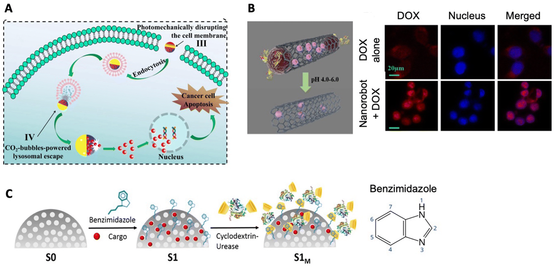 | ||
| Fig. 7 Endosomal escape of drug DOX carried by nanorobots. (A) DOX drug-carrying calcium carbonate Janus micromotors use CO2 gas propulsion for lysosomal escape to the cytoplasm. Reproduced and modified with permission.223 (B) Fe3O4 magnetic nanoparticles serve as caps for carbon nanotube-based nanorobots, which get uncapped at the low pH of lysosomes and release drug DOX (left scheme). Free DOX drug is effluxed from the nucleus back to the cytoplasm while DOX carried by nanorobots is contained in the nucleus. DOX is in red and nuclei are stained blue. Reproduced and modified with permission.202 (C) Left: DOX-drug-loaded urease powered nanomotors feature pH-responsive nanovalves that open and release DOX drug in the acidic pH of lysosome. Reproduced and modified with permission.234 Right: Schematic representation of the structure of benzimidazole. | ||
Another notable example of endolysosomal escape is the DOX delivery to cells by carbon nanotubes with Fe3O4 magnetic nanoparticles used as caps for openings (Fig. 7B). Nanoparticles were attached to the DOX-loaded nanotube using a glutathione linker and the EDC coupling method. The vehicles were further functionalized with transferrin, leveraging the common overexpression of TfR on various cancer cell types for targeted delivery.202
These nanobots were fueled by Fe3O4-mediated decomposition of hydrogen peroxide, which can be found in excess in the microenvironment. The design of nanobots also offered the possibility of magnetic guidance. In vitro experiments involved incubating colon cancer cells with the nanobots and control-free DOX and nanorobots without transferrin. Interestingly, the rapid diffusion of free DOX into cells within the first hour outperformed the internalization efficiency of DOX transported via nanobots. The Nanobots internalized through endocytosis and direct penetration of the plasma membrane, reaching the cytoplasm and lysosomes. In lysosomes the low pH of 5 triggered the degradation of amide linkage, which was followed by the disattachment of Fe3O4 nanoparticles from the carbon nanotube, facilitating the release of DOX into the intracellular space. At 4 h and 24 h time points, the concentration of DOX in the nucleus was significantly higher (8-times and 35-times, respectively), when DOX was delivered by nanorobots. Free DOX, on the other hand, exhibited strong efflux from the nucleus, which should be its final destination, back to cytoplasm: a common mechanism of drug resistance (Fig. 7B). Nanobots lacking transferrin showed reduced efficacy in DOX delivery compared to transferrin-equipped nanobots, which facilitated receptor-mediated endocytosis of the vehicle.202
As highlighted earlier, small molecules may not always require a vehicle for crossing the plasma membrane. However, the present study emphasizes the significance of research into delivery systems, even for small molecules. The nuclear accumulation of DOX delivered by nanobots proved to be considerably more efficient and sustained compared to free DOX, which entered the cell independently. This underscores the nuanced advantages and potential breakthroughs that specialized delivery systems can offer, even for small molecules, in optimizing therapeutic outcomes.
Intracellular fate has been tracked also in DOX delivery by asymmetric hydrogel nanomotors. These nanomotors were assembled using particles composed of a Fe3O4 magnetic core and a Cu9S8 shell enclosed within a hydrogel nanodroplet together with DOX and dopamine using a flow-focusing configured microfluidic chip. Dopamine played a role in anti-tumor immune activation. These nanomotors were further coated with a lipid bilayer containing folic acid for cancer cell-specific targeting and leveraged self-thermophoresis achieved through NIR II irradiation. The nanomotors exhibited intracellular uptake under the influence of NIR II light, achieving a significant degree of internalization within just 25 minutes. In contrast, nanomotors without irradiation required 180 minutes to reach the same level of internalization. Intracellular fate studies revealed that, at the 60-minute mark, nanomotors primarily resided in lysosomes. However, by the 180-minute mark, they successfully escaped the lysosome, when their fluorescence no longer overlapped with fluorescently marked lysosomes. The intracellular release of DOX was observed at 6-hour mark when the fluorescent signal of DOX stopped overlapping with fluorescently labeled nanomotors. The hydrogel nanomotors were further applied in in vivo cancer immunochemotherapy of a breast cancer mouse model.134
The last discussed study of this chapter is the case of endolysosomal escape of enzymatic nanomotors with pH-responsive nanovalves gating the DOX cargo. Mesoporous silica nanoparticles were loaded with either dye or DOX and functionalized with benzimidazole (see structure in Fig. 7C). Subsequent conjugation to cyclodextrin-urease through the formation of inclusion complexes between the cyclodextrin-urease and benzimidazole capped the cargo-loaded pores. The addition of cyclodextrin urease facilitated enhanced diffusion in urea solution (Fig. 7C), an enzymatic reaction producing ammonia. At a neutral pH of 7.5 the nanovalves remained closed. Once in acidic pH of 5 mimicking the environment of the lysosome, the benzimidazole groups protonated and the supramolecular unit unraveled. This triggered the release of the caps and subsequent release of the cargo. Incubating nanomotors loaded with dye with cancer cells revealed significantly more efficient cellular internalization at all time-points in the presence of fuel compared to fuel-free conditions. Additionally, 1-h incubation of cancer cells and nanomotors loaded with DOX showed a 2.4 times larger amount of DOX inside the cells under urea-fueled conditions than fuel-free conditions. Furthermore, DOX was mainly present in the nucleus, indicating successful escape from the acidic lysosomes234 (Fig. 7C).
Future outlook
As we envision the future, the horizon broadens with possibilities. At the forefront of nanorobotics lies the ambitious pursuit of developing smart, motorized, and biocompatible nanoconstructs. These nanoscale entities should be capable of autonomous movement, navigating the bloodstream, and sensing changing pH levels or chemical gradients of various molecules. Their ability should extend to the seamless crossing of multiple biological membranes, delivering a diverse range of therapeutic cargo types to the target tissue with minimal accumulation in off-target organs and without causing any damage. Therefore, we should strive for creating innovative strategies for successful membrane crossing as this aspect is the key ability of nanorobots that have the potential to be used for drug delivery in the human body. This transformative mission calls for synergistic partnership and effective collaboration between material scientists and biologists, pooling their expertise to craft nanorobots for meaningful applications with profound translational potential, propelling the field towards a future of exceptional precision in medical nanorobotics. As we contemplate “Where to move from here ?” the journey ahead beckons us to chart the nanorobotic frontier with innovation and strategic insight.Experimental and translational challenges
While the existing findings shed light on the immense potential of nanorobotics, there are still challenges to address. Many nanorobots are still being crafted from materials unsuitable for use within the human body or rely on fuel concentrations absent in mammalian organisms. Additionally, some designs overlook the need for crossing biological barriers in vivo and therefore conclude their nanorobotic journey with an in vitro application. Recognizing and addressing these hurdles is vital for advancing the practicality and effectiveness of nanorobotic systems in real-world therapeutic scenarios.In the context of in vivo applications, numerous studies have used mouse models with subcutaneously induced tumors, conveniently protruding from the body of the mouse, allowing for the straightforward placement of stationary magnets to guide nanorobots. Despite the efficiency of such magnetic guidance into tumor tissue, it is important to acknowledge that this approach may have limitations in terms of clinical applicability, with relatively few scenarios beyond skin cancer or certain types of head and neck cancers readily benefiting from this technique in its current form. In clinical settings, tumors are often located deep within tissues. Implementing more clinically relevant actuation mechanisms such as a rotating magnetic field becomes crucial for navigating nanorobots effectively in deeper tissue layers. Moreover, the efficiency of commonly used NIR irradiation may differ significantly between subcutaneous tumors and those set deep within other tissues. The translation of these innovative approaches to more clinically relevant scenarios requires a careful examination of the animal models, actuation mechanisms, and the optimization of delivery systems to ensure their effectiveness in diverse tumor locations. As advancements continue, it becomes imperative to tailor these strategies to match the intricacies of real-world clinical scenarios.
There is an equal need for the exploration of more diverse therapeutic cargo options to investigate the full powers of nanorobotic delivery for drugs of various types and characteristics. While initial tests often rely on widely used small molecules like DOX, it is essential to recognize that this represents just one category of therapeutic molecules. DOX, with its traceability, serves as an excellent starting point for in vivo testing, yet it only scratches the surface of the potential therapeutic payloads. To unlock the full capabilities of nanorobotic drug delivery, efforts should be directed toward investigating a broader spectrum of therapeutics. This includes delving into the realms of protein therapeutics, nucleic acids, and genome-editing tools. By embracing this broader perspective, we can uncover the nuanced possibilities that nanorobotics holds in the delivery of a wide array of therapeutic agents, paving the way for more comprehensive and effective drug delivery strategies.
When translating any technology from bench to bedside, a strict set of rules must be adhered to during the manufacturing process to ensure efficient scale-up and regulatory approval. An in-depth review addressing challenges in the translation of nanomedicine, including practical advice has been published and is applicable to nanorobots as well.235 To establish the safety of the nanoformulation, extensive preclinical studies are essential, evaluating the biodistribution and pharmacokinetic properties of the nanocarrier. It is important to note that the laboratory conditions involved in manufacturing nanoformulations for in vivo experiments on animal models differ significantly from the much stricter Good Manufacturing Practice (GMP) standards required for therapeutics heading toward clinical use. Ensuring batch-to-batch consistency and high reproducibility is essential and attention must be also given to achieving storage stability, which is an integral aspect in the development of nanoformulations with clinical potential.
Artificial intelligence (AI) and machine learning (ML) have transformed various aspects of our lives and hold great promise for advancing numerous fields.236 In the realm of robotics AI and ML is enhancing robots’ intelligence, efficiency and adaptability.3 On the microscale, ML can be utilized for motion control237 and path planning in complex environments.238 It also aids in predicting the toxicity of nanomaterials239–241 and protein corona formation on nanoparticles,242,243 potentially advancing material discovery and the design of nanoconstructs for translational applications.244 AI can be also used for predicting the interactions between drugs and nanoconstructs with biological structures, and for instance, predict the permeability of BBB,245–247 which could greatly improve the design of nanorobots for drug delivery.248 However, integrating AI into nanorobots to enable autonomous navigation, decision-making, or complex task execution within the human body remains a futuristic challenge. Given the dimensions of these machines, the creation of nanoscale AI components that are both functional and biocompatible presents a significant technological hurdle. Nonetheless, AI and ML are undeniably transforming translational nanomedicine, potentially accelerating the development of drug delivery systems and enhancing their biocompatibility and functionality.
Ethical concerns regarding the use of nanorobots for translational purposes must be carefully addressed. The safety of nanorobots in human applications requires thorough evaluation before advancing to clinical trials, ensuring compliance with legal regulations. If AI and ML are integrated into nanorobots, collecting health data, the patient privacy must be safeguarded with legislative measures clearly outlining data protection. If nanorobot-based treatments prove to be expensive, issues of equitable access must be tackled to ensure that patients from diverse social and geographic backgrounds can benefit from these advancements.249–251
Commercial viability challenges
The realm of nanorobotics for drug delivery presents a visionary yet formidable landscape, where scientific innovations meet the harsh realities of business. The financial enormity involved in the research and development of a drug spanning from drug discovery through preclinical and clinical research to FDA approval and further safety monitoring emphasizes the challenges faced by drug candidates. Costs soaring into hundreds of millions to several billions of US dollars impose a strict selection process for clinical drug candidates.252The allure of a novel delivery system for pharmaceutical companies and investors hinges on myriads of factors. Commercially viable nanoformulations not only rival, but also surpass the efficiency of established treatments, or offer reduced side effects, improved biodistribution, or other enhancements. The attractiveness is further dependent on the potential patient population benefiting from these novel nanoformulations. Investors seek the intersection of therapeutic efficacy, market demand, and commercial viability. The manufacturing process is also taken into consideration in these decisions. The possible need for specialized manufacturing equipment, the complexity of the manufacturing process and the cost of materials all play a role. Multicomponent systems with intricate multi-step synthesis tend to be more expensive to manufacture and to receive regulatory approval especially adds a layer of expense and regulatory scrutiny particularly when satisfactory therapies exist.
Therapeutics designed for diseases lacking a curative treatment may find leeway in experimental treatments and allow for flexibility in manufacturing costs, as they emerge as the sole treatment available. In these instances, the absence of alternatives not only encourages innovation but also introduces a degree of adaptability to the financial intricacies associated with manufacturing. The current research should reflect and address all these factors, taking into consideration potential manufacturing costs, complexity of the production process and, and whether existing curative treatments for the targeted disease are available. The novel nanoformulation must offer improvements beyond these existing treatments.
Even though the discussed challenges are undeniably intimidating, indicating the long journey nanorobotics has yet to undertake, the initial glimpses of nanorobotic applications provide a beacon of hope. These glimpses hint at a future where these motorized nanodevices could revolutionize medicine, offering innovative solutions to longstanding problems.
Environmental challenges
It is clear that nanorobots must exhibit exceptional biocompatibility with the human body to be viable for drug delivery. Beyond the human scale, a pivotal aspect is ensuring the environmental inertness of nanorobots once they finish their task and are excreted from the body to prevent any unintended interactions with other organisms. Potential negative impact on the environment during the nanorobots manufacturing process also must be taken into consideration. Surprisingly little information is currently available on the topic of environmental challenges posed by nanorobots253 possibly due to the nascent nature of the field, with biological applications still confined to laboratory settings. However, in-depth reviews on the environmental impact of nanomaterials have been published, shedding light on the risks associated with each material254 and discussing the broader impact of nanoparticles on the environment throughout their life cycle including production, use, disposal, and recycling.255 The risks and potential consequences of nanorobots must be meticulously evaluated before propelling nanorobotic technology to the stages of scale-up and clinical applications. In doing so, we sustain not only the promise of transformative medical technology but also a steadfast commitment to environmental protection.Data availability
Data sharing is not applicable to this article as no datasets were generated or analysed during the current study.Conflicts of interest
There are no conflicts to declare.Acknowledgements
This work was supported by funds from the Ministry of Health of the Czech Republic (NW24-08-00276). ZH acknowledges financial support from the Czech Science Foundation (project no. 23-04740M) and project no. CZ.02.01.01/00/22_008/0004631 Materials and technologies for sustainable development within the Jan Amos Komensky Operational Program financed by the European Union and from the state budget of the Czech Republic. The work was supported by ERDF/ESF project TECHSCALE (no. CZ.02.01.01/00/22_008/0004587). This research was co-funded by the European Union under the REFRESH -Research Excellence For REgion Sustainability and High-tech Industries project number CZ.10.03.01/00/22_003/0000048 via the Operational Programme Just Transition.References
- K. Čapek, Rossum's Universal Robots, Aventinum, Prague, 7th edn, 1926, p. 89 Search PubMed.
- L. F. P. Oliveira, A. P. Moreira and M. F. Silva, Advances in Agriculture Robotics: A State-of-the-Art Review and Challenges Ahead, Robotics, 2021, 10(2), 52 CrossRef.
- M. Soori, B. Arezoo and R. Dastres, Artificial intelligence, machine learning and deep learning in advanced robotics, a review, Cognit. Rob., 2023, 3, 54 CrossRef.
- H. Ravichandar, A. S. Polydoros, S. Chernova and A. Billard, Recent Advances in Robot Learning from Demonstration, Annu. Rev. Control Robot. Auton. Syst., 2020, 3, 297 CrossRef.
- A. Dzedzickis, J. Subačiūtė-Žemaitienė, E. Šutinys, U. Samukaitė-Bubnienė and V. Bučinskas, Advanced Applications of Industrial Robotics: New Trends and Possibilities, Appl. Sci., 2022, 12(1), 135 CrossRef.
- A. Loganathan and N. S. Ahmad, A systematic review on recent advances in autonomous mobile robot navigation, Eng. Sci. Technol. Int. J., 2023, 40, 101343 Search PubMed.
- J. Li, I. Rozen and J. Wang, Rocket Science at the Nanoscale, ACS Nano, 2016, 10(6), 5619 CrossRef PubMed.
- H. Wang and M. Pumera, Fabrication of Micro/Nanoscale Motors, Chem. Rev., 2015, 115(16), 8704 CrossRef PubMed.
- R. Liu and A. Sen, Autonomous Nanomotor Based on Copper–Platinum Segmented Nanobattery, J. Am. Chem. Soc., 2011, 133(50), 20064 CrossRef PubMed.
- W. Gao, A. Uygun and J. Wang, Hydrogen-Bubble-Propelled Zinc-Based Microrockets in Strongly Acidic Media, J. Am. Chem. Soc., 2012, 134(2), 897 CrossRef PubMed.
- W. Gao, A. Pei and J. Wang, Water-Driven Micromotors, ACS Nano, 2012, 6(9), 8432 CrossRef PubMed.
- F. Soto, E. Karshalev, F. Zhang, B. Esteban Fernandez de Avila, A. Nourhani and J. Wang, Smart Materials for Microrobots, Chem. Rev., 2022, 122(5), 5365 CrossRef PubMed.
- C. Chen, S. Ding and J. Wang, Materials consideration for the design, fabrication and operation of microscale robots, Nat. Rev. Mater., 2024, 9(3), 159 CrossRef.
- J. Kim, P. Mayorga-Burrezo, S. J. Song, C. C. Mayorga-Martinez, M. Medina-Sánchez and S. Pané, et al., Advanced materials for micro/nanorobotics, Chem. Soc. Rev., 2024, 53, 9190 RSC.
- P. L. Venugopalan, B. Esteban-Fernández de Ávila, M. Pal, A. Ghosh and J. Wang, Fantastic Voyage of Nanomotors into the Cell, ACS Nano, 2020, 14(8), 9423 CrossRef PubMed.
- F. Zhang, R. Mundaca-Uribe, N. Askarinam, Z. Li, W. Gao and L. Zhang, et al., Biomembrane-Functionalized Micromotors: Biocompatible Active Devices for Diverse Biomedical Applications, Adv. Mater., 2022, 34(5), 2107177 CrossRef CAS PubMed.
- J. Li, L. Dekanovsky, B. Khezri, B. Wu, H. Zhou and Z. Sofer, Biohybrid Micro- and Nanorobots for Intelligent Drug Delivery, Cyborg Bionic Syst., 2022, 9824057 CAS.
- B. Zhang, H. Pan, Z. Chen, T. Yin, M. Zheng and L. Cai, Twin-bioengine self-adaptive micro/nanorobots using enzyme actuation and macrophage relay for gastrointestinal inflammation therapy, Sci. Adv., 2023, 9(8), eadc8978 CrossRef CAS PubMed.
- H. Zhang, Z. Li, C. Gao, X. Fan, Y. Pang and T. Li, et al., Dual-responsive biohybrid neutrobots for active target delivery, Sci. Rob., 2021, 6(52), eaaz9519 CrossRef PubMed.
- V. D. Nguyen, H. K. Min, H. Y. Kim, J. Han, Y. H. Choi and C. S. Kim, et al., Primary Macrophage-Based Microrobots: An Effective Tumor Therapy In Vivo by Dual-Targeting Function and Near-Infrared-Triggered Drug Release, ACS Nano, 2021, 15(5), 8492 CrossRef CAS PubMed.
- H. Xu, M. Medina-Sánchez, W. Zhang, M. P. H. Seaton, D. R. Brison and R. J. Edmondson, et al., Human spermbots for patient-representative 3D ovarian cancer cell treatment, Nanoscale, 2020, 12(39), 20467 RSC.
- R. Yang, T. Wei, H. Goldberg, W. Wang, K. Cullion and D. S. Kohane, Getting Drugs across Biological Barriers, Adv. Mater., 2017, 29(37), 1606596 CrossRef PubMed.
- D. Wu, Q. Chen, X. Chen, F. Han, Z. Chen and Y. Wang, The blood–brain barrier: structure, regulation, and drug delivery, Signal Transduction Targeted Ther., 2023, 8(1), 1 CrossRef.
- M. Laksitorini, V. D. Prasasty, P. K. Kiptoo and T. J. Siahaan, Pathways and Progress in Improving Drug Delivery through the Intestinal Mucosa and Blood-Brain Barriers, Ther. Delivery, 2014, 5(10), 1143 CrossRef.
- S. A. Smith, L. I. Selby, A. P. R. Johnston and G. K. Such, The Endosomal Escape of Nanoparticles: Toward More Efficient Cellular Delivery, Bioconjugate Chem., 2019, 30(2), 263 CrossRef.
- S. Wilhelm, A. J. Tavares, Q. Dai, S. Ohta, J. Audet and H. F. Dvorak, et al., Analysis of nanoparticle delivery to tumours, Nat. Rev. Mater., 2016, 1(5), 16014 CrossRef.
- B. Ouyang, W. Poon, Y. N. Zhang, Z. P. Lin, B. R. Kingston and A. J. Tavares, et al., The dose threshold for nanoparticle tumour delivery, Nat. Mater., 2020, 19(12), 1362 CrossRef.
- A. Ressnerova, F. Novotny, H. Michalkova, M. Pumera, V. Adam and Z. Heger, Efficient Protein Transfection by Swarms of Chemically Powered Plasmonic Virus-Sized Nanorobots, ACS Nano, 2021, 15(8), 12899 CrossRef.
- M. Hansen-Bruhn, B. E. F. de Ávila, M. Beltrán-Gastélum, J. Zhao, D. E. Ramírez-Herrera and P. Angsantikul, et al., Active Intracellular Delivery of a Cas9/sgRNA Complex Using Ultrasound-Propelled Nanomotors, Angew. Chem., Int. Ed., 2018, 57(10), 2657 CrossRef PubMed.
- Y. Zhu, Y. Song, Z. Cao, L. Dong, Y. Lu and X. Yang, et al., Magnetically Actuated Active Deep Tumor Penetration of Deformable Large Nanocarriers for Enhanced Cancer Therapy, Adv. Funct. Mater., 2021, 31(35), 2103655 CrossRef.
- B. Esteban-Fernández de Ávila, C. Angell, F. Soto, M. A. Lopez-Ramirez, D. F. Báez and S. Xie, et al., Acoustically Propelled Nanomotors for Intracellular siRNA Delivery, ACS Nano, 2016, 10(5), 4997 CrossRef.
- S. Naahidi, M. Jafari, M. Logan, Y. Wang, Y. Yuan and H. Bae, et al., Biocompatibility of hydrogel-based scaffolds for tissue engineering applications, Biotechnol. Adv., 2017, 35(5), 530 CrossRef.
- D. Iannazzo, C. Espro, C. Celesti, A. Ferlazzo and G. Neri, Smart Biosensors for Cancer Diagnosis Based on Graphene Quantum Dots, Cancers, 2021, 13(13), 3194 CrossRef.
- M. J. Mitchell, M. M. Billingsley, R. M. Haley, M. E. Wechsler, N. A. Peppas and R. Langer, Engineering precision nanoparticles for drug delivery, Nat. Rev. Drug Discovery, 2021, 20(2), 101 CrossRef.
- E. Fang, X. Liu, M. Li, Z. Zhang, L. Song and B. Zhu, et al., Advances in COVID-19 mRNA vaccine development, Signal Transduction Targeted Ther., 2022, 7, 94 CrossRef PubMed.
- K. Okamura, Interdisciplinarity revisited: evidence for research impact and dynamism, Palgrave Commun., 2019, 5(1), 1 CrossRef.
- G. Capponi, A. Martinelli and A. Nuvolari, Breakthrough innovations and where to find them, Res. Policy, 2022, 51(1), 104376 CrossRef.
- D. Bird and N. M. Ravindra, Transdermal drug delivery and patches—An overview, Med. Devices Sens., 2020, 3(6), e10069 CrossRef.
- F. Erdő, N. Hashimoto, G. Karvaly, N. Nakamichi and Y. Kato, Critical evaluation and methodological positioning of the transdermal microdialysis technique. A review, J. Controlled Release, 2016, 233, 147 CrossRef PubMed.
- C. A. Janeway Jr, P. Travers, M. Walport and M. J. Shlomchik, in The mucosal immune system, Immunobiology: The Immune System in Health and Disease, Garland Science, 5th edn, 2001 Search PubMed.
- J. K. W. Lam, C. C. K. Cheung, M. Y. T. Chow, E. Harrop, S. Lapwood and S. I. G. Barclay, et al., Transmucosal drug administration as an alternative route in palliative and end-of-life care during the COVID-19 pandemic, Adv. Drug Delivery Rev., 2020, 160, 234 CrossRef.
- M. Huang, M. Zhang, H. Zhu, X. Du and J. Wang, Mucosal vaccine delivery: A focus on the breakthrough of specific barriers, Acta Pharm. Sin. B, 2022, 12(9), 3456 CrossRef PubMed.
- F. P. Canale, C. Basso, G. Antonini, M. Perotti, N. Li and A. Sokolovska, et al., Metabolic modulation of tumours with engineered bacteria for immunotherapy, Nature, 2021, 598(7882), 662 CrossRef PubMed.
- A. Marabelle, L. Tselikas, T. de Baere and R. Houot, Intratumoral immunotherapy: using the tumor as the remedy, Ann. Oncol., 2017, 28, 33 CrossRef PubMed.
- W. C. Aird, Endothelial Cell Heterogeneity, Cold Spring Harbor Perspect. Med., 2012, 2(1), a006429 Search PubMed.
- W. C. Aird, Phenotypic Heterogeneity of the Endothelium, Circ. Res., 2007, 100(2), 158 CrossRef PubMed.
- Y. Matsumura and H. Maeda, A new concept for macromolecular therapeutics in cancer chemotherapy: mechanism of tumoritropic accumulation of proteins and the antitumor agent smancs, Cancer Res., 1986, 46, 6387 Search PubMed.
- S. K. Hobbs, W. L. Monsky, F. Yuan, W. G. Roberts, L. Griffith and V. P. Torchilin, et al., Regulation of transport pathways in tumor vessels: role of tumor type and microenvironment, Proc. Natl. Acad. Sci. U. S. A., 1998, 95(8), 4607 CrossRef.
- S. Sindhwani, A. M. Syed, J. Ngai, B. R. Kingston, L. Maiorino and J. Rothschild, et al., The entry of nanoparticles into solid tumours, Nat. Mater., 2020, 19(5), 566 CrossRef PubMed.
- D. M. McDonald, Angiogenesis and remodeling of airway vasculature in chronic inflammation, Am. J. Respir. Crit. Care Med., 2001, 164, S39 CrossRef PubMed.
- K. E. M. Hellenthal, L. Brabenec and N. M. Wagner, Regulation and Dysregulation of Endothelial Permeability during Systemic Inflammation, Cells, 2022, 11(12), 1935 CrossRef.
- S. Torosean, B. Flynn, J. Axelsson, J. Gunn, K. S. Samkoe and T. Hasan, et al., Nanoparticle uptake in tumors is mediated by the interplay of vascular and collagendensity with interstitial pressure, Nanomedicine, 2013, 9(2), 151 CrossRef PubMed.
- K. R. Levental, H. Yu, L. Kass, J. N. Lakins, M. Egeblad and J. T. Erler, et al., Matrix Crosslinking Forces Tumor Progression by Enhancing Integrin signaling, Cell, 2009, 139(5), 891 CrossRef PubMed.
- P. A. Netti, D. A. Berk, M. A. Swartz, A. J. Grodzinsky and R. K. Jain, Role of extracellular matrix assembly in interstitial transport in solid tumors, Cancer Res., 2000, 60(9), 2497 Search PubMed.
- J. Choi, K. Credit, K. Henderson, R. Deverkadra, Z. He and H. Wiig, et al., Intraperitoneal immunotherapy for metastatic ovarian carcinoma: Resistance of intratumoral collagen to antibody penetration, Clin. Cancer Res., 2006, 12(6), 1906 CrossRef PubMed.
- C. H. Heldin, K. Rubin, K. Pietras and A. Ostman, High interstitial fluid pressure - an obstacle in cancer therapy, Nat. Rev. Cancer, 2004, 4(10), 806 CrossRef.
- Y. Gao, Y. Shi, M. Fu, Y. Feng, G. Lin and D. Kong, et al., Simulation study of the effects of interstitial fluid pressure and blood flow velocity on transvascular transport of nanoparticles in tumor microenvironment, Comput Methods Programs Biomed., 2020, 193, 105493 CrossRef PubMed.
- V. P. Chauhan, T. Stylianopoulos, Y. Boucher and R. K. Jain, Delivery of molecular and nanoscale medicine to tumors: transport barriers and strategies, Annu. Rev. Chem. Biomol. Eng., 2011, 2, 281 CrossRef PubMed.
- B. Muz, P. Puente, F. de la, Azab and A. K. Azab, The role of hypoxia in cancer progression, angiogenesis, metastasis, and resistance to therapy, Hypoxia, 2015, 3, 83 CrossRef PubMed.
- W. R. Wilson and M. P. Hay, Targeting hypoxia in cancer therapy, Nat. Rev. Cancer, 2011, 11(6), 393 CrossRef PubMed.
- M. Yan, Q. Chen, T. Liu, X. Li, P. Pei and L. Zhou, et al., Site-selective superassembly of biomimetic nanorobots enabling deep penetration into tumor with stiff stroma, Nat. Commun., 2023, 14, 4628 CrossRef.
- S. Xie, C. Mo, W. Cao, S. Xie, S. Li and Z. Zhang, et al., Bacteria-propelled microtubular motors for efficient penetration and targeting delivery of thrombolytic agents, Acta Biomater., 2022, 142, 49 CrossRef.
- H. Kadry, B. Noorani and L. Cucullo, A blood–brain barrier overview on structure, function, impairment, and biomarkers of integrity, Fluids Barriers CNS, 2020, 17(1), 69 CrossRef.
- A. Abrahao, Y. Meng, M. Llinas, Y. Huang, C. Hamani and T. Mainprize, et al., First-in-human trial of blood–brain barrier opening in amyotrophic lateral sclerosis using MR-guided focused ultrasound, Nat. Commun., 2019, 10(1), 4373 CrossRef PubMed.
- A. Conti, H. A. S. Kamimura, A. Novell, A. Duggento and N. Toschi, Magnetic Resonance Methods for Focused Ultrasound-Induced Blood-Brain Barrier Opening, Front. Phys., 2020, 8 Search PubMed.
- Y. Meng, C. B. Pople, H. Lea-Banks, A. Abrahao, B. Davidson and S. Suppiah, et al., Safety and efficacy of focused ultrasound induced blood-brain barrier opening, an integrative review of animal and human studies, J. Controlled Release, 2019, 309, 25 CrossRef PubMed.
- C. H. Fan, H. L. Liu, C. Y. Huang, Y. J. Ma, T. C. Yen and C. K. Yeh, Detection of Intracerebral Hemorrhage and Transient Blood-Supply Shortage in Focused-Ultrasound-Induced Blood–Brain Barrier Disruption by Ultrasound Imaging, Ultrasound Med. Biol., 2012, 38(8), 1372 CrossRef PubMed.
- Z. I. Kovacs, S. Kim, N. Jikaria, F. Qureshi, B. Milo and B. K. Lewis, et al., Disrupting the blood-brain barrier by focused ultrasound induces sterile inflammation, Proc. Natl. Acad. Sci. U. S. A., 2017, 114(1), E75 CrossRef PubMed.
- S. H. Jeong, J. H. Jang and Y. B. Lee, Drug delivery to the brain via the nasal route of administration: exploration of key targets and major consideration factors, J. Pharm. Investig., 2023, 53(1), 119 CrossRef PubMed.
- V. Pandey, A. Gadeval, S. Asati, P. Jain, N. Jain and R. K. Roy, et al., Formulation strategies for nose-to-brain delivery of therapeutic molecules, Drug Delivery Systems, Academic Press, ch. 7, 2020, pp. 291–332 Search PubMed.
- G. C. Terstappen, A. H. Meyer, R. D. Bell and W. Zhang, Strategies for delivering therapeutics across the blood–brain barrier, Nat. Rev. Drug Discovery, 2021, 20(5), 362 CrossRef.
- Q. Huang, A. T. Chen, K. Y. Chan, H. Sorensen, A. J. Barry and B. Azari, et al., Targeting AAV vectors to the central nervous system by engineering capsid–receptor interactions that enable crossing of the blood–brain barrier, PLoS Biol., 2023, 7, e3002112 CrossRef.
- X. He, J. Xie, J. Zhang, X. Wang, X. Jia and H. Yin, et al., Acid-Responsive Dual-Targeted Nanoparticles Encapsulated Aspirin Rescue the Immune Activation and Phenotype in Autism Spectrum Disorder, Adv. Sci., 2022, 9(14), 2104286 CrossRef.
- R. G. Thorne, G. J. Pronk, V. Padmanabhan and W. H. Frey, Delivery of insulin-like growth factor-I to the rat brain and spinal cord along olfactory and trigeminal pathways following intranasal administration, Neuroscience, 2004, 127(2), 481 CrossRef.
- J. L. Shirley, Y. P. Jong, C. de, Terhorst and R. W. Herzog, Immune Responses to Viral Gene Therapy Vectors, Mol. Ther., 2020, 28(3), 709 CrossRef PubMed.
- T. Yang, M. Braun, W. Lembke, F. McBlane, J. Kamerud and S. DeWall, et al., Immunogenicity assessment of AAV-based gene therapies: An IQ consortium industry white paper, Mol. Ther.--Methods Clin. Dev., 2022, 26, 471 CrossRef.
- Y. Zou, Y. Liu, Z. Yang, D. Zhang, Y. Lu and M. Zheng, et al., Effective and Targeted Human Orthotopic Glioblastoma Xenograft Therapy via a Multifunctional Biomimetic Nanomedicine, Adv. Mater., 2018, 30(51), 1803717 CrossRef PubMed.
- T. Yin, Q. Fan, F. Hu, X. Ma, Y. Yin and B. Wang, et al., Engineered Macrophage-Membrane-Coated Nanoparticles with Enhanced PD-1 Expression Induce Immunomodulation for a Synergistic and Targeted Antiglioblastoma Activity, Nano Lett., 2022, 22(16), 6606 CrossRef.
- G. Deng, X. Peng, Z. Sun, W. Zheng, J. Yu and L. Du, et al., Natural-Killer-Cell-Inspired Nanorobots with Aggregation-Induced Emission Characteristics for Near-Infrared-II Fluorescence-Guided Glioma Theranostics, ACS Nano, 2020, 14(9), 11452 CrossRef PubMed.
- E. Nance, S. H. Pun, R. Saigal and D. L. Sellers, Drug delivery to the central nervous system, Nat. Rev. Mater., 2022, 7(4), 314 CrossRef PubMed.
- J. L. Kavanau, Structure and Functions of Biological Membranes, Nature, 1963, 198(4880), 525 CrossRef.
- J. D. Robertson, The molecular structure and contact relationships of cell membranes, Prog. Biophys. Mol. Biol., 1960, 10, 343–418 Search PubMed.
- J. L. Johnson and L. A. Johnson, Homeostasis of Lipid Metabolism in Disorders of the Brain, in Encyclopedia of Behavioral Neuroscience, ed. S. Della Sala, Elsevier, Oxford, 2nd edn, 2022, p. 372 Search PubMed.
- M. H. Gordon, FATS Classification, in Encyclopedia of Food Sciences and Nutrition, ed. B. Caballero, Academic Press, Oxford, 2nd edn, 2003, p. 2287 Search PubMed.
- J. B. Helms and C. Zurzolo, Lipids as Targeting Signals: Lipid Rafts and Intracellular Trafficking, Traffic, 2004, 5(4), 247 CrossRef PubMed.
- K. Simons and E. Ikonen, Functional rafts in cell membranes, Nature, 1997, 387(6633), 569 CrossRef CAS PubMed.
- R. Zhang, X. Qin, F. Kong, P. Chen and G. Pan, Improving cellular uptake of therapeutic entities through interaction with components of cell membrane, Drug Delivery, 2019, 26(1), 328 CrossRef CAS.
- L. Zhong, Y. Li, L. Xiong, W. Wang, M. Wu and T. Yuan, et al., Small molecules in targeted cancer therapy: advances, challenges, and future perspectives, Signal Transduction Targeted Ther., 2021, 6(1), 1–48 CrossRef.
- S. Verma, D. Miles, L. Gianni, I. E. Krop, M. Welslau and J. Baselga, et al., Trastuzumab Emtansine for HER2-Positive Advanced Breast Cancer, N. Engl. J. Med., 2012, 367(19), 1783 CrossRef CAS PubMed.
- S. J. Keam, Trastuzumab Deruxtecan: First Approval, Drugs, 2020, 80(5), 501 CrossRef CAS PubMed.
- B. E. F. de Ávila, H. Gong, J. Wang, N. S. Olmo, P. del, Ortega and F. J. de la Mata, et al., Combination of Ruthenium Dendrimers and Acoustically Propelled Gold Nanowires as a Platform for Active Intracellular Drug Delivery Towards Breast Cancer Therapy, Clin. Oncol. Res., 2019,(4), 1–5 Search PubMed.
- R. Urade, Y. Hayashi and M. Kito, Endosomes differ from plasma membranes in the phospholipid molecular species composition, Biochim. Biophys. Acta, 1988, 946(1), 151 CrossRef CAS PubMed.
- M. S. Almeida, E. de, Susnik, B. Drasler, P. Taladriz-Blanco, A. Petri-Fink and B. Rothen-Rutishauser, Understanding nanoparticle endocytosis to improve targeting strategies in nanomedicine, Chem. Soc. Rev., 2021, 50(9), 5397 RSC.
- J. J. Rennick, A. P. R. Johnston and R. G. Parton, Key principles and methods for studying the endocytosis of biological and nanoparticle therapeutics, Nat. Nanotechnol., 2021, 16(3), 266 CrossRef CAS PubMed.
- I. Mellman, R. Fuchs and A. Helenius, Acidification of the Endocytic and Exocytic Pathways, Annu. Rev. Biochem., 1986, 55(1), 663–700 CrossRef CAS.
- I. Canton and G. Battaglia, Endocytosis at the nanoscale, Chem. Soc. Rev., 2012, 41(7), 2718 RSC.
- P. Saftig and J. Klumperman, Lysosome biogenesis and lysosomal membrane proteins: trafficking meets function, Nat. Rev. Mol. Cell Biol., 2009, 10(9), 623 CrossRef CAS PubMed.
- J. Gilleron, W. Querbes, A. Zeigerer, A. Borodovsky, G. Marsico and U. Schubert, et al., Image-based analysis of lipid nanoparticle–mediated siRNA delivery, intracellular trafficking and endosomal escape, Nat. Biotechnol., 2013, 31(7), 638 CrossRef CAS PubMed.
- P. Lönn, A. D. Kacsinta, X. S. Cui, A. S. Hamil, M. Kaulich and K. Gogoi, et al., Enhancing Endosomal Escape for Intracellular Delivery of Macromolecular Biologic Therapeutics, Sci. Rep., 2016, 6(1), 32301 CrossRef PubMed.
- A. Mittal, A. S. Raber, U. F. Schaefer, S. Weissmann, T. Ebensen and K. Schulze, et al., Non-invasive delivery of nanoparticles to hair follicles: a perspective for transcutaneous immunization, Vaccine, 2013, 31(34), 3442 CrossRef CAS.
- Y. Deng, R. Mathaes, G. Winter and J. Engert, Encapsulation of antigen-loaded silica nanoparticles into microparticles for intradermal powder injection, Eur. J. Pharm. Sci., 2014, 63, 154 CrossRef CAS.
- A. Thakur and C. Foged, Nanoparticles for mucosal vaccine delivery, in Nanoengineered Biomaterials for Advanced Drug Delivery, ed. M. Mozafari, Elsevier, 2020, ch. 25, p. 603 Search PubMed.
- J. M. V. Makabenta, A. Nabawy, C. H. Li, S. Schmidt-Malan, R. Patel and V. M. Rotello, Nanomaterial-based therapeutics for antibiotic-resistant bacterial infections, Nat. Rev. Microbiol., 2021, 19(1), 23–36 CrossRef CAS.
- E. V. Lengert, E. E. Talnikova, V. V. Tuchin and Y. I. Svenskaya, Prospective Nanotechnology-Based Strategies for Enhanced Intra- and Transdermal Delivery of Antifungal Drugs, Skin Pharmacol. Physiol., 2020, 33(5), 261 CrossRef CAS PubMed.
- J. Ou, K. Liu, J. Jiang, D. A. Wilson, L. Liu and F. Wang, et al., Micro-/Nanomotors toward Biomedical Applications: The Recent Progress in Biocompatibility, Small, 2020, 16(27), 1906184 CrossRef CAS PubMed.
- Z. Wu, J. Troll, H. H. Jeong, Q. Wei, M. Stang and F. Ziemssen, et al., A swarm of slippery micropropellers penetrates the vitreous body of the eye, Sci. Adv., 2018, 4(11), eaat4388 CrossRef CAS.
- Y. Dai, X. Bai, L. Jia, H. Sun, Y. Feng and L. Wang, et al., Precise Control of Customized Macrophage Cell Robot for Targeted Therapy of Solid Tumors with Minimal Invasion, Small, 2021, 17(41), 2103986 CrossRef CAS.
- S. Jeon, S. Kim, S. Ha, S. Lee, E. Kim and S. Y. Kim, et al., Magnetically actuated microrobots as a platform for stem cell transplantation, Sci. Rob., 2019, 4(30), eaav4317 CrossRef PubMed.
- L. Wang, J. Wang, J. Hao, Z. Dong, J. Wu and G. Shen, et al., Guiding Drug Through Interrupted Bloodstream for Potentiated Thrombolysis by C-Shaped Magnetic Actuation System In Vivo, Adv. Mater., 2021, 33(51), 2105351 CrossRef CAS.
- O. Felfoul, M. Mohammadi, S. Taherkhani, D. de Lanauze, Y. Zhong Xu and D. Loghin, et al., Magneto-aerotactic bacteria deliver drug-containing nanoliposomes to tumour hypoxic regions, Nat. Nanotechnol., 2016, 11(11), 941 CrossRef CAS PubMed.
- H. Chen, Y. Li, Y. Wang, P. Ning, Y. Shen and X. Wei, et al., An Engineered Bacteria-Hybrid Microrobot with the Magnetothermal Bioswitch for Remotely Collective Perception and Imaging-Guided Cancer Treatment, ACS Nano, 2022, 16(4), 6118 CrossRef CAS PubMed.
- H. Zhou, C. C. Mayorga-Martinez, S. Pané, L. Zhang and M. Pumera, Magnetically Driven Micro and Nanorobots, Chem. Rev., 2021, 121(8), 4999–5041 CrossRef CAS.
- Y. Alapan, U. Bozuyuk, P. Erkoc, A. C. Karacakol and M. Sitti, Multifunctional surface microrollers for targeted cargo delivery in physiological blood flow, Sci Rob., 2020, 5(42), eaba5726 CrossRef.
- P. Wrede, O. Degtyaruk, S. K. Kalva, X. L. Deán-Ben, U. Bozuyuk and A. Aghakhani, et al., Real-time 3D optoacoustic tracking of cell-sized magnetic microrobots circulating in the mouse brain vasculature, Sci. Adv., 2022, 8(19), eabm9132 CrossRef CAS PubMed.
- Q. Wang, K. F. Chan, K. Schweizer, X. Du, D. Jin and S. C. H. Yu, et al., Ultrasound Doppler-guided real-time navigation of a magnetic microswarm for active endovascular delivery, Sci. Adv., 2021, 7(9), eabe5914 CrossRef CAS PubMed.
- N. Singh, G. J. S. Jenkins, R. Asadi and S. H. Doak, Potential toxicity of superparamagnetic iron oxide nanoparticles (SPION), Nano Rev., 2010, 1, DOI:10.3402/nano.v1i0.5358.
- V. Iacovacci, I. Naselli, A. Rita Salgarella, F. Clemente, L. Ricotti and C. Cipriani, Stability and in vivo safety of gold, titanium nitride and parylene C coatings on NdFeB magnets implanted in muscles towards a new generation of myokinetic prosthetic limbs, RSC Adv., 2021, 11(12), 6766 RSC.
- M. Ahamed, Toxic response of nickel nanoparticles in human lung epithelial A549 cells, Toxicol. In Vitro, 2011, 25(4), 930 CrossRef CAS PubMed.
- M. Ermolli, C. Menné, G. Pozzi, M. Á. Serra and L. A. Clerici, Nickel, cobalt and chromium-induced cytotoxicity and intracellular accumulation in human hacat keratinocytes, Toxicology, 2001, 159(1), 23–31 CrossRef CAS PubMed.
- M. Sitti and D. S. Wiersma, Pros and Cons: Magnetic versus Optical Microrobots, Adv. Mater., 2020, 32(20), 1906766 CrossRef CAS PubMed.
- S. Su and P. M. Kang, Systemic Review of Biodegradable Nanomaterials in Nanomedicine, Nanomaterials, 2020, 10(4), 656 CrossRef CAS PubMed.
- K. Villa and M. Pumera, Fuel-free light-driven micro/nanomachines: artificial active matter mimicking nature, Chem. Soc. Rev., 2019, 48(19), 4966 RSC.
- B. Jang, A. Hong, H. E. Kang, C. Alcantara, S. Charreyron and F. Mushtaq, et al., Multiwavelength Light-Responsive Au/B-TiO2 Janus Micromotors, ACS Nano, 2017, 11(6), 6146 CrossRef CAS PubMed.
- X. Wang, V. Sridhar, S. Guo, N. Talebi, A. Miguel-López and K. Hahn, et al., Fuel-Free Nanocap-Like Motors Actuated Under Visible Light, Adv. Funct. Mater., 2018, 28(25), 1705862 CrossRef.
- C. E. Tedford, S. DeLapp, S. Jacques and J. Anders, Quantitative analysis of transcranial and intraparenchymal light penetration in human cadaver brain tissue, Lasers Surg. Med., 2015, 47(4), 312 CrossRef PubMed.
- R. Zein, W. Selting and M. R. Hamblin, Review of light parameters and photobiomodulation efficacy: dive into complexity, J. Biomed. Opt., 2018, 23(12), 1–17 Search PubMed.
- A. N. Bashkatov, E. A. Genina, V. I. Kochubey and V. V. Tuchin, Optical properties of human skin, subcutaneous and mucous tissues in the wavelength range from 400 to 2000 nm, J. Phys. Appl. Phys., 2005, 38(15), 2543 CrossRef CAS.
- G. Hong, A. L. Antaris and H. Dai, Near-infrared fluorophores for biomedical imaging, Nat. Biomed. Eng., 2017, 1(1), 1–22 Search PubMed.
- S. He, J. Song, J. Qu and Z. Cheng, Crucial breakthrough of second near-infrared biological window fluorophores: design and synthesis toward multimodal imaging and theranostics, Chem. Soc. Rev., 2018, 47(12), 4258 RSC.
- S. Diao, J. L. Blackburn, G. Hong, A. L. Antaris, J. Chang and J. Z. Wu, et al., Fluorescence Imaging In Vivo at Wavelengths beyond 1500 nm, Angew. Chem., Int. Ed., 2015, 54(49), 14758 CrossRef CAS PubMed.
- S. Roy, N. Bag, S. Bardhan, I. Hasan and B. Guo, Recent progress in NIR-II fluorescence imaging-guided drug delivery for cancer theranostics, Adv. Drug Delivery Rev., 2023, 197, 114821 CrossRef CAS.
- T. Zhou, S. Xie, C. Zhou, Y. Chen, H. Li and P. Liu, et al., All-In-One Second Near-Infrared Light-Responsive Drug Delivery System for Synergistic Chemo-Photothermal Therapy, ACS Appl. Bio Mater., 2022, 5(8), 3841 CrossRef CAS.
- V. D. Nguyen, H. K. Min, D. H. Kim, C. S. Kim, J. Han and J. O. Park, et al., Macrophage-Mediated Delivery of Multifunctional Nanotherapeutics for Synergistic Chemo–Photothermal Therapy of Solid Tumors, ACS Appl. Mater. Interfaces, 2020, 12(9), 10130 CrossRef CAS PubMed.
- Y. Wang, W. Chen, Z. Wang, Y. Zhu, H. Zhao and K. Wu, et al., NIR-II Light Powered Asymmetric Hydrogel Nanomotors for Enhanced Immunochemotherapy, Angew. Chem., Int. Ed., 2023, 62(3), e202212866 CrossRef CAS.
- K. Villa and M. Pumera, Fuel-free light-driven micro/nanomachines: artificial active matter mimicking nature, Chem. Soc. Rev., 2019, 48(19), 4966 RSC.
- D. L. Miller, A. Abo, J. S. Abramowicz, T. A. Bigelow, D. Dalecki and E. Dickman, et al., Diagnostic Ultrasound Safety Review for Point-of-Care Ultrasound Practitioners, J Ultrasound Med., 2020, 39(6), 1069 CrossRef PubMed.
- D. Kagan, M. J. Benchimol, J. C. Claussen, E. Chuluun-Erdene, S. Esener and J. Wang, Acoustic Droplet Vaporization and Propulsion of Perfluorocarbon-Loaded Microbullets for Targeted Tissue Penetration and Deformation, Angew. Chem., Int. Ed., 2012, 51(30), 7519 CrossRef CAS PubMed.
- J. Voß and R. Wittkowski, Acoustic Propulsion of Nano- and Microcones: Dependence on the Viscosity of the Surrounding Fluid, Langmuir, 2022, 38(35), 10736 CrossRef PubMed.
- W. Wang, S. Li, L. Mair, S. Ahmed, T. J. Huang and T. E. Mallouk, Acoustic Propulsion of Nanorod Motors Inside Living Cells, Angew. Chem., Int. Ed., 2014, 53(12), 3201 CrossRef CAS.
- J. Li, C. C. Mayorga-Martinez, C. D. Ohl and M. Pumera, Ultrasonically Propelled Micro- and Nanorobots, Adv. Funct. Mater., 2022, 32(5), 2102265 CrossRef CAS.
- R. F. Ismagilov, A. Schwartz, N. Bowden and G. M. Whitesides, Autonomous Movement and Self-Assembly, Angew. Chem., Int. Ed., 2002, 41(4), 652 CrossRef CAS.
- W. F. Paxton, K. C. Kistler, C. C. Olmeda, A. Sen, S. K. Angelo and Y. Cao,
et al., Catalytic Nanomotors:
![[thin space (1/6-em)]](https://www.rsc.org/images/entities/char_2009.gif) Autonomous Movement of Striped Nanorods, J. Am. Chem. Soc., 2004, 126(41), 13424 CrossRef CAS PubMed.
Autonomous Movement of Striped Nanorods, J. Am. Chem. Soc., 2004, 126(41), 13424 CrossRef CAS PubMed. - W. Z. Teo, H. Wang and M. Pumera, Beyond platinum: silver-catalyst based bubble-propelled tubular micromotors, Chem. Commun., 2016, 52(23), 4333 RSC.
- Y. Wang, R. M. Hernandez, D. J. Bartlett, J. M. Bingham, T. R. Kline and A. Sen, et al., Bipolar Electrochemical Mechanism for the Propulsion of Catalytic Nanomotors in Hydrogen Peroxide Solutions, Langmuir, 2006, 22(25), 10451 CrossRef CAS PubMed.
- Y. Wu, X. Lin, Z. Wu, H. Möhwald and Q. He, Self-propelled polymer multilayer Janus capsules for effective drug delivery and light-triggered release, ACS Appl. Mater. Interfaces, 2014, 6(13), 10476 CrossRef CAS PubMed.
- S. Gao, J. Hou, J. Zeng, J. J. Richardson, Z. Gu and X. Gao, et al., Superassembled Biocatalytic Porous Framework Micromotors with Reversible and Sensitive pH-Speed Regulation at Ultralow Physiological H2O2 Concentration, Adv. Funct. Mater., 2019, 29(18), 1808900 CrossRef.
- M. Mathesh, J. Sun and D. A. Wilson, Enzyme catalysis powered micro/nanomotors for biomedical applications, J. Mater. Chem. B, 2020, 8(33), 7319 RSC.
- M. Mathesh, J. Sun and D. A. Wilson, Enzyme catalysis powered micro/nanomotors for biomedical applications, J. Mater. Chem. B, 2020, 8(33), 7319 RSC.
- X. Ma, A. Jannasch, U. R. Albrecht, K. Hahn, A. Miguel-López and E. Schäffer, et al., Enzyme-Powered Hollow Mesoporous Janus Nanomotors, Nano Lett., 2015, 15(10), 7043 CrossRef PubMed.
- P. Schattling, B. Thingholm and B. Städler, Enhanced Diffusion of Glucose-Fueled Janus Particles, Chem. Mater., 2015, 27(21), 7412 CrossRef CAS.
- Q. Wang, R. Dong, C. Wang, S. Xu, D. Chen and Y. Liang, et al., Glucose-Fueled Micromotors with Highly Efficient Visible-Light Photocatalytic Propulsion, ACS Appl. Mater. Interfaces, 2019, 11(6), 6201 CrossRef CAS PubMed.
- T. Kwon, N. Kumari, A. Kumar, J. Lim, C. Y. Son and I. S. Lee, Au/Pt-Egg-in-Nest Nanomotor for Glucose-Powered Catalytic Motion and Enhanced Molecular Transport to Living Cells, Angew. Chem., Int. Ed., 2021, 60(32), 17579 CrossRef CAS PubMed.
- Nijemeisland M. Abdelmohsen LKEA, G. M. Pawar, G. J. A. Janssen, R. J. M. Nolte and J. C. M. van Hest, et al., Dynamic Loading and Unloading of Proteins in Polymeric Stomatocytes: Formation of an Enzyme-Loaded Supramolecular Nanomotor, ACS Nano, 2016, 10(2), 2652 CrossRef PubMed.
- D. Xu, J. Hu, X. Pan, S. Sánchez, X. Yan and X. Ma, Enzyme-Powered Liquid Metal Nanobots Endowed with Multiple Biomedical Functions, ACS Nano, 2021, 15(7), 11543 CrossRef CAS PubMed.
- C. Simó, M. Serra-Casablancas, A. C. Hortelao, V. Di Carlo, S. Guallar-Garrido and S. Plaza-García, et al., Urease-powered nanobots for radionuclide bladder cancer therapy, Nat. Nanotechnol., 2024, 1–11 Search PubMed.
- A. C. Hortelão, R. Carrascosa, N. Murillo-Cremaes, T. Patiño and S. Sánchez, Targeting 3D Bladder Cancer Spheroids with Urease-Powered Nanomotors, ACS Nano, 2019, 13(1), 429 CrossRef PubMed.
- B. Esteban-Fernández de Ávila, P. Angsantikul, D. E. Ramírez-Herrera, F. Soto, H. Teymourian and D. Dehaini, et al., Hybrid biomembrane–functionalized nanorobots for concurrent removal of pathogenic bacteria and toxins, Sci. Rob., 2018, 3(18), eaat0485 CrossRef PubMed.
- Z. Wu, T. Li, J. Li, W. Gao, T. Xu and C. Christianson, et al., Turning Erythrocytes into Functional Micromotors, ACS Nano, 2014, 8(12), 12041 CrossRef CAS PubMed.
- S. Tang, F. Zhang, H. Gong, F. Wei, J. Zhuang and E. Karshalev, et al., Enzyme-powered Janus platelet cell robots for active and targeted drug delivery, Sci. Rob., 2020, 5(43), eaba6137 CrossRef PubMed.
- M. Wan, Q. Wang, R. Wang, R. Wu, T. Li and D. Fang, et al., Platelet-derived porous nanomotor for thrombus therapy, Sci. Adv., 2020, 6(22), eaaz9014 CrossRef CAS PubMed.
- H. Zhang, Z. Li, Z. Wu and Q. He, Cancer Cell Membrane-Camouflaged Micromotor, Adv. Ther., 2019, 2(12), 1900096 CrossRef CAS.
- M. Zhou, Y. Xing, X. Li, X. Du, T. Xu and X. Zhang, Cancer Cell Membrane Camouflaged Semi-Yolk@Spiky-Shell Nanomotor for Enhanced Cell Adhesion and Synergistic Therapy, Small, 2020, 16(39), 2003834 CrossRef CAS PubMed.
- Q. Wang, S. Yang and L. Zhang, Untethered Micro/Nanorobots for Remote Sensing: Toward Intelligent Platform, Nano-Micro Lett., 2023, 16(1), 40 CrossRef PubMed.
- C. Chen, X. Chang, P. Angsantikul, J. Li, B. Esteban-Fernández de Ávila and E. Karshalev, et al., Chemotactic Guidance of Synthetic Organic/Inorganic Payloads Functionalized Sperm Micromotors, Adv. Biosyst., 2018, 2(1), 1700160 CrossRef.
- M. M. Stanton, J. Simmchen, X. Ma, A. Miguel-López and S. Sánchez, Biohybrid Janus Motors Driven by Escherichia coli, Adv. Mater. Interfaces, 2016, 3(2), 1500505 CrossRef.
- B. Mostaghaci, O. Yasa, J. Zhuang and M. Sitti, Bioadhesive Bacterial Microswimmers for Targeted Drug Delivery in the Urinary and Gastrointestinal Tracts, Adv. Sci., 2017, 4(6), 1700058 CrossRef PubMed.
- M. Elsabahy and K. L. Wooley, Design of polymeric nanoparticles for biomedical delivery applications, Chem. Soc. Rev., 2012, 41(7), 2545 RSC.
- W. Jiang, B. Y. S. Kim, J. T. Rutka and W. C. W. Chan, Nanoparticle-mediated cellular response is size-dependent, Nat. Nanotechnol., 2008, 3(3), 145 CrossRef CAS PubMed.
- L. Ding, C. Yao, X. Yin, C. Li, Y. Huang and M. Wu, et al., Size, Shape, and Protein Corona Determine Cellular Uptake and Removal Mechanisms of Gold Nanoparticles, Small, 2018, 14(42), 1801451 CrossRef PubMed.
- A. C. Wong and D. W. Wright, Size-Dependent Cellular Uptake of DNA Functionalized Gold Nanoparticles, Small, 2016, 12(40), 5592 CrossRef CAS PubMed.
- E. Blanco, H. Shen and M. Ferrari, Principles of nanoparticle design for overcoming biological barriers to drug delivery, Nat. Biotechnol., 2015, 33(9), 941 CrossRef CAS PubMed.
- C. Kinnear, T. L. Moore, L. Rodriguez-Lorenzo, B. Rothen-Rutishauser and A. Petri-Fink, Form Follows Function: Nanoparticle Shape and Its Implications for Nanomedicine, Chem. Rev., 2017, 117(17), 11476 CrossRef CAS PubMed.
- L. Huang, W. Yan, B. Cai, Y. Song, Q. Lv and G. Wang, et al., Dual-engineered, “Trojanized” macrophages bio-modally eradicate tumors through biologically and photothermally deconstructing cancer cells in an on-demand, NIR-commanded, self-explosive manner, Biomaterials, 2020, 250, 120021 CrossRef CAS PubMed.
- H. S. Choi, W. Liu, P. Misra, E. Tanaka, J. P. Zimmer and B. Itty Ipe, et al., Renal clearance of quantum dots, Nat. Biotechnol., 2007, 25(10), 1165 CrossRef CAS PubMed.
- M. Longmire, P. L. Choyke and H. Kobayashi, Clearance Properties of Nano-sized Particles and Molecules as Imaging Agents: Considerations and Caveats, Nanomedicine, 2008, 3(5), 703 CrossRef CAS PubMed.
- K. Szafranska, L. D. Kruse, C. F. Holte, P. McCourt and B. Zapotoczny, The wHole Story About Fenestrations in LSEC, Front. Physiol., 2021, 12 Search PubMed.
- L. T. Chen and L. Weiss, The Role of the Sinus Wall in the Passage of Erythrocytes Through the Spleen, Blood, 1973, 41(4), 529 CrossRef CAS PubMed.
- M. Cataldi, C. Vigliotti, T. Mosca, M. Cammarota and D. Capone, Emerging Role of the Spleen in the Pharmacokinetics of Monoclonal Antibodies, Nanoparticles and Exosomes, Int. J. Mol. Sci., 2017, 18(6), 1249 CrossRef PubMed.
- N. Doshi and S. Mitragotri, Macrophages Recognize Size and Shape of Their Targets, PLoS One, 2010, 5(4), e10051 CrossRef PubMed.
- D. Paul, S. Achouri, Y. Z. Yoon, J. Herre, C. E. Bryant and P. Cicuta, Phagocytosis Dynamics Depends on Target Shape, Biophys. J., 2013, 105(5), 1143 CrossRef CAS PubMed.
- J. A. Champion and S. Mitragotri, Shape Induced Inhibition of Phagocytosis of Polymer Particles, Pharm. Res., 2009, 26(1), 244 CrossRef CAS PubMed.
- H. Herd, N. Daum, A. T. Jones, H. Huwer, H. Ghandehari and C. M. Lehr, Nanoparticle Geometry and Surface Orientation Influence Mode of Cellular Uptake, ACS Nano, 2013, 7(3), 1961 CrossRef CAS PubMed.
- P. P. Wibroe, A. C. Anselmo, P. H. Nilsson, A. Sarode, V. Gupta and R. Urbanics, et al., Bypassing adverse injection reactions to nanoparticles through shape modification and attachment to erythrocytes, Nat. Nanotechnol., 2017, 12(6), 589 CrossRef CAS.
- M. Bartneck, H. A. Keul, S. Singh, K. Czaja, J. Bornemann and M. Bockstaller, et al., Rapid Uptake of Gold Nanorods by Primary Human Blood Phagocytes and Immunomodulatory Effects of Surface Chemistry, ACS Nano, 2010, 4(6), 3073 CrossRef CAS PubMed.
- K. Xiao, Y. Li, J. Luo, J. S. Lee, W. Xiao and A. M. Gonik, et al., The effect of surface charge on in vivo biodistribution of PEG-oligocholic acid based micellar nanoparticles, Biomaterials, 2011, 32(13), 3435 CrossRef CAS PubMed.
- R. R. Arvizo, O. R. Miranda, D. F. Moyano, C. A. Walden, K. Giri and R. Bhattacharya, et al., Modulating Pharmacokinetics, Tumor Uptake and Biodistribution by Engineered Nanoparticles, PLoS One, 2011, 6(9), e24374 CrossRef CAS.
- L. Vroman, A. L. Adams, G. C. Fischer and P. C. Munoz, Interaction of high molecular weight kininogen, factor XII, and fibrinogen in plasma at interfaces, Blood, 1980, 55(1), 156 CrossRef CAS.
- L. Vroman and A. Lukosevicius, Ellipsometer Recordings of Changes in Optical Thickness of Adsorbed Films associated with Surface Activation of Blood Clotting, Nature, 1964, 204(4959), 701 CrossRef CAS.
- C. D. Walkey, J. B. Olsen, H. Guo, A. Emili and W. C. W. Chan, Nanoparticle Size and Surface Chemistry Determine Serum Protein Adsorption and Macrophage Uptake, J. Am. Chem. Soc., 2012, 134(4), 2139 CrossRef CAS PubMed.
- Y. Qie, H. Yuan, C. A. von Roemeling, Y. Chen, X. Liu and K. D. Shih, et al., Surface modification of nanoparticles enables selective evasion of phagocytic clearance by distinct macrophage phenotypes, Sci. Rep., 2016, 6(1), 26269 CrossRef CAS.
- F. Wang, L. Yu, M. P. Monopoli, P. Sandin, E. Mahon and A. Salvati, et al., The biomolecular corona is retained during nanoparticle uptake and protects the cells from the damage induced by cationic nanoparticles until degraded in the lysosomes, Nanomedicine, 2013, 9(8), 1159 CrossRef CAS PubMed.
- G. Maiorano, S. Sabella, B. Sorce, V. Brunetti, M. A. Malvindi and R. Cingolani, et al., Effects of cell culture media on the dynamic formation of protein-nanoparticle complexes and influence on the cellular response, ACS Nano, 2010, 4(12), 7481 CrossRef CAS.
- V. Mirshafiee, R. Kim, S. Park, M. Mahmoudi and M. L. Kraft, Impact of protein pre-coating on the protein corona composition and nanoparticle cellular uptake, Biomaterials, 2016, 75, 295–304 CrossRef CAS.
- G. Caracciolo, F. Cardarelli, D. Pozzi, F. Salomone, G. Maccari and G. Bardi, et al., Selective targeting capability acquired with a protein corona adsorbed on the surface of 1,2-dioleoyl-3-trimethylammonium propane/DNA nanoparticles, ACS Appl. Mater. Interfaces, 2013, 5(24), 13171 CrossRef CAS PubMed.
- F. Chen, G. Wang, J. I. Griffin, B. Brenneman, N. K. Banda and V. M. Holers, et al., Complement proteins bind to nanoparticle protein corona and undergo dynamic exchange in vivo, Nat. Nanotechnol., 2017, 12(4), 387 CrossRef CAS.
- W. C. Chou and Z. Lin, Impact of protein coronas on nanoparticle interactions with tissues and targeted delivery, Curr. Opin. Biotechnol, 2024, 85, 103046 CrossRef CAS.
- C. Cattoglio, G. Facchini, D. Sartori, A. Antonelli, A. Miccio and B. Cassani, et al., Hot spots of retroviral integration in human CD34+ hematopoietic cells, Blood, 2007, 110(6), 1770 CrossRef CAS.
- S. J. Howe, M. R. Mansour, K. Schwarzwaelder, C. Bartholomae, M. Hubank and H. Kempski, et al., Insertional mutagenesis combined with acquired somatic mutations causes leukemogenesis following gene therapy of SCID-X1 patients, J. Clin. Invest., 2008, 118(9), 3143 CrossRef CAS PubMed.
- S. Hacein-Bey-Abina, A. Garrigue, G. P. Wang, J. Soulier, A. Lim and E. Morillon, et al., Insertional oncogenesis in 4 patients after retrovirus-mediated gene therapy of SCID-X1, J. Clin. Invest., 2008, 118(9), 3132 CrossRef CAS PubMed.
- A. Lek, B. Wong, A. Keeler, M. Blackwood, K. Ma and S. Huang, et al., Death after High-Dose rAAV9 Gene Therapy in a Patient with Duchenne's Muscular Dystrophy, N. Engl. J. Med., 2023, 389(13), 1203 CrossRef CAS.
- W. M. Pardridge, J. L. Buciak and P. M. Friden, Selective transport of an anti-transferrin receptor antibody through the blood-brain barrier in vivo, J. Pharmacol. Exp. Ther., 1991, 259(1), 66–70 CAS.
- S. S. Andhari, R. D. Wavhale, K. D. Dhobale, B. V. Tawade, G. P. Chate and Y. N. Patil, et al., Self-Propelling Targeted Magneto-Nanobots for Deep Tumor Penetration and pH-Responsive Intracellular Drug Delivery, Sci. Rep., 2020, 10(1), 4703 CrossRef CAS.
- A. Joseph, C. Contini, D. Cecchin, S. Nyberg, L. Ruiz-Perez and J. Gaitzsch, et al., Chemotactic synthetic vesicles: Design and applications in blood-brain barrier crossing, Sci. Adv., 2017, 3(8), e1700362 CrossRef PubMed.
- Z. Zhao, A. Ukidve, J. Kim and S. Mitragotri, Targeting Strategies for Tissue-Specific Drug Delivery, Cell, 2020, 181(1), 151 CrossRef CAS PubMed.
- J. A. Ledermann, S. Canevari and T. Thigpen, Targeting the folate receptor: diagnostic and therapeutic approaches to personalize cancer treatments, Ann. Oncol., 2015, 26(10), 2034 CrossRef CAS.
- G. Zhu, G. Niu and X. Chen, Aptamer–Drug Conjugates, Bioconjugate Chem., 2015, 26(11), 2186 CrossRef CAS.
- L. T. Senbanjo and M. A. Chellaiah, CD44: A Multifunctional Cell Surface Adhesion Receptor Is a Regulator of Progression and Metastasis of Cancer Cells, Front. Cell Dev. Biol., 2017, 5, 18 Search PubMed.
- S. Misra, V. C. Hascall, R. R. Markwald and S. Ghatak, Interactions between Hyaluronan and Its Receptors (CD44, RHAMM) Regulate the Activities of Inflammation and Cancer, Front. Immunol., 2015, 6, 201 Search PubMed.
- S. G. Patel, E. J. Sayers, L. He, R. Narayan, T. L. Williams and E. M. Mills, et al., Cell-penetrating peptide sequence and modification dependent uptake and subcellular distribution of green florescent protein in different cell lines, Sci. Rep., 2019, 9(1), 6298 CrossRef.
- Z. G. Yue, W. Wei, P. P. Lv, H. Yue, L. Y. Wang and Z. G. Su, et al., Surface Charge Affects Cellular Uptake and Intracellular Trafficking of Chitosan-Based Nanoparticles, Biomacromolecules, 2011, 12(7), 2440 CrossRef CAS.
- C. R. Miller, B. Bondurant, S. D. McLean, K. A. McGovern and D. F. O’Brien, Liposome-cell interactions in vitro: effect of liposome surface charge on the binding and endocytosis of conventional and sterically stabilized liposomes, Biochemistry, 1998, 37(37), 12875 CrossRef CAS PubMed.
- A. Albanese, P. S. Tang and W. C. W. Chan, The Effect of Nanoparticle Size, Shape, and Surface Chemistry on Biological Systems, Annu. Rev. Biomed. Eng., 2012, 14(1), 1–16 CrossRef CAS.
- S. Khatoon, H. S. Han, M. Lee, H. Lee, D. W. Jung and T. Thambi, et al., Zwitterionic mesoporous nanoparticles with a bioresponsive gatekeeper for cancer therapy, Acta Biomater., 2016, 40, 282 CrossRef CAS PubMed.
- Y. Y. Yuan, C. Q. Mao, X. J. Du, J. Z. Du, F. Wang and J. Wang, Surface Charge Switchable Nanoparticles Based on Zwitterionic Polymer for Enhanced Drug Delivery to Tumor, Adv. Mater., 2012, 24(40), 5476 CrossRef CAS.
- B. D. Chithrani, A. A. Ghazani and W. C. W. Chan, Determining the Size and Shape Dependence of Gold Nanoparticle Uptake into Mammalian Cells, Nano Lett., 2006, 6(4), 662 CrossRef CAS.
- Y. Sun, Y. Sha, G. Cui, F. Meng and Z. Zhong, Lysosomal-mediated drug release and activation for cancer therapy and immunotherapy, Adv. Drug Delivery Rev., 2023, 192, 114624 CrossRef CAS PubMed.
- L. I. Selby, C. M. Cortez-Jugo, G. K. Such and A. P. R. Johnston, Nanoescapology: progress toward understanding the endosomal escape of polymeric nanoparticles, Wiley Interdiscip. Rev. Nanomed. Nanobiotechnol., 2017, 9(5), e1452 CrossRef.
- D. Kirpotin, K. Hong, N. Mullah, D. Papahadjopoulos and S. Zalipsky, Liposomes with detachable polymer coating: destabilization and fusion of dioleoylphosphatidylethanolamine vesicles triggered by cleavage of surface-grafted poly(ethylene glycol), FEBS Lett., 1996, 388(2–3), 115 CrossRef CAS PubMed.
- J. P. Behr, The Proton Sponge: a Trick to Enter Cells the Viruses Did Not Exploit, Chimia, 1997, 51(1–2), 34 CrossRef CAS.
- L. Zheng, S. R. Bandara, Z. Tan and C. Leal, Lipid nanoparticle topology regulates endosomal escape and delivery of RNA to the cytoplasm, Proc. Natl. Acad. Sci. U. S. A., 2023, 120(27), e2301067120 CrossRef CAS.
- A. Erazo-Oliveras, K. Najjar, L. Dayani, T. Y. Wang, G. A. Johnson and J. P. Pellois, Protein delivery into live cells by incubation with an endosomolytic agent, Nat. Methods, 2014, 11(8), 861 CrossRef CAS PubMed.
- M. Zhao and R. Weissleder, Intracellular cargo delivery using tat peptide and derivatives, Med. Res. Rev., 2004, 24(1), 1–12 CrossRef CAS PubMed.
- X. Zhou, X. Huang, B. Wang, L. Tan, Y. Zhang and Y. Jiao, Light/gas cascade-propelled Janus micromotors that actively overcome sequential and multi-staged biological barriers for precise drug delivery, Chem. Eng. J., 2021, 408, 127897 CrossRef CAS.
- J. Shao, S. Cao, D. S. Williams, L. K. E. A. Abdelmohsen and J. C. M. van Hest, Photoactivated Polymersome Nanomotors: Traversing Biological Barriers, Angew. Chem., Int. Ed., 2020, 59(39), 16918 CrossRef CAS PubMed.
- M. Khattab, F. Wang and A. H. A. Clayton, A pH-induced conformational switch in a tyrosine kinase inhibitor identified by electronic spectroscopy and quantum chemical calculations, Sci. Rep., 2017, 7(1), 16271 CrossRef PubMed.
- J. Hraběta, M. Belhajová, H. Šubrtová, M. A. Merlos Rodrigo, Z. Heger and T. Eckschlager, Drug Sequestration in Lysosomes as One of the Mechanisms of Chemoresistance of Cancer Cells and the Possibilities of Its Inhibition, Int. J. Mol. Sci., 2020, 21(12), 4392 CrossRef.
- N. Seebacher, D. J. R. Lane, D. R. Richardson and P. J. Jansson, Turning the gun on cancer: Utilizing lysosomal P-glycoprotein as a new strategy to overcome multi-drug resistance, Free Radical Biol. Med., 2016, 96, 432 CrossRef CAS.
- J. Li, B. E. F. Ávila, W. de, Gao, L. Zhang and J. Wang, Micro/Nanorobots for Biomedicine: Delivery, Surgery, Sensing, and Detoxification, Sci. Rob., 2017, 2(4), eaam6431 CrossRef.
- H. Zhang, L. Wang, Z. Li, Y. Ji, Z. Wu and Q. He, Biosafety evaluation of dual-responsive neutrobots, J. Mater. Chem. B, 2022, 10(37), 7556 RSC.
- B. C. Palmer and L. A. DeLouise, Nanoparticle-Enabled Transdermal Drug Delivery Systems for Enhanced Dose Control and Tissue Targeting, Molecules, 2016, 21(12), 1719 CrossRef.
- M. Y. Leong, Y. L. Kong, K. Burgess, W. F. Wong, G. Sethi and C. Y. Looi, Recent Development of Nanomaterials for Transdermal Drug Delivery, Biomedicines, 2023, 11(4), 1124 CrossRef CAS.
- E. A. Chowdhury, B. Noorani, F. Alqahtani, A. Bhalerao, S. Raut and F. Sivandzade, et al., Understanding the brain uptake and permeability of small molecules through the BBB: A technical overview, J. Cereb. Blood Flow Metab., 2021, 41(8), 1797 CrossRef.
- M. E. Davis, Glioblastoma: Overview of Disease and Treatment, Clin. J. Oncol. Nurs., 2016, 20(5), S2 CrossRef PubMed.
- A. Llopis-Lorente, A. García-Fernández, N. Murillo-Cremaes, A. C. Hortelão, T. Patiño and R. Villalonga, et al., Enzyme-Powered Gated Mesoporous Silica Nanomotors for On-Command Intracellular Payload Delivery, ACS Nano, 2019, 13(10), 12171 CrossRef CAS.
- J. M. Metselaar and T. Lammers, Challenges in nanomedicine clinical translation, Drug Delivery Transl. Res., 2020, 10(3), 721 CrossRef PubMed.
- A. V. Singh, G. Bansod, M. Mahajan, P. Dietrich, S. P. Singh and K. Rav, et al., Digital Transformation in Toxicology: Improving Communication and Efficiency in Risk Assessment, ACS Omega, 2023, 8(24), 21377 CrossRef CAS.
- S. A. Abbasi, A. Ahmed, S. Noh, N. L. Gharamaleki, S. Kim and A. M. M. B. Chowdhury, et al., Autonomous 3D positional control of a magnetic microrobot using reinforcement learning, Nat. Mach. Intell., 2024, 6(1), 92–105 CrossRef.
- J. K. Alageshan, A. K. Verma, J. Bec and R. Pandit, Machine learning strategies for path-planning microswimmers in turbulent flows, Phys. Rev. E, 2020, 101(4–1), 043110 CrossRef CAS.
- A. K. Halder, A. Melo and M. N. D. S. Cordeiro, A unified in silico model based on perturbation theory for assessing the genotoxicity of metal oxide nanoparticles, Chemosphere, 2020, 244, 125489 CrossRef CAS PubMed.
- Z. Ji, W. Guo, E. L. Wood, J. Liu, S. Sakkiah and X. Xu, et al., Machine Learning Models for Predicting Cytotoxicity of Nanomaterials, Chem. Res. Toxicol., 2022, 35(2), 125 Search PubMed.
- I. Furxhi and F. Murphy, Predicting In Vitro Neurotoxicity Induced by Nanoparticles Using Machine Learning, Int. J. Mol. Sci., 2020, 21(15), 5280 Search PubMed.
- M. R. Findlay, D. N. Freitas, M. Mobed-Miremadi and K. E. Wheeler, Machine learning provides predictive analysis into silver nanoparticle protein corona formation from physicochemical properties, Environ. Sci.: Nano, 2018, 5(1), 64–71 RSC.
- Z. Ban, P. Yuan, F. Yu, T. Peng, Q. Zhou and X. Hu, Machine learning predicts the functional composition of the protein corona and the cellular recognition of nanoparticles, Proc. Natl. Acad. Sci. U. S. A., 2020, 117(19), 10492 CrossRef CAS.
- Y. Jia, X. Hou, Z. Wang and X. Hu, Machine Learning Boosts the Design and Discovery of Nanomaterials, ACS Sustainable Chem. Eng., 2021, 9(18), 6130 CrossRef CAS.
- A. A. Toropov, A. P. Toropova, M. Beeg, M. Gobbi and M. Salmona, QSAR model for blood-brain barrier permeation, J. Pharmacol. Toxicol. Methods, 2017, 88, 7–18 CrossRef CAS.
- B. Shaker, J. Lee, Y. Lee, M. S. Yu, H. M. Lee and E. Lee, et al., A machine learning-based quantitative model (LogBB_Pred) to predict the blood-brain barrier permeability (logBB value) of drug compounds, Bioinformatics, 2023, 39(10), btad577 CrossRef CAS PubMed.
- A. Yousfan, M. J. Al Rahwanji, A. Hanano and H. Al-Obaidi, A Comprehensive Study on Nanoparticle Drug Delivery to the Brain: Application of Machine Learning Techniques, Mol. Pharm., 2024, 21(1), 333 CrossRef CAS.
- A. V. Singh, V. Chandrasekar, P. Janapareddy, D. E. Mathews, P. Laux and A. Luch, et al., Emerging Application of Nanorobotics and Artificial Intelligence To Cross the BBB: Advances in Design, Controlled Maneuvering, and Targeting of the Barriers, ACS Chem. Neurosci., 2021, 12(11), 1835 CrossRef CAS.
- S. Wasti, I. H. Lee, S. Kim, J. H. Lee and H. Kim, Ethical and legal challenges in nanomedical innovations: a scoping review, Front. Genet., 2023, 14, 1163392 CrossRef PubMed.
- M. A. M. Ferreira and J. A. Filipe, Ethical Considerations on Nanotechnology, arXiv, 2022, preprint, arXiv:2202.01063 DOI:10.48550/arXiv.2202.01063.
- A. V. Singh, P. Bhardwaj, A. K. Upadhyay, A. Pagani, J. Upadhyay and J. Bhadra, et al., Navigating regulatory challenges in molecularly tailored nanomedicine, Explor. BioMat-X, 2024, 1(2), 124 Search PubMed.
- M. Schlander, K. Hernandez-Villafuerte, C. Y. Cheng, J. Mestre-Ferrandiz and M. Baumann, How Much Does It Cost to Research and Develop a New Drug? A Systematic Review and Assessment, PharmacoEconomics, 2021, 39(11), 1243 CrossRef PubMed.
- R. Arvidsson and S. Foss Hansen, Environmental and health risks of nanorobots: an early review, Environ. Sci.: Nano, 2020, 7(10), 2875 RSC.
- P. C. Ray, H. Yu and P. P. Fu, Toxicity and Environmental Risks of Nanomaterials: Challenges and Future Needs, J. Environ. Sci. Health, Part C: Environ. Carcinog. Ecotoxicol. Rev., 2009, 27(1), 1–35 CrossRef CAS.
- G. Martínez, M. Merinero, M. Pérez-Aranda, E. M. Pérez-Soriano, T. Ortiz and B. Begines, et al., Environmental Impact of Nanoparticles’ Application as an Emerging Technology: A Review, Materials, 2020, 14(1), 166 CrossRef.
| This journal is © The Royal Society of Chemistry 2025 |