 Open Access Article
Growth, anisotropy, and spectroscopy of Tm3+ and Yb3+ doped MgWO4 crystals
Ghassen
Zin Elabedine
 a,
Rosa Maria
Solé
 a,
Sami
Slimi
a,
Magdalena
Aguiló
a,
Francesc
Díaz
a,
Weidong
Chen
bc,
Valentin
Petrov
b and
Xavier
Mateos†
a,
Sami
Slimi
a,
Magdalena
Aguiló
a,
Francesc
Díaz
a,
Weidong
Chen
bc,
Valentin
Petrov
b and
Xavier
Mateos†
 *a
*a
aFísica i Cristal·lografia de Materials (FiCMA), Universitat Rovira i Virgili (URV), 43007 Tarragona, Spain. E-mail: xavier.mateos@urv.cat
bMax Born Institute for Nonlinear Optics and Short Pulse Spectroscopy, Max-Born-Str. 2a, 12489 Berlin, Germany
cFujian Institute of Research on the Structure of Matter, Chinese Academy of Sciences, Fuzhou, 350002 Fujian, China
First published on 13th February 2025
Abstract
We report on an improved crystal growth process, reassessment of the orientation of the optical ellipsoid, and polarized spectroscopy of doped monoclinic magnesium monotungstate (MgWO4) in a new dielectric frame. A set of crystals, including undoped, Yb3+-doped, and Tm3+-doped MgWO4, were grown by the top-seeded solution growth (TSSG) method with K2W2O7 as a solvent. This approach resulted in high-quality crystals with a significantly reduced growth time compared to those grown using Na2WO4. The crystal structures were confirmed by powder X-ray diffraction, and the lattice parameters were determined using Le Bail fitting. We review the growth methodology and emphasize the revision of the principal optical axes orientation in this biaxial crystal which differs substantially from previous reports. Polarized Raman spectroscopy was conducted based on this revised orientation. The absorption and stimulated emission cross-sections of the studied ions were derived for the principal light polarizations, comparing these findings with existing results to validate the new dielectric frame orientation.
1. Introduction
The divalent metal monotungstates, denoted as M2+WO4, find applications as scintillators1 and, when doped with rare-earth ions (RE3+), in solid-state lighting and lasers.2–4 Historically the primary focus has been on their tetragonal scheelite-type phase, which occurs when M2+ = Ca2+, Sr2+, Ba2+, or Pb2+. Besides as laser hosts such crystals have proven to be highly suitable for Raman shifters. Their thermal conductivity (κ) is moderate, approximately 3 W m−1 K−1.5 In the tetragonal MWO4 crystals, the RE3+ dopant ions replace the M2+ cations, however, the doping level is limited due to the mismatch of ionic radii and the need for charge compensation. Unfortunately, the tetragonal structure does not offer substantial transition cross-sections of the RE3+ ions for efficient lasing. Furthermore, the negative thermo-optic coefficients (dn/dt) for tetragonal M2+WO4 crystals typically cannot be offset by thermal expansion. Consequently, RE3+-doped tetragonal M2+WO4 monotungstates garnered more attention for applications as phosphors, nano- and microcrystals.6Besides the tetragonal scheelite structure family of divalent metal monotungstates, there has been a growing interest in recent years in laser applications of the monoclinic structure, which occurs for M2+ = Mg2+, Zn2+, Cd2+, etc.7 These compounds feature a crystal symmetry known as wolframite [(Fe,Mn)WO4] type, which falls within the monoclinic class with the space group P2/c.8 They exhibit a high degree of order with a single crystallographic site for the M2+ cations of six-fold oxygen coordination (site symmetry: C2).9 Two noteworthy representatives of this crystal family are magnesium monotungstate (MgWO4), recognized as huanzalaite in its natural mineral state, and zinc monotungstate (ZnWO4), alternatively named sanmartinite.
These crystals are optically biaxial, offering strong optical anisotropy. Due to their appealing optical and thermal properties, introducing RE3+ ions to enable laser operation has been under consideration. The crystal structure of the undoped MgWO4 has been documented in ref. 10. As a host material, it is characterized by a relatively high thermal conductivity of κ ∼8.7 W m−1 K−1,11 as measured for a random crystal orientation. In recent years, significant efforts have been directed towards synthesizing nanocrystals and ceramics based on monoclinic M2+WO4 phases.12–14 Initially, single-crystal MgWO4 was explored as a scintillator.14–16 Later, it was studied as a promising host for transition-metal ions such as Cr3+, showing broad emission spectra in the near-infrared spectral range.17 However, achieving laser operation with Cr3+:MgWO4 remains an ongoing challenge.
Previous research with RE3+ dopants primarily concentrated on the 1 μm spectral range by incorporating ytterbium (Yb3+)3,18–20 and the 2 μm spectral range by incorporating thulium (Tm3+).21–26 Notably, a diode-pumped high-power laser based on Yb3+:MgWO4 was demonstrated,20 delivering impressive 18.2 W near 1056 nm in a linearly polarized output with a slope efficiency of ∼89%. Pulses as short as 125 fs at 1065 nm were obtained by semiconductor saturable absorber (SESAM) mode-locking (ML) of a Yb3+:MgWO4 laser at 117 Hz.19 A continuous-wave (CW) diode-pumped Tm3+:MgWO4 laser generated an output power of 3.09 W in the 2022–2034 nm wavelength range with a slope efficiency of 50%.20 Employing graphene for ML such a laser at a repetition rate of 76 MHz produced 86 fs pulses at 2017 nm with a bandwidth of 53 nm (ref. 21) while using single-walled carbon-nanotubes (SWCNTs), pulses as short as 76 fs could be generated at 2037 nm with a bandwidth of 64 nm for a repetition rate of 86.5 MHz.25
Previous studies have revealed various favorable spectroscopic characteristics of MgWO4 crystals doped with RE3+ ions. These properties include significant anisotropy of the transition cross-sections for polarized light, relatively large Stark splitting of the ground states, and the presence of unevenly broadened spectral bands. These characteristics result from the low symmetry of the RE3+ site, where dopant ions replace the Mg2+ host-forming cations, and the significant difference in ionic radii between Mg2+ (0.72 Å)27 and the RE3+ dopant (e.g., the ionic radii of Tm3+ and Yb3+ for VI-fold coordination amount to 0.88 and 0.868 Å, respectively27), as well as the valence state difference between the dopant and host-forming cations, leading to distortion in the local crystal field. Charge compensation occurs by M2+ vacations or various valence impurity cations entering the interstitial positions.28,29 Additionally, it was suggested that incorporation of monovalent alkali-metal cations (e.g., Na+ from the flux in the case of MgWO4), play a crucial role in the modification of the spectroscopic properties of these crystals.7,23
Growing MgWO4 crystals using the conventional Czochralski method is challenging due to their high-temperature phase transition. Instead, the top-seeded solution growth (TSSG) method is a well-documented and effective alternative for obtaining high-quality MgWO4 crystals.3,11 Early studies have shown that MgWO4 crystals may exhibit notable defects affecting their optical properties.3,30 These color centers and other defects may prevent the desired laser operation with Yb3+ and Tm3+ doped MgWO4 crystals. Additionally, the growth duration of these crystals is a critical consideration (30 days of growth3). It can be improved by fine-tuning the growth parameters such as the choice of solvent (with lower viscosity, offering sufficient solubility of MgWO4 and a wide enough crystallization region of monoclinic MgWO4), solute/solvent ratio, seed orientation, cooling rate, and angular rotation velocity of the growing crystal.
In this work, high quality undoped MgWO4 and Tm3+ and Yb3+ doped crystals were grown by the TSSG method with optimized growth time. For the first time to our knowledge, K2W2O7 was used as a flux instead of Na2WO4. No additional post-growth annealing was applied. We provide a comprehensive analysis of the orientation of the optical indicatrix (dielectric frame) with respect to the crystallographic frame, measurements of the principal refractive indices and precise polarization-resolved spectroscopy. The present study revealed an unexpected deviation from the previously reported orientation of the optical ellipsoid used in earlier spectroscopic characterization of doped MgWO4. The polarization-resolved spectroscopy of Tm3+ and Yb3+ doped MgWO4 samples and the polarized Raman spectroscopy were performed according to the newly determined orientation of the principal optical axes.
2. Experimental
2.1 Crystal growth
Single crystals of MgWO4 were grown using the TSSG method under similar conditions. The starting growth involved undoped MgWO4 crystals, followed by two doped crystals with different RE3+ ion dopants, Tm3+ and Yb3+.All samples were grown in a vertical tubular furnace, using a Kanthal wire as the heating element and an Eurotherm 903P temperature controller/programmer connected to a thyristor. A type S thermocouple, located near the heater, was used to control the furnace temperature. For the undoped crystal, K2W2O7 was used as a solvent with a molar ratio of the solution composition K2W2O7/MgWO4 = 75/25. The starting materials, K2CO3, K2O, MgO and WO3, were mixed in the appropriate ratios, weighting about 240 g, and placed in a platinum (Pt) crucible with a diameter of 40 mm and a height of 52 mm. The crucible was then positioned in the center of the furnace to maintain an axial temperature gradient of 1 °C cm−1, ensuring that the bottom was hotter than the surface. The mixture was homogenized by maintaining the solution at 50 °C above the expected saturation temperature (TSA) for 9 h. The TSA was determined with a b-oriented MgWO4 seed in contact with the solution surface. After accurately determining TSA by monitoring the growth and dissolution of the seed, the growth process began with the same b-oriented MgWO4 seed, while the solution temperature was gradually decreased by 40 °C at a rate of 0.12 °C h−1. The crystal was rotated at 60 rpm. Subsequently, it was removed from the solution and cooled slowly to room temperature (RT) at a rate of 40 °C h−1. The obtained crystals were transparent and free from defects, with typical dimensions of 11.3 × 10.6 × 14.9 mm3 along the a* × b × c directions, see Fig. 1(a). Several identical crystal growth attempts were carried out, resulting in crystals with very similar dimensions and quality.
 | ||
| Fig. 1 Photographs of the crystals grown along the [010] direction: (a) undoped MgWO4, (b) Tm3+:MgWO4, and (c) Yb3+:MgWO4. | ||
For the doped crystals, a Pt crucible with a length of 47 mm and a diameter of 45 mm was used due to availability. The same starting materials were mixed. For the Tm-doped crystal, Tm2O3 was added according to the formula Tm0.1Mg0.9WO4, with the mixture weighting 150 g. A growth approach similar to that used for the undoped crystal was applied, including growth direction, rotation speed, cooling rate, thermal gradient, and speed. The dimensions of the obtained crystals were 9.1 × 14.8 × 10.3 mm3 (a* × b × c). The as-grown crystals were transparent, as shown in Fig. 1(b). Similarly, the Yb-doped crystals were grown by adding Yb2O3 to the starting material, assuring a starting concentration of 10 at% of Yb3+ relative to Mg2+ in the solution. The 10% dopant concentration in the solution was chosen to reproduce the crystals reported in the literature.3,24 The resulting crystals typically measured 6.1 × 7.7 × 12.9 mm3 (a* × b × c). These crystals were transparent, free of cracks and inclusions as shown in Fig. 1(c). All the crystals exhibited a wine-brown coloration. We attribute the coloration in our crystals to the presence of oxygen vacancies. In order to remove them, we have treated the crystals thermally in air, under some conditions of temperature and time. Because of the large volume of the crystals, the oxygen did not penetrate significantly to remove those vacancies. For that reason, after the thermal treatment we still observe the coloration.
Growing the crystals took less than 15 days each, which is twice as fast compared to previously reported growth rates for the Yb-doped MgWO4 crystals, amounting to 30 days to achieve similar dimensions.3 This improved growth rate is attributed to the use of K2W2O7 as the solvent, which facilitates faster mass and heat transport for nucleation and growth due to the lower viscosity of the solution, resulting from the higher WO3 content.
Microprobe analysis using wavelength dispersive spectroscopy, conducted with a Cameca Camebax SX-100 analyzer, determined the actual doping levels in the crystals. The Tm3+ doping level was found to be 0.78 at% (ion density: NTm = 1.2 × 1020 cm−3), resulting in a segregation coefficient KTm = Ccrystal/Csolution of 0.078. For the Yb3+ ion, the doping level was determined to be 1.11 at% (ion density: NYb = 1.69 × 1020 cm−3) with a segregation coefficient KYb of 0.111.
2.2 Characterization methods
To study the changes in unit cell parameters due to Yb3+ and Tm3+ doping and to calculate the linear thermal expansion tensor, small MgWO4 single crystals were grown on a Pt rod immersed in the solution. After the crystals were cleaned, they were ground in an agate mortar and pestle for X-ray diffraction (XRD) measurements.The unit cell parameters of the three crystals were determined from powder XRD analysis using a Bruker-AXS D8 Advance diffractometer equipped with a vertical θ–θ goniometer and Cu Kα radiation. Diffracted X-ray were detected using a LynxEye-XE-T position-sensitive detector (PSD) featuring an opening angle of 2.94°. Data were acquired over the 2θ range of 10° to 80°, with a step size of 0.02° and a step time of 2 s. The obtained XRD patterns were analyzed using Le Bail fitting, facilitated through Topas V6 software. The initial model for the Le Bail fitting was based on the crystal structure of undoped MgWO4 (Powder Diffraction File™ (PDF®), PDF card #73-0562, The International Centre for Diffraction Data (ICDD®), ICSD code 22357).
To determine the linear thermal expansion tensor of MgWO4, powder XRD measurements were conducted at temperatures ranging from 30 to 600 °C. The heating rate was 10 °C min−1, with a delay time of 300 s before each measurement. The powder sample was placed inside an MCT-HIGHTEMP chamber with a Pt heating element. Data were collected for the same settings except for the step time which was 1 s.
In order to compare the crystalline quality when doping, a 2θ/ω scan of the (030) reflection (rocking curve) of the MgWO4 and Yb:MgWO4 single crystals was recorded with the same X-ray equipment used for the previous measurements. The rocking curves were obtained by scanning within a range of ±1.2° ω width by taking 120 frames at a step size of 0.02° and 10 s per frame.
The refractive indices were systematically determined for polarization along the three principal optical axes for each crystal investigated using a prism-film coupler, specifically a Metricon 2010 instrument. The samples used were 2 mm thick in the b direction, enabling the measurement of the refractive index for the three principal polarizations.
A Varian CARY 5000 spectrophotometer equipped with a Glan–Taylor polarizer was used to measure the polarized absorption spectra of cubic samples oriented in the dielectric frame for the 3H6 → 3H4 Tm3+ electronic transition and the 2F7/2 → 2F5/2 Yb3+ electronic transition.
Luminescence measurements were performed by exciting the same samples with a laser diode emitting at 966 nm for Yb3+ and 793 nm for Tm3+. The emitted light was collected in a 90° configuration. A Glan–Taylor polarizer was employed to discriminate the polarization of the luminescence.
The decay time of the luminescence was quantified using an optical spectrum analyzer (OSA, Yokogawa, model AQ6375B) in conjunction with a 2 GHz digital oscilloscope (Tektronix DPO5204B). The excitation was performed at very low power and directed towards the edge of the sample to mitigate reabsorption effects.
The Raman spectra were acquired using polarized light in conjunction with a Renishaw Via Raman confocal microscope equipped with a 50× objective. The excitation wavelength used was 633 nm, generated by a He–Ne laser, and the spectral resolution was approximately 1 cm−1.
3. Results and discussion
3.1 Phase structure and lattice parameters
The crystal structure and phase were confirmed through XRD analysis. The RT XRD patterns for the 0.78% Tm:MgWO4, 1.11% Yb:MgWO4 and undoped MgWO4 are presented in Fig. 2. The diffraction peaks exhibited by the crystals grown in this work, in terms of both relative intensity and position, closely align with those outlined in the crystallographic database for undoped MgWO4, see Fig. 2. The crystal structure of MgWO4 is monoclinic (centrosymmetric point group 2/m, space group – P2/c, No. 13). It is important to emphasize that for the crystallographic frame we adopt the a < c setting with b corresponding to the monoclinic axis according to the P2/c space group, and in addition the convention of β > 90° for the monoclinic angle8,10 as well as righthandedness of the frame for univocal correspondence with other reference frames.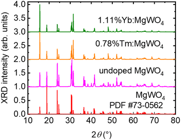 | ||
| Fig. 2 Measured powder X-ray diffraction (XRD) results for the Tm-doped, Yb-doped, and undoped MgWO4, and theoretical pattern for undoped MgWO4 based on PDF card #73-0562. | ||
In the pursuit of comprehending the structural modifications, the unit cell parameters of the three crystals were determined using the Le Bail method, Fig. 3.31 Initial unit cell parameters for the Le Bail refinement were extracted from.32 As anticipated, the obtained unit cell parameters and volume in Table 1 were larger for the doped crystals due to the larger ionic radii of the doping ions compared to Mg2+.27
| Crystal | a (Å) | b (Å) | c (Å) | β (°) | Volume (Å3) |
|---|---|---|---|---|---|
| MgWO4 | 4.686(7) | 5.674(1) | 4.927(8) | 90.710(7) | 131.036(5) |
| 0.78%Tm:MgWO4 | 4.693(1) | 5.677(1) | 4.931(6) | 90.741(6) | 131.382(9) |
| 1.11%Yb:MgWO4 | 4.695(1) | 5.677(9) | 4.932(7) | 90.753(7) | 131.492(3) |
The analysis of the rocking curves of the (030) reflection of MgWO4 and Yb:MgWO4 single crystals consists of two peaks for each crystal, corresponding to the Kα1 and Kα2 radiation from the X-ray source. For the evaluation of the crystals, we fitted both peaks to pseudo-Voigt functions. We have focused on Kα1 to extract conclusions from the measurements because of its higher intensity and found a full width at half maximum (FWHM) of the reflection to be 0.036° for MgWO4 and 0.031° for Yb:MgWO4, indicating the absence of significant difference between them and, due to the very small values, the high crystals quality.
3.2 Linear thermal expansion tensor
The thermal expansion of undoped MgWO4 was studied by recording XRD patterns at temperatures of 30, 50, 100, 200, 300, 400, 500 and 600 °C. The unit cell parameters (a, b, c, and β) were determined for each temperature using the Le Bail method. All lattice constants increased linearly with temperature (see Fig. 4). The volume of the unit cell (V) showed the same behaviour of increasing with temperature. | ||
| Fig. 4 (a–e) Linear dependence of lattice constants a, b, c, and β, and the volume of the unit-cell V of undoped MgWO4 crystal on temperature. | ||
The coefficients of the linear thermal expansion tensor of MgWO4 in the crystallophysic frame were obtained from the slope of the linear fits of the thermal evolution of the normalized unit cell parameters of this phase (see Fig. 5). This tensor is presented below:
 parallel to a axis,
  parallel to b axis and
  parallel to c* direction (c* = a × b), defining an orthogonal auxiliary frame.
 | ||
| Fig. 5 Normalized unit cell parameters of MgWO4versus temperature, (L = a, b, c, and c × cos(β − 90°)). | ||
By diagonalizing the previous tensor, the linear thermal expansion tensor of MgWO4 in the eigenframe is obtained:
The above values can be compared with our previous results obtained for Ho:MgWO4:7 the corresponding principal values were very close, 14.8, 8.09, and 5.07 × 10⁻⁶ K−1, respectively, and the rotation angle for the X1 axis was 37.35° for a monoclinic angle of β = 90.74°. This good agreement shades more doubt in the accuracy of the thermal expansion results reported in ref. 11 for Cr3+:MgWO4 using a dilatometer.
3.3 Orientation of the optical indicatrix
The monoclinic MgWO4 crystal is optically biaxial and as such possesses three principal optical directions (axes of the optical indicatrix) forming an orthogonal frame, which are known as Ng, Nm and Np, according to the notation of the corresponding refractive indexes ng > nm > np, where the subscript g denotes the highest refractive index, and p denotes the lowest one. One of the principal optical axes (in this case Nm) aligns with the C2 symmetry axis (the b axis), while the other two axes (Np and Ng) lie in the orthogonal mirror plane (the a–c plane). These principal axes define the principal planes and light polarized in or perpendicular to these planes does not experience polarization rotation upon propagation.Initially, the orientation of the principal optical axes for 1.25 at% Yb:MgWO4 was studied using transmission measurements with a polarizing microscope without information on the refractive index, hence they were denoted in a neutral manner, e.g. as XYZ.3 In this work, the rotation of the two principal optical axes lying in the a–c plane around the b-axis of the quasi-orthogonal abc frame was measured to be in the 36.4–37.1° range with an obvious error seen comparing with the monoclinic angle β given (see Fig. 2 in ref. 3). This error was rectified in ref. 23 in terms of rotation of the dielectric frame relative to the abc frame in the opposite direction with the two angles in the a–c plane being the same. The refractive indices were estimated then in this new frame from transmission measurements near 1.2 μm: np = 1.97, nm = 2.03, and ng = 2.13 with an error of ± 0.02 which enabled the assignment of Ng, Nm and Np.
Apart from the above controversial assignment of the orthogonal dielectric frame NpNmNg, notable disparities surfaced between the respective spectroscopic studies, encompassing variations not only in the absolute values of absorption and stimulated emission (SE) cross-sections for the three polarizations but also in the spectral shape, with similarities observed in absorption cross-sections and marked dissimilarity in SE cross-sections.3,20,23,24 The uncertainties led to the study and presentation of the Ho- and Er-doped MgWO4 spectroscopy in the crystallographic frame abc.7,30 For laser applications, however, spectroscopy in the orthogonal dielectric frame is desirable, because although this does not guarantee that cross-sections are maximized, more important is to ensure that polarization–rotation (waveplate) effects are absent in a laser emitting polarized radiation. The above discrepancies raised concerns, prompting a reassessment of the orientation of our doped MgWO4 crystals. Initial attempts using the dielectric frame orientation from23 yielded inaccurate results for our samples, Tm:MgWO4 and Yb:MgWO4, further justifying a thorough investigation into the relative frame orientation.
Subsequent to orienting our samples in the crystallographic abc frame through XRD, an examination of the principal optical axes was conducted using a crossed polarizers setup. The 2 mm thick samples, precisely cut perpendicular to the b direction, were positioned between two crossed Glan–Taylor polarizers. Through systematic rotation (with a goniometer with a precision of better than 1°), we successfully identified the principal optical axes in the a–c plane. Remarkably, one principal optical axis deviated by 6 ± 1° from the c direction (easily detectable as a natural edge of the grown crystal and confirmed by XRD), while the other was found to be exactly at 90° from the first zero transmission position. This consistent observation was replicated across all examined samples. The substantial deviation from the previously reported positions of these axes3,23 solidified our suspicions, creating the need for an additional confirmation.
To substantiate our findings, the next investigative step involved determining the refractive index at various angles using the same goniometer relative to the c crystallographic axis for our undoped MgWO4. The refractive index was measured with the prism coupler instrument using linearly polarized light at 633 nm and rotating the 2 mm thick polished plate around the b-axis. As shown in Fig. 7, knowing that Ng aligns with the highest refractive index while Np corresponds to the lowest refractive index, our measurements corroborated that Ng is located at 5° from the c direction when rotating the crystal clockwise, as depicted in Fig. 7(a) and (b). Importantly, these refractive index measurements align closely with the results obtained through the crossed polarizers setup, providing further confirmation for the revised orientation of the dielectric frame of our crystal.
Based on the reproducible results obtained with different samples, we confidently assert that the dielectric frame orientations reported in earlier studies on MgWO4-based crystals were inaccurate. Our proposed orientation, supported by a comprehensive study, diverges from previous reports. Earlier assertions placed Ng at 36.4° from the a direction,3,23 whereas our findings conclusively determine that Ng is located at 5 ± 1° from the c direction as shown in Fig. 8. Similarly, Np was previously reported at 37.1° from the a direction, but our results definitively place Np at 6 ± 1° from it. Thus, the previously reported substantial angles for MgWO4 between the crystallographic and optical axes are likely inaccurate. Fig. 8 illustrates the correct orientation we propose for the monoclinic MgWO4 crystal.
 | ||
| Fig. 8 Orientation of the optical indicatrix axes (Np, Nm, Ng) forming the orthogonal dielectric frame with respect to the crystallographic frame (a, b, c) in monoclinic MgWO4. | ||
Since laser elements are cut for propagation within one of the principal planes in order to avoid waveplate like polarization rotation effects, it is of practical interest to calculate the thermal expansion along the three dielectric axes of the newly determined frame. Matrix transformation gives αp = 12.71 × 10⁻⁶ K−1, αm = 8.26 × 10⁻⁶ K−1, and αg = 7.62 × 10⁻⁶ K−1.
3.4 Refractive index measurements
The refractive indices of the three different samples were measured with the prism coupler device at various angles relative to the crystallographic axes to confirm the consistency of the angle between the principal optical and crystallographic axes.As previously mentioned, using a 633 nm He–Ne laser, we verified that the Ng axis, which is associated with the highest refractive index, is located at an angle of 5 ± 1° from the c direction. Similarly, the Np axis, with the lowest refractive index, is situated at an angle of 95 ± 1° from the c direction, while the Nm axis is aligned parallel to the b direction. Table 2 presents the refractive indices along the three principal optical axes for the grown crystals at 633 nm.
| Crystal | n p | n m | n g |
|---|---|---|---|
| MgWO4 | 1.9880 | 2.0478 | 2.2027 |
| Tm:MgWO4 | 1.9889 | 2.0478 | 2.2002 |
| Yb:MgWO4 | 1.9894 | 2.0476 | 2.2011 |
3.5 Polarized Raman spectroscopy
Polarized Raman spectra of MgWO4 were previously presented in ref. 23 for Tm doping and in ref. 20 for Yb doping. To further characterize the doped crystals in the newly determined dielectric frame, we measured the polarized Raman spectra for both Tm3+:MgWO4 and Yb3+:MgWO4 at RT, as illustrated in Fig. 9. We used Porto's notation m(nk)![[l with combining macron]](https://www.rsc.org/images/entities/i_char_006c_0304.gif) , where ‘m’ and ‘
, where ‘m’ and ‘![[l with combining macron]](https://www.rsc.org/images/entities/i_char_006c_0304.gif) ’ indicate the propagation directions of the excitation and scattered light, respectively, while ‘n’ and ‘k’ represent the corresponding polarization states.
’ indicate the propagation directions of the excitation and scattered light, respectively, while ‘n’ and ‘k’ represent the corresponding polarization states.
 | ||
Fig. 9 Polarized Raman spectra for g(_)ḡ, m(_)![[m with combining macron]](https://www.rsc.org/images/entities/i_char_006d_0304.gif) , and p(_) , and p(_)![[p with combining macron]](https://www.rsc.org/images/entities/i_char_0070_0304.gif) of (a–c) Tm doped MgWO4 crystal and (d–f) Yb doped MgWO4 crystal. The excitation wavelength was 633 nm. of (a–c) Tm doped MgWO4 crystal and (d–f) Yb doped MgWO4 crystal. The excitation wavelength was 633 nm. | ||
In the monoclinic structure of MgWO4, there are two unit formulas per unit cell (Z = 2). According to factor group theory, 36 lattice modes are predicted at the centre of the Brillouin zone: Γ(k = 0) = 8Ag + 10Bg + 8Au + 10Bu. Of these, 18 modes are Raman-active (even modes Ag and Bg), and 18 are IR-active (odd modes Au and Bu). All the modes for MgWO4 were assigned in ref. 33.
In the Tm3+:MgWO4 and Yb3+:MgWO4 crystals, the most intense mode, occurring around 918 cm−1, corresponds to symmetric stretching W–O vibrations (ν1 mode, A1g symmetry) within the WO6 octahedra, while modes around 813 cm−1 and 711 cm−1 are assigned to asymmetric stretching (ν2 mode, Eg symmetry). Notably, a trend in peak intensities is evident. For instance, the peak at approximately 918 cm−1 (an Ag mode) is prominent in the pp, mm, and gg polarizations for different directions of excitation and scattered light, while it is less pronounced in other polarizations. This pattern is observed for several Ag mode peaks. Conversely, Bg modes, such as those around 813 cm−1 and 102 cm−1, are less pronounced in the pp, mm, and gg polarizations and clearer in other polarizations.
When comparing our Raman spectra of Yb:MgWO4 crystal with those reported in ref. 20 for m(_)![[m with combining macron]](https://www.rsc.org/images/entities/i_char_006d_0304.gif) configuration, notable differences in peak intensities are observed, particularly for the m(pg)
configuration, notable differences in peak intensities are observed, particularly for the m(pg)![[m with combining macron]](https://www.rsc.org/images/entities/i_char_006d_0304.gif) polarization configuration. In this work, m(pg)
polarization configuration. In this work, m(pg)![[m with combining macron]](https://www.rsc.org/images/entities/i_char_006d_0304.gif) shows unexpectedly high intensities across several Ag modes, comparable to m(pp)
shows unexpectedly high intensities across several Ag modes, comparable to m(pp)![[m with combining macron]](https://www.rsc.org/images/entities/i_char_006d_0304.gif) , while in the present measurements, m(pg)
, while in the present measurements, m(pg)![[m with combining macron]](https://www.rsc.org/images/entities/i_char_006d_0304.gif) yields much lower intensities. This discrepancy may indicate possible differences in crystal orientation but one cannot rule out the effect of different actual doping levels as well.
yields much lower intensities. This discrepancy may indicate possible differences in crystal orientation but one cannot rule out the effect of different actual doping levels as well.
3.6 Optical spectroscopy
Polarized optical absorption measurements of Tm3+:MgWO4 and Yb3+:MgWO4 at RT were performed in the newly determined dielectric frame. Fig. 10 depicts the obtained absorption cross-sections (σabs) for the Tm3+ and Yb3+ doped MgWO4 crystals, derived as σabs = αabs/NTm/Yb, where αabs is the measured absorption coefficient, and NTm and NYb are the actual Tm3+ and Yb3+ ion density respectively. The polished cubic samples had dimensions of 4.74 × 6.04 × 5.34 mm3 for Tm:MgWO4 and 2.86 × 3.57 × 2.56 mm3 for Yb:MgWO4, both aligned along the principal optical directions Np, Nm, and Ng, respectively.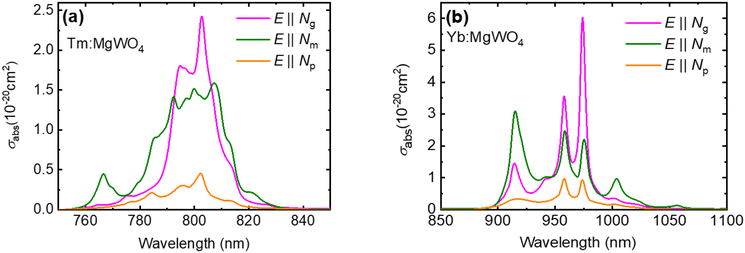 | ||
| Fig. 10 Absorption cross-sections, σabs, for light polarizations E||Ng, Nm, and Np for: (a) Tm3+ doped MgWO4 crystal and (b) Yb3+ doped MgWO4 crystal. | ||
High-brightness Ti–sapphire lasers or high-power AlGaAs diode lasers, emitting at approximately 800 nm, are suitable for pumping Tm3+-doped crystals due to the 3H6 → 3H4 transition of the Tm3+ ion. The maximum absorption cross-section (σabs) of Tm3+:MgWO4 amounts to 2.42 × 10⁻²⁰ cm2 at 803 nm, for light polarization E||Ng, and the corresponding absorption bandwidth (FWHM) is 15.5 nm, see Fig. 10(a). The σabs values are lower for the other principal polarizations. For E||Np, σabs = 0.45 × 10⁻²⁰ cm2 at 802 nm, and for E||Nm, σabs = 1.59 × 10⁻²⁰ cm2 at 807 nm. Notably for E||Nm, the absorption bandwidth is superior, FWHM = 20 nm. The absorption bands around 800 nm are in general significantly wider than what is typically observed in monoclinic double tungstates such as Tm3+:KLu(WO4)2. In the latter, for E||Nm the absorption cross-section (σabs) reaches a value of 5.95 × 10⁻²⁰ cm2 at 802 nm, yet the absorption bandwidth is 4 nm.34 Such broadband absorption behavior significantly simplifies the task of wavelength stabilization for AlGaAs pump laser diodes.
Fig. 10(b) shows the polarized absorption cross-section of Yb3+:MgWO4 around 1 μm, associated with the 2F7/2 → 2F5/2 electronic transition. Yb3+:MgWO4 features strongly anisotropic absorption properties. The maximum σabs amounts to 6.1 × 10⁻²⁰ cm2 at 974 nm, corresponding to a FWHM of 6 nm, for light polarization E||Ng, and this peak is associated with the so-called zero-phonon-line (ZPL) in absorption for the Yb3+ ion. When compared to Yb:KLu(WO4)2 with its σabs = 1.47 × 10⁻¹⁹ cm2 at 981.1 nm (E||Nm), the absorption in Yb3+:MgWO4 is roughly two times lower but has a broader FWHM (6 vs. 3.5 nm).32 For the other two polarizations, the absorption at this wavelength is lower: σabs = 2.24 × 10⁻²⁰ cm2 for E||Nm, and σabs = 0.94 × 10⁻²⁰ cm2 for E||Np. Notably, these maximum σabs values are almost twice as high as those reported for the isostructural Yb3+:ZnWO4, for which a maximum σabs of 2.73 × 10⁻²⁰ cm2 at 973.5 nm was measured with a FWHM = 6 nm, for E||Ng.35
In the case of Tm-doping, the results obtained in the XYZ frame22,24 are very close to our present results. Comparing with previously published absorption spectra, the difference is stronger for Yb-doping: the cross-sections obtained in ref. 3 in the XYZ frame are much lower although the spectral shapes look similar. The results in ref. 20 are very close to the present ones since they were obtained after an independent alignment in the dielectric frame NpNmNg between crossed polarizers and were thus independent of the exact frame location relative to the crystallographic frame.
The SE cross-section, σSE, was calculated using a combination of two methods. The first method employed the Füchtbauer–Ladenburg (F–L) equation36 based on the measured luminescence intensity W(λ):
The second approach used was based on the reciprocity method (RM),37 using the Stark splitting data determined in ref. 23 for the Tm3+ ion and in ref. 20 for the Yb3+ ion:
The maximum SE cross-section (σSE) for Tm3+:MgWO4 is observed at 1876 nm for E||Nm, amounting to 3.33 × 10⁻²⁰ cm2, see Fig. 11(a). For Yb3+:MgWO4, the maximum σSE reaches 6.63 × 10⁻²⁰ cm2 at 1058 nm, again for E||Nm, while for E||Ng and E||Np, σSE at the same wavelength drops to 1.98 × 10⁻²⁰ and 0.54 × 10⁻²⁰ cm2, respectively. Notably, for E||Ng, maximum σSE occurs at 974 nm, reaching 5.93 × 10⁻²⁰ cm2, which is close to the maximum value observed for E||Nm, see Fig. 11(b). For both dopant ions, a strong anisotropy of the SE cross-section is observed, implying a natural selection of a linear polarization for laser emission.
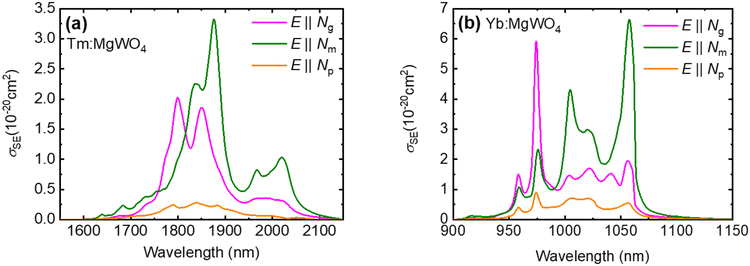 | ||
| Fig. 11 Stimulated emission cross-sections, σSE, for the principal light polarizations E||Np, Nm, and Ng of (a) Tm3+ doped MgWO4 crystal and (b) Yb3+ doped MgWO4 crystal. | ||
The obtained SE cross-sections for Tm3+:MgWO4, are higher for the two dominating polarizations (E||Ng and E||Nm) than the previously obtained values in what was thought to be the dielectric frame, both when denoted as XYZ24 and as NpNmNg.23 For Yb3+:MgWO4, the difference compared to the XYZ assumption3 is much larger, showing in roughly two times higher values and very different shapes, whereas minor discrepancies are observed comparing with results reported in ref. 20 in the NpNmNg frame.
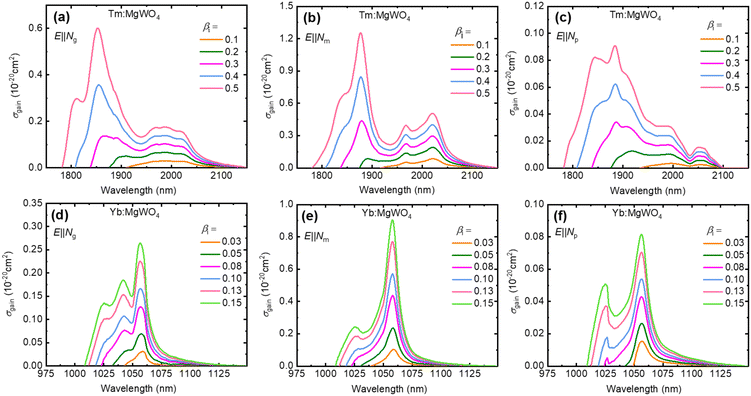
For quasi-3-level laser systems the expected oscillation wavelength can be estimated from the corresponding gain cross-sections, σgain. The gain profiles for Tm3+ and Yb3+ doped MgWO4 were calculated using the relation σgain = βiσSE − (1 − βi)σabs, where βi = N2/NRE represents the population inversion ratio, i.e. the ratio of dopant ions in the excited state, N2, to the total ion density NRE. Here, N2 corresponds to the upper laser states 3F4 for Tm3+ and 2F5/2 for Yb3+.
The gain spectra associated with the 3F4 → 3H6 transition in Tm3+:MgWO4 are displayed in Fig. 12(a)–(c) for the three principal light polarizations E||Ng, Nm, Np. The maximum σgain is observed for the E||Nm polarization, with a local maximum at ∼2020 nm at lower inversion ratios (βi < 0.3), which suggest a linearly polarized laser output along Nm at wavelengths exceeding 2 μm. This wavelength range is accessible due to the pronounced Stark splitting of the Tm3+ ground-state (3H6) in MgWO4, ΔE(3H6) = 633 cm−1,23 an important advantage over the monoclinic double tungstates such as Tm3+:KLu(WO4)2 when it comes to mode-locking in the sub-100 fs regime in relation to structured water vapor air absorption below 2 μm.21,25 No such local maximum is seen for the other strong polarization E||Ng. One of these two polarizations will be naturally selected for any principal cut of the laser element. All previous laser studies have been based on the strongest E||Nm output polarization (sometimes designated as E||Y).
The polarized σgain spectra corresponding to the 2F5/2 → 2F7/2 transition in Yb3+:MgWO4 are shown in Fig. 12(d)–(f). Again, the E||Nm polarization yields the highest σgain centered around 1057 nm. This is consistent with previously reported linear output polarization from Yb:MgWO4 lasers. The only occasion when E||Ng was utilized was in a mode-locked laser,21 just to confirm that its performance is inferior notwithstanding the superior absorption efficiency, cf.Fig. 11(b). Note that the weakest polarization E||Np has never been studied in a Tm or Yb lasers based on MgWO4. It can be only imposed by a Brewster angle of the laser element surfaces and is potentially interesting only in Q-switched lasers to obtain higher output energies and peak powers.
Compared to previously reported σgain calculations for both Tm3+ and Yb3+ doped MgWO4 crystals, our results are higher20,22,24 or much higher and different compared to ref. 3 where the XYZ frame was used; note also that there shall be typographical errors in Fig. 3 of ref. 20 where these cross-sections for Yb3+:MgWO4 shall be actually ten times higher.
The luminescence lifetimes (τlum) of the 3F4 state of Tm3+ and the 2F5/2 state of Yb3+ in MgWO4 were determined through temporal decay measurements. For Tm3+ ions excited at 793 nm, the luminescence decay at 1878 nm was monitored, see Fig. 13(a), revealing a lifetime of 1.81 ms. This value is comparable to the previously reported lifetime for Tm3+:MgWO4, τlum = 1.93 ms (ref. 24) and Tm3+,Li+:ZnWO4, τlum = 2.08 ms.38 For Yb3+:MgWO4, the lifetime was derived by exciting Yb3+ at 966 nm and monitoring the luminescence at 1057 nm, see Fig. 13(b), yielding a decay constant of 388 μs. This result aligns with earlier measurements for Yb3+:MgWO4 (366 μs),3 and Yb3+,Li+:ZnWO4 (367 μs).39 Notably, the decay curves exhibited a single exponential pattern, consistent with the presence of a single site for both Tm3+ and Yb3+ ions in MgWO4.
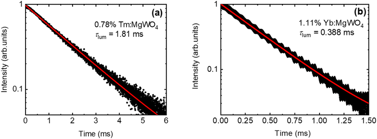 | ||
| Fig. 13 Decay curves of (a) the Tm3+ 3F4 state and (b) the Yb3+ 2F5/2 state in MgWO4: symbols – experimental data, solid lines – single-exponential fits. | ||
Conclusions
In summary, we have successfully grown monoclinic MgWO4 crystals, both undoped and doped with Tm3+ and Yb3+ ions, using K2W2O7 as a solvent for the first time. This method significantly reduced the growth time to 15 days, which is half the duration reported in previous studies for crystals of similar size. We also determined the orientation of the principal optical axes relative to the crystallographic axes and obtained polarized absorption, stimulated emission, and gain cross-section spectra for the relevant transitions of the Tm3+ and Yb3+ ions, as well as polarized Raman spectra.Our analysis revealed that the angle between the principal optical axis Ng, corresponding to the highest refractive index, and the crystallographic axis c is only 5 ± 1°. This finding corrects a recurring error in prior studies regarding the orientation of MgWO4 crystals. The partial consistency of our absorption and stimulated emission cross-section data with some of the literature further supports that previous misidentifications were likely due to incorrect crystallographic axes orientation. However, it is difficult to evaluate the impact of wrong active element orientation in previous laser experiments carried out with Tm3+:MgWO4 and Yb3+:MgWO4 crystals. They were all based on the best E||Nm polarization.3,7,18–26 The direction Nm is in fact easy to identify since in MgWO4 it is perpendicular to a natural crystal surface, however, the crystal cut utilizing this polarization might be still erroneous. A wrong cut for the propagation direction might result in lower gain cross-sections even though the propagating beam in the crystal is an eigen polarization, when this happens in a principal plane containing the Nm principal optical axis.
Summarizing, the doped MgWO4 crystals exhibit broad spectral bands and pronounced polarization anisotropy in their transition cross-sections, and both with Tm and Yb active ions are very promising for high-power sub-100 fs pulse generation from mode-locked lasers.
Data availability
The data supporting our findings are available from the corresponding author upon reasonable request.Conflicts of interest
There are no conflicts to declare.Acknowledgements
This work was financially supported by project PID2022-141499OB-100, funded by MICIU/AEI/10.13039/501100011033/ and by FEDER/UE.References
- J. Zhang, J. Pan, J. Yin, J. Wang, J. Pan, H. Chen and R. Mao, Structural investigation and scintillation properties of Cd1−xZnxWO4 solid solution single crystals, CrystEngComm, 2015, 17, 3503–3508 RSC.
- X. Wang, Z. Fan, H. Yu, H. Zhang and J. Wang, Characterization of ZnWO4 Raman crystal, Opt. Mater. Express, 2017, 7, 1732–1744 Search PubMed.
- L. Zhang, W. Chen, J. Lu, H. Lin, L. Li, G. Wang, G. Zhang and Z. Lin, Characterization of growth, optical properties, and laser performance of monoclinic Yb:MgWO4 crystal, Opt. Mater. Express, 2016, 6, 1627–1634 Search PubMed.
- J. Chen, L. Dong, F. Liu, H. Xua and J. Liu, Investigation of Yb:CaWO4 as a potential new self-Raman laser crystal, CrystEngComm, 2021, 23, 427–435 Search PubMed.
- P. A. Popov, S. A. Skrobov, E. V. Zharikov, D. A. Lis, K. A. Subbotin, L. I. Ivleva, V. N. Shlegel', M. B. Kosmyna and A. N. Shekhovtsov, Investigation of the thermal conductivity of tungstate crystals, Crystallogr. Rep., 2018, 63, 111–116 CrossRef CAS.
- D. Kumar, B. P. Singh, M. Srivastava, A. Srivastava, P. Singh, A. Srivastava and S. K. Srivastava, Structural and photoluminescence properties of thermally stable Eu3+ activated CaWO4 nanophosphor via Li+ incorporation, J. Lumin., 2018, 203, 507–514 CrossRef CAS.
- L. Zhang, P. Loiko, J. M. Serres, E. Kifle, H. Lin, G. Zhang, E. Vilejshikova, E. Dunina, A. Kornienko, L. Fomicheva, U. Griebner, V. Petrov, Z. Lin, W. Chen, K. Subbotin, M. Aguiló, F. Díaz and X. Mateos, Growth, spectroscopy and first laser operation of monoclinic Ho3+:MgWO4 crystal, J. Lumin., 2019, 213, 316–325 Search PubMed.
- E. Cavalli, A. Belletti and M. G. Brik, Optical spectra and energy levels of the Cr3+ ions in MWO4 (M=Mg, Zn, Cd) and MgMoO4 crystals, J. Phys. Chem. Solids, 2008, 69, 29–34 CrossRef CAS.
- P. F. Schofield, K. S. Knight and G. Cressey, Neutron powder diffraction study of the scintillator material ZnWO4, J. Mater. Sci., 1996, 31, 2873–2877 CrossRef CAS.
- V. B. Kravchenko, Crystal structure of the monoclinic form of magnesium tungstate MgWO4, J. Struct. Chem., 1969, 10, 139–140 Search PubMed.
- L. Zhang, Y. Huang, S. Sun, F. Yuan, Z. Lin and G. Wang, Thermal and spectral characterization of Cr3+:MgWO4—a promising tunable laser material, J. Lumin., 2016, 169, 161–164 CrossRef CAS.
- E. N. Sota, F. Che Ros and J. Hassan, Synthesis and characterisation of AWO4 (A = Mg, Zn) tungstate ceramics, J. Phys.: Conf. Ser., 2018, 1083, 012002 Search PubMed.
- S. Wannapop, T. Thongtem and S. Thongtem, Photoemission and energy gap of MgWO4 particles connecting as nanofibers synthesized by electrospinning–calcination combinations, Appl. Surf. Sci., 2012, 258, 4971–4976 CrossRef CAS.
- J. Meng, T. Chen, X. Wei, J. Li and Z. Zhang, Template-free hydrothermal synthesis of MgWO4 nanoplates and their application as photocatalysts, RSC Adv., 2019, 9, 2567–2571 RSC.
- F. A. Danevich, D. M. Chernyak, A. M. Dubovik, B. V. Grinyov, S. Henry, H. Kraus, V. M. Kudovbenko, V. B. Mikhailik, L. L. Nagornaya, R. B. Podviyanuk, O. G. Polischuk, I. A. Tupitsyna and Y. Y. Vostretsov, MgWO4–A new crystal scintillator, Nucl. Instrum. Methods Phys. Res., Sect. A, 2009, 608, 107–115 CrossRef CAS.
- V. B. Mikhailik, H. Kraus, V. Kapustyanyk, M. Panasyuk, Y. Prots, V. Tsybulskyi and L. Vasylechko, Structure, luminescence and scintillation properties of the MgWO4–MgMoO4 system, J. Phys.: Condens. Matter, 2008, 20, 365219 CrossRef.
- L. Li, Y. Yu, G. Wang, L. Zhang and Z. Lin, Crystal growth, spectral properties and crystal field analysis of Cr3+:MgWO4, CrystEngComm, 2013, 15, 6083–6089 RSC.
- J. Lu, H. Lin, G. Zhang, B. Li, L. Zhang, Z. Lin, Y.-F. Chen, V. Petrov and W. Chen, Direct generation of an optical vortex beam from a diode-pumped Yb:MgWO4 laser, Laser Phys. Lett., 2017, 14, 085807 CrossRef.
- H. Lin, G. Zhang, L. Zhang, Z. Lin, F. Pirzio, A. Agnesi, V. Petrov and W. Chen, Continuous-wave and SESAM mode-locked femtosecond operation of a Yb:MgWO4 laser, Opt. Express, 2017, 25, 11827–11832 CrossRef CAS PubMed.
- P. Loiko, M. Chen, J. M. Serres, M. Aguiló, F. Díaz, H. Lin, G. Zhang, L. Zhang, Z. Lin, P. Camy, S.-B. Dai, Z. Chen, Y. Zhao, L. Wang, W. Chen, U. Griebner, V. Petrov and X. Mateos, Spectroscopy and high-power laser operation of a monoclinic Yb3+:MgWO4 crystal, Opt. Lett., 2020, 45, 1770–1773 CrossRef PubMed.
- Y. Wang, W. Chen, M. Mero, L. Zhang, H. Lin, Z. Lin, G. Zhang, F. Rotermund, Y. J. Cho, P. Loiko, X. Mateos, U. Griebner and V. Petrov, Sub-100 fs Tm:MgWO4 laser at 2017 nm mode locked by a graphene saturable absorber, Opt. Lett., 2017, 42, 3076–3079 CrossRef CAS PubMed.
- P. Loiko, J. M. Serres, X. Mateos, M. Aguilo, F. Diaz, L. Zhang, Z. Lin, H. Lin, G. Zhang, K. Yumashev, V. Petrov, U. Griebner, Y. Wang, S. Y. Choi, F. Rotermund and W. Chen, Monoclinic Tm3+:MgWO4: a promising crystal for continuous-wave and passively Q-switched lasers at ∼2 μm, Opt. Lett., 2017, 42, 1177–1180 CrossRef CAS PubMed.
- P. Loiko, Y. Wang, J. M. Serres, X. Mateos, M. Aguiló, F. Díaz, L. Zhang, Z. Lin, H. Lin, G. Zhang, E. Vilejshikova, E. Dunina, A. Kornienko, L. Fomicheva, V. Petrov, U. Griebner and W. Chen, Monoclinic Tm:MgWO4 crystal: Crystal-field analysis, tunable and vibronic laser demonstration, J. Alloys Compd., 2018, 763, 581–591 CrossRef CAS.
- L. Zhang, H. Lin, G. Zhang, X. Mateos, J. M. Serres, M. Aguiló, F. Díaz, U. Griebner, V. Petrov, Y. Wang, P. Loiko, E. Vilejshikova, K. Yumashev, Z. Lin and W. Chen, Crystal growth, optical spectroscopy and laser action of Tm3+-doped monoclinic magnesium tungstate, Opt. Express, 2017, 25, 3682–3693 Search PubMed.
- L. Wang, W. Chen, Y. Zhao, Y. Wang, Z. Pan, H. Lin, G. Zhang, L. Zhang, Z. Lin, J. E. Bae, T. G. Park, F. Rotermund, P. Loiko, X. Mateos, M. Mero, U. Griebner and V. Petrov, Single-walled carbon-nanotube saturable absorber assisted Kerr-lens mode-locked Tm:MgWO4 laser, Opt. Lett., 2020, 45, 6142–6145 CrossRef CAS PubMed.
- E. Kifle, P. Loiko, J. R. Vazquez De Aldana, C. Romero, V. Llamas, J. M. Serres, M. Aguilo, F. Diaz, L. Zhang, Z. Lin, H. Lin, G. Zhang, V. Zakharov, A. Veniaminov, V. Petrov, U. Griebner, X. Mateos and W. Chen, Low-loss fs-laser-written surface waveguide lasers at >2 μm in monoclinic Tm3+:MgWO4, Opt. Lett., 2020, 45, 4060–4063 Search PubMed.
- R. D. Shannon and C. T. Prewitt, Revised values of effective ionic radii, Acta Crystallogr., Sect. B, 1970, 26, 1046–1048 CrossRef CAS.
- A. Lupei, V. Lupei, C. Gheorghe, L. Gheorghe and A. Achim, Multicenter structure of the optical spectra and the charge-compensation mechanisms in Nd: SrWO4 laser crystals, J. Appl. Phys., 2008, 104, 083102 Search PubMed.
- W. Kolbe, K. Petermann and G. Huber, Broadband emission and laser action of Cr3+ doped zinc tungstate at 1 μm wavelength, IEEE J. Quantum Electron., 1985, 21, 1596–1599 Search PubMed.
- L. Zhang, L. Basyrova, P. Loiko, P. Camy, Z. Lin, G. Zhang, S. Slimi, R. M. Solé, X. Mateos, M. Aguiló, F. Díaz, E. Dunina, A. Kornienko, U. Griebner, V. Petrov, L. Wang and W. Chen, Growth, structure, and polarized spectroscopy of monoclinic Er3+:MgWO4 crystal, Opt. Mater. Express, 2022, 12, 2028–2040 Search PubMed.
- A. Le Bail, Whole powder pattern decomposition methods and applications: A retrospection, Powder Diffr., 2005, 20, 316–326 Search PubMed.
- N. J. Dunning and H. D. Megaw, The crystal structure of magnesium tungstate, Trans. Faraday Soc., 1946, 42, 705–709 Search PubMed.
- J. Ruiz-Fuertes, D. Errandonea, S. López-Moreno, J. González, O. Gomis, R. Vilaplana, F. J. Manjón, A. Muñoz, P. Rodríguez-Hernández, A. Friedrich, I. A. Tupitsyna and L. L. Nagornaya, High-pressure Raman spectroscopy and lattice-dynamics calculations on scintillating MgWO4: Comparison with isomorphic compounds, Phys. Rev. B: Condens. Matter Mater. Phys., 2011, 83, 214112 Search PubMed.
- V. Petrov, M. C. Pujol, X. Mateos, Ò. Silvestre, S. Rivier, M. Aguiló, R. M. Solé, J. Liu, U. Griebner and F. Díaz, Growth and properties of KLu(WO4)2, and novel ytterbium and thulium lasers based on this monoclinic crystalline host, Laser Photonics Rev., 2007, 1, 179–212 CrossRef CAS.
- G. Z. Elabedine, K. Subbotin, P. Loiko, Y. Zimina, S. Pavlov, A. Titov, P. Camy, A. Braud, R. M. Solé, M. Aguiló, F. Díaz, W. Chen, X. Mateos and V. Petrov, Monoclinic Yb3+,Li+:ZnWO4 – efficient broadly emitting laser material, Proc. SPIE, 2024, 12864, 128640K Search PubMed.
- B. F. Aull and H. P. Jenssen, Vibronic interactions in Nd:YAG resulting in nonreciprocity of absorption and stimulated emission cross sections, IEEE J. Quantum Electron., 1982, 18, 925–930 Search PubMed.
- S. A. Payne, L. L. Chase, L. K. Smith, W. L. Kway and W. F. Wyers, Infrared cross-section measurements for crystals doped with Er3+, Tm3+, and Ho3+, IEEE J. Quantum Electron., 1992, 28, 2619–2630 Search PubMed.
- G. Z. Elabedine, K. Subbotin, P. Loiko, Z. Pan, Y. Zimina, K. Kuleshova, A. Titov, A. Nady, P. Camy, A. Braud, R. M. Solé, M. Aguiló, F. Díaz, W. Chen, W. Chen, X. Mateos, X. Mateos and V. Petrov, Monoclinic Tm3+:ZnWO4: Novel 2 μm laser crystal, Advanced Solid State Lasers Conference, Tacoma (WA), USA, Oct 8, 2023, vol. 12, OPTICA Online Technical Digest, paper AW1A.5 Search PubMed.
- A. Volokitina, S. P. David, P. Loiko, K. Subbotin, A. Titov, D. Lis, R. M. Solé, V. Jambunathan, A. Lucianetti, T. Mocek, P. Camy, W. Chen, U. Griebner, V. Petrov, M. Aguiló, F. Díaz and X. Mateos, Monoclinic zinc monotungstate Yb3+,Li+:ZnWO4: Part II. Polarized spectroscopy and laser operation, J. Lumin., 2021, 231, 117811 CrossRef CAS.
Footnote |
| † Serra Húnter Fellow, Spain. |
| This journal is © The Royal Society of Chemistry 2025 |


