
 Open Access Article
 This Open Access Article is licensed under a
 Creative Commons Attribution 3.0 Unported Licence
Zn(II)-metallo-photoantibiotics: experimental and computational approach identifying a therapeutic role for antibacterial and antibiofilm applications†
Rajesh
Kushwaha‡
 a,
Sangeeta
Kumari‡
b,
Arya
Mishra
a,
Anjali
Upadhyay
b,
Archana
Rai
c,
Malay
Nayak
b,
Sudip
Mukherjee
*b and
Samya
Banerjee
*a
a,
Sangeeta
Kumari‡
b,
Arya
Mishra
a,
Anjali
Upadhyay
b,
Archana
Rai
c,
Malay
Nayak
b,
Sudip
Mukherjee
*b and
Samya
Banerjee
*a
aDepartment of Chemistry, Indian Institute of Technology (BHU), Varanasi, Uttar Pradesh 221005, India. E-mail: samya.chy@itbhu.ac.in
bSchool of Biomedical Engineering, Indian Institute of Technology (BHU), Varanasi, Uttar Pradesh 221005, India. E-mail: sudip.bme@iitbhu.ac.in
cDivision of Organic Chemistry, CSIR-National Chemical Laboratory, Pune 411008, India
First published on 10th June 2025
Abstract
The antibacterial profiles of curcumin-based novel Zn(II)-metallo-photoantibiotics against E. coli and B. subtilis are reported. In silico studies indicated their ROS generation capacity and binding interaction with bacterial proteins. Therapeutic results indicated the advantages of these Zn(II)-metallo-photoantibiotics in antibacterial photodynamic therapy.
Antimicrobial resistance (AMR) is one of this century's most pressing global health challenges.1 Current antibiotics, predominantly based on organic molecules, are increasingly failing due to the rapid evolution of resistant bacterial strains.2,3 Traditional approaches, including clinical antibiotic modification or developing new organic compounds, often fail due to their single-target mechanisms.2,3 Henceforth, researchers have been investigating next-generation antibiotics and novel antibacterial therapies to tackle these challenges. Recently, antibacterial photodynamic therapy (aPDT) has shown great potential to overcome drug resistance problems due to its different mechanisms of action.4,5 Due to the multi-targeting ability of the generated ROS, aPDT can potentially damage a wide range of bacterial strains regardless of their drug resistance.4,5 Also, developing resistance to aPDT is unlikely due to shorter drug-light interaction, minimal dark toxicity, extensive cellular damage preventing cross-generational adaptation, etc.5,6 Moreover, aPDT selectively targets bacterial infection sites without harming healthy tissues.5,6 Recently, metal complexes have caught attention as antibacterial agents. The Community for Open Antimicrobial Drug Discovery recently reported that metal complexes have outstanding hit rates (10×) against critical bacteria compared to organic molecules and can overcome the AMR problem.3,7 Therefore, various transition metal complexes, including Ir(III), Ru(II), Os(II), Pt(II), and Re(I), have caught significant attention as promising aPDT agents.5,7 However, concerns about the inherent toxicity of heavy metals have prompted a shift toward exploring 3d metal-based alternatives.8 Among 3d metals, zinc is essential, required for several physiological and intracellular biochemical functions.9 Although Zn(II) is highly biocompatible and shows strong antibacterial potential with phthalocyanine- and porphyrins, Zn(II) complexes remain scarcely explored for aPDT applications.10 Herein, we developed novel photo-responsive Zn(II) complexes, [Zn(cur)(pybi)Cl] (Zn1), [Zn(cur)(Me-pybi)Cl] (Zn2), and [Zn(cur)(pen-pybi)Cl] (Zn3), where acac = acetyleacetonate; cur = curcumin anion; pybi = 2-(pyridin-2-yl)-1H-benzo[d]imidazole; Me-pybi = 1-methyl-2-(pyridin-2-yl)-1H-benzo[d]imidazole; pen-pybi = 1-pentyl-2-(pyridin-2-yl)-1H-benzo[d]imidazole (Fig. 1a) aimed at harnessing visible light for antibacterial therapy. Curcumin has been utilized due to visible light absorption/emission properties and its ability to produce ROS upon light irradiation, causing oxidative stress and damaging bacterial components.11 Benzimidazole has been used for its well-established antimicrobial properties.12 The alkyl substitutions at the benzimidazole moiety were strategically introduced to modulate lipophilicity and photophysical behavior.12
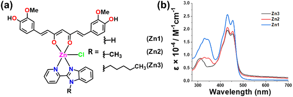 | ||
Fig. 1 (a) Chemical structures of Zn1–Zn3. (b) UV-Vis spectra of Zn1–Zn3 in DMSO![[thin space (1/6-em)]](https://www.rsc.org/images/entities/char_2009.gif) :water (1:9 v:v) solution. :water (1:9 v:v) solution. | ||
Zn1–Zn3 were synthesized (Scheme S1, ESI†) and characterized thoroughly (Fig. S1–S10, ESI†). HPLC analysis of Zn1–Zn3 ensured >97% purity (Fig. S11–S13, ESI†). The UV-Vis spectra of Zn1–Zn3 showed a strong curcumin-based absorption near 430 nm (Fig. 1b), indicating their potential to sense visible light for antibacterial responses.11Zn1–Zn3 displayed a curcumin-based emission maximum at ca. 545 nm (λex = 430 nm) (Fig. S14, ESI†).11 The octanol–water partition coefficients (logPo/w value) of Zn1–Zn3 were ca. +1.6 to +2.5, indicating their lipophilic characteristics. Zn1–Zn3 showed excellent photostability under light exposure, with no notable spectral changes up to 1 h in DMSO:water (1:9, v/v) (Fig. S15, ESI†).
DFT calculations were performed on Zn1–Zn3 to rationalize their electronic structures and photophysical properties. The optimized structures of Zn1–Zn3 showed a distorted square pyramidal structure (Fig. 2a and Fig. S16, ESI†). Furthermore, the electronic structures were characterized based on their FMOs (Fig. S17, ESI†). The analysis of the FMOs revealed that the HOMO−1 was localized on the Zn(II) center and Cl. The HOMO and LUMO+1 were distributed on curcumin with slight involvement of Cl. The LUMO was on the benzimidazole moieties. The Eg = ELUMO − EHOMO remained similar for Zn1–Zn3 (Table S1 and Fig. S18a, ESI†). The calculated UV-Vis spectra of Zn1–Zn3 were found to be in accordance with the experimental data (Fig. S18b, ESI†). SOMO plots and spin density plots at the triplet excited state of Zn1–Zn3 (Fig. 2b and Fig. S19, S20, ESI†) revealed that the unpaired electrons are localized on the curcumin mainly.
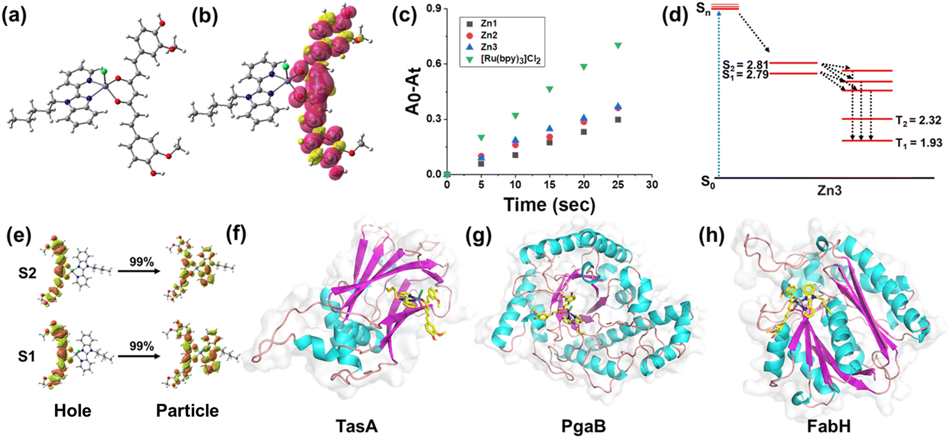 | ||
| Fig. 2 (a) Optimized structure of Zn3. (b) Spin density distribution on Zn3 at the adiabatic triplet state. (c) 1O2 generation by Zn1–Zn3 (5 μM) with respect to time in DMSO:water (2:98 v:v) solution. (d) Calculated excited state energy and possible ISC channels of Zn3 (energy in eV). (e) NTOs for S0 → S1/S2 transition for Zn3. (e)–(h) Domain architecture of the Zn3-docked structure with (f) TasA; (g) PgaB; and (h) FabH. | ||
The absorbance within the visible range and high photostability of Zn1–Zn3 inspired us to investigate them as aPDT agents. In aPDT, 1O2 production plays a critical role in causing oxidative stress, disrupting membranes, denaturing proteins, and damaging DNA.4,5 The 1O2 generation ability of Zn1–Zn3 was determined using diphenyl isobenzofuran (DPBF) as a 1O2 probe.12 The absorbance of DPBF remained unchanged in the presence of Zn1–Zn3 (Fig. S21, ESI†) under dark conditions, indicating no detectable 1O2 generation. However, there was a gradual decrease in the DPBF-based absorption peaks when light was exposed, exhibiting light-triggered 1O2 generation (Fig. S22, ESI†). The 1O2 quantum yield (ΦΔ) of Zn1–Zn3 was 0.07–0.11 with [Ru(bpy)3]Cl2 as standard (ΦΔ = 0.22) (Fig. 2c and Fig. S23, ESI†).12 To gain insight into the underlying PDT mechanism, we further investigated the excited singlet/triplet state properties of Zn3 (Tables S2 and S3, ESI†). The obtained result revealed several triplet states with higher energy than 0.98 eV, the energy required to convert cellular 3O2 into reactive 1O2 (Table S3, ESI†).13 Usually, the intersystem crossing (ISC) efficiently occurs when there is a small energy gap (ΔES1–Tn < 0.3 eV) between the S1 and Tn states.13 Thus, based on excitation energy analysis, the possible channels for ISC transitions of Zn3 are given in Fig. 2d. The NTO analysis of these transitions indicated the involvement of 1LLCT to 3LLCT transitions (Fig. 2e and Fig. S24, ESI†). It is essential to mention that Zn(II) complexes possess a d10 configuration that restricts the excited states’ internal quenching by low-lying d–d* states.13 Thus, low-cost and biocompatible Zn(II) complexes might be attractive aPDT agents.
Molecular docking (MD) has emerged as a powerful tool for identifying the therapeutic role of drugs in their antibacterial activity. We performed molecular docking with Zn1–Zn3 and three crucial target proteins, such as TasA (PDB ID: 5OF2) of gram-positive (B. subtilis) and PgaB (PDB ID: 4P7O) and FabH (PDB ID: 1EBL) of gram-negative (E. coli) bacteria (Fig. 2f–h and Fig. S25, ESI†).14 TasA is a major structural protein in the biofilms of B. subtilis, contributing to stability and resilience.14 PgaB plays a crucial role in the biofilm formation process by deacetylating poly-β-1,6-N-acetyl-D-glucosamine.14 FabH is a key enzyme in bacterial fatty acid biosynthesis, essential for cell membrane formation. Its inhibition can disrupt lipid metabolism.14 Thus, the complexes displaying favourable binding interactions against these crucial proteins may show promising antibacterial and antibiofilm activity. The MD result of Zn1–Zn3 with these target proteins underlined their notable binding interactions with the key amino acids via hydrogen bonding, pi interaction, van der Waals interaction, etc. (Fig. S26–28, ESI†). The docking results are summarized in Table S4, ESI.† Against PgaB and TasA, Zn3 exhibited higher binding efficacy than Zn1 and Zn2 due to more H-bonding. In the case of FabH, Zn1 showed better binding efficacy than Zn2 and Zn3. The interactions of Zn1–Zn3 with different receptor surfaces of these proteins are depicted in Fig. S29–S37, ESI.† Overall, Zn1–Zn3 exhibited notable binding affinities (−8.61 kcal mol−1) and favourable interactions with these key bacterial proteins, surpassing the accepted threshold (−6.0 kcal mol−1) for drug-like binding in docking studies, suggesting their potential antibacterial and antibiofilm effects.15
The encouraging visible light-triggered 1O2 generation ability and molecular docking results of Zn1–Zn3 pushed us to determine their light-activated antibacterial properties against E. coli and B. subtilis. The time-dependent growth-inhibition kinetics of Zn1–Zn3 against E. coli and B. subtilis indicated that Zn3 significantly inhibited the bacterial growth at 25 μM and 50 μM up to 24 h under light exposure (400–700 nm, 10 J cm−2) (Fig. 3a, b and Fig. S38, ESI†). In the dark, Zn1–Zn3 showed no inhibition of bacterial growth. Preliminary screening studies revealed that Zn1 and Zn2 did not show comparable antibacterial efficacy to Zn3. Hence, Zn3 was selected for further in vitro and in vivo antibacterial studies.
 | ||
| Fig. 3 (a) and (b) Kinetics growth curve of Zn3-treated E. coli (a) and B. subtilis (b) under light and dark conditions. (c) and (d) In-cell ROS production in (c) E. coli and (d) B. subtilis induced by Zn3 (50 μM) under light and dark conditions. | ||
Zn3 showed minimum inhibitory concentration value, i.e., MIC90 = 21.89 μM in E. coli and MIC90 = 45.5 μM in B. subtilis (Fig. S39 ESI†). In a zone inhibition study, upon light irradiation, Zn3 demonstrated a larger bacterial zone inhibition against E. coli (at 25 μM, 50 mm, and at 50 μM, 50 mm) and B. subtilis (at 25 μM, 23 mm and at 50 μM, 27 mm) (Fig. S40, ESI†). However, in the dark, Zn3 showed no bacterial zone inhibition at 25 μM and 50 μM (Fig. S40, ESI†). Moreover, Fig. S41a and b ESI† shows that Zn3 inhibited E. coli and B. subtilis colony formation under light exposure compared to the dark.
DCFH-DA assay assessed in-cell ROS generation by Zn3, where nonfluorescent DCFH-DA was converted to green-emissive DCF upon reacting with ROS.12 The obtained result, shown in Fig. 3c and d, revealed that the green fluorescence intensity in the Zn3 (50 μM) + light-treated groups was found to be 2- to 3-fold higher than under dark conditions in both E. coli and B. subtilis (Fig. 3c and d). This result revealed that Zn3 induced significantly higher ROS within bacteria than the control and positive control groups under light exposure.
As a defense strategy against antimicrobial agents, pathogenic bacteria frequently develop biofilms.15 To validate antibiofilm efficacy, Zn3 was coated on polydimethylsiloxane (PDMS) disks (used in medical devices and biomedical applications), and the biofilm formation was evaluated using crystal violet staining.16Zn3-coated PDMS exhibited the strongest biofilm inhibition under light treatment compared to untreated controls and the positive control group, including the dark conditions (Fig. 4a). FE-SEM was used to verify the morphological change of E. coli biofilms under both treatment conditions. As shown in Fig. 4b and Fig. S42 ESI,† the control group under dark and light conditions demonstrated the best integrity of the bacterial membrane with a higher number of closely attached bacteria, forming an intact biofilm. However, Zn3 coating under dark and light conditions drastically reduced total bacterial attachment, causing severe rupture on the surface of the bacterial wall with a notable area of shrinkage. The effect of Zn3 on the destruction of biofilms was also investigated using confocal laser scanning microscopy. The Zn3-coated group effectively eliminated an E. coli biofilm under light exposure, similar to the ciprofloxacin (positive control) group (Fig. 4c and Fig. S43, ESI†). In contrast, the uncoated and dark groups showed no such reduction in biofilms. Quantification using ImageJ software revealed a higher number of GFP-E. coli in the control group compared to the positive control (P.C.) and Zn3-light-treated groups (Fig. S43, ESI†). The fluorescence intensity of dead cells was significantly higher in both the positive control and Zn3-light-treated groups (Fig. S43, ESI†). In addition, EPS (extracellular polymeric substances) weight studies also suggested that the Zn3 under light exposure can inhibit the EPS formation (Fig. S44, ESI†). These findings suggested that Zn3 effectively destroyed biofilm formation under light irradiation.
 | ||
| Fig. 4 (a) CV staining assay to quantify the antibiofilm activity of Zn3-coated PDMS disks treated with E. coli. (b) SEM images of Zn3-coated and uncoated PDMS disks (control) incubated with E. coli for biofilm formation; scale bar: CT = 2 μm, Zn3 = 10 μm. (c) Confocal images of the Zn3-dark and light-treated group of E. coli, CT = control, scale bar: 50 μm. | ||
Bacterial skin infections caused by E. coli are a leading cause of concern in healthcare.17 These infections vary from mild to more severe illnesses, and the development of AMR for several strains of E. coli makes it challenging to treat the infection.16 The effect of Zn3 for eradicating bacterial infections was studied in a rat model (Fig. S45, ESI†) by applying Zn3 (50 μM) on the infected skin under light exposure. The images of the colony plates showed that the treatment with Zn3+Light, or ciprofloxacin (P.C.), significantly reduces bacterial colonies compared to the untreated groups at day 3 (Fig. S45, ESI†). However, Zn3 treatment in the dark did not inhibit the bacterial colonies. Cytotoxicity and haemolysis evaluation of any antibacterial drug is a crucial step to ensure its safety for therapeutic applications.18 An MTT assay was used to evaluate the toxicity of Zn3 in HEK-293 (human embryonic kidney) cells. The results revealed that ca. 80% of cells were viable at 50 μM (Fig. S46, ESI†), indicating that this dose of Zn3 is safe for biomedical applications. The haemolysis study of Zn3 displayed insignificant haemolysis (<5%) in the red blood cells of Wistar rats, ensuring its high biosafety (Fig. S47, ESI†). It is important to highlight that zinc and zinc-associated complexes are biocompatible. In addition, many reports have demonstrated the faster metabolism and degradation of zinc complexes in cellular conditions, with adequate systematic excretion, reducing the risk of long-term toxicity.19
In this work, three novel curcumin-based Zn(II)-metallo-photoantibiotics, Zn1–Zn3, were developed and screened as aPDT agents. In silico studies revealed the potential of Zn1–Zn3 for 1O2 generation and high binding efficacy with crucial target bacterial proteins such as FabH, PgaB of gram-negative (E. coli) and TasA of gram-positive (B. subtilis) bacteria. Zn3, the most active one, eliminated E. coli infection in a rat model after visible light activation, while showing good biosafety characteristics. This work expands the scope of Zn(II)-curcumin photoantibiotics development for efficient antibiofilm eradication and in vivo bacterial infection treatment. Ongoing efforts are directed to developing red light-sensitive Zn(II)-complexes with enhanced ISC, and potent antibacterial activity, similar to highly active heavy-metal or porphyrin-based aPDT agents. Compared to other photoactive metal complexes such as Ru, Au, Pt, Os, Fe etc., Zn-complexes stand out due to the inherent anti-microbial properties of zinc ions. Moreover, the developing antibacterial Zn-complexes might be a more cost-effective option.
This work was supported by the SERB (now ANRF), India (SRG/2022/000030) and (BT/PR49530/MED/32/839/2023) from DBT, India. R. K. thanks the GOI for the PMRF.
Conflicts of interest
There are no conflicts to declare.Data availability
The data supporting this article have been included as part of the ESI†References
- S. K. Ahmed, S. Hussein, K. Qurbani, R. H. Ibrahim, A. Fareeq, K. A. Mahmood and M. G. Mohamed, J. Med. Surg. Public Health, 2024, 2, 100081 CrossRef.
- G. Muteeb, M. T. Rehman, M. Shahwan and M. Aatif, Pharmaceuticals, 2023, 16, 1615 CrossRef PubMed.
- A. Frei, A. D. Verderosa, A. G. Elliott, J. Zuegg and M. A. T. Blaskovich, Nat. Rev. Chem., 2023, 7, 202–224 CrossRef PubMed.
- M. Piksa, C. Lian, I. C. Samuel, K. J. Pawlik, I. D. W. Samuel and K. Matczyszyn, Chem. Soc. Rev., 2023, 52, 1697–1722 RSC.
- T. W. Rees, P.-Y. Ho and J. Hess, ChemBioChem, 2023, 24, e202200796 CrossRef PubMed.
- X. Hu, H. Zhang, Y. Wang, B.-C. Shiu, J.-H. Lin, S. Zhang, C.-W. Lou and T.-T. Li, Chem. Eng. J., 2022, 450, 138129 CrossRef.
- A. Frei, J. Zuegg, A. G. Elliott, M. Baker, S. Braese, C. Brown, F. Chen, C. G. Dowson, G. Dujardin, N. Jung, A. P. King, A. M. Mansour, M. Massi, J. Moat, H. A. Mohamed, A. K. Renfrew, P. J. Rutledge, P. J. Sadler, M. H. Todd, C. E. Willans, J. J. Wilson, M. A. Cooper and M. A. T. Blaskovich, Chem. Sci., 2020, 11, 2627–2639 RSC.
- C. Wegeberg and O. S. Wenger, JACS Au, 2021, 1, 1860–1876 CrossRef CAS PubMed.
- J. P. McClung and A. G. Scrimgeour, Mil. Med., 2005, 170, 1048–1052 CrossRef PubMed.
- T. H. S. Souza, J. F. Sarmento-Neto, S. O. Souza, B. L. Raposo, B. P. Silva, C. P. F. Borges, B. S. Santos, P. E. C. Filho, J. S. Reboucas and A. Fontes, J. Photochem. Photobiol., C, 2021, 49, 100454 CrossRef CAS.
- S. Banerjee and A. R. Chakravarty, Acc. Chem. Res., 2015, 48, 2075–2083 CrossRef CAS PubMed.
- (a) E. Vitaku, D. T. Smith and J. T. Njardarson, J. Med. Chem., 2014, 57, 10257–10274 CrossRef CAS PubMed; (b) L. Wanka, K. Iqbal and P. R. Schreiner, Chem. Rev., 2013, 113, 3516–3604 CrossRef CAS PubMed.
- (a) R. Kushwaha, V. Singh, S. Peters, A. K. Yadav, D. Dolui, S. Saha, S. Sarkar, A. Dutta, B. Koch, T. Sadhukhan and S. Banerjee, J. Phys. Chem. B, 2023, 127, 10266–10278 CrossRef CAS PubMed; (b) G. Xu, C. Li, C. Chi, L. Wu, Y. Sun, J. Zhao, X.-H. Xia and S. Gou, Nat. Commun., 2022, 13, 3064 CrossRef CAS PubMed.
- (a) X. Lu, J. Tang, Z. Zhang and K. Ding, Curr. Med. Chem., 2015, 22, 651–667 CrossRef CAS PubMed; (b) X. Wang, J. F. Preston and T. Romeo, J. Bacteriol., 2004, 186, 2724–2734 CrossRef CAS PubMed; (c) D. Romero, C. Aguilar, R. Losick and R. Kolter, Proc. Natl. Acad. Sci. U. S. A., 2010, 107, 2230–2234 CrossRef CAS PubMed.
- (a) M. García-Ortegón, G. N. C. Simm, A. J. Tripp, J. M. Hernández-Lobato, A. Bender and S. Bacallado, J. Chem. Inf. Model., 2022, 62, 3486–3502 CrossRef; (b) L. Ivanova and M. Karelson, Molecules, 2022, 27, 9041 CrossRef CAS PubMed.
- (a) P. D. Martino, AIMS Microbiol., 2018, 4, 274–288 Search PubMed; (b) I. Miranda, A. Souza, P. Sousa, J. Ribeiro, E. M. S. Castanheira, R. Lima and G. Minas, J. Funct. Biomater., 2021, 13, 2 CrossRef PubMed.
- M. Croxen and B. Finlay, Nat. Rev. Microbiol., 2010, 8, 26–38 CrossRef CAS PubMed.
- I. Bacskay, D. Nemes, F. Fenyvesi, J. Varadi, G. Vasvari, P. Feher, M. Vecsernyes and Z. Ujhelyi, Cytotoxicity, 2017, 978-1-78923-431-2 Search PubMed.
- B. Chen, P. Yu, W. N. Chan, F. Xie, Y. Zhang, L. Liang, K. T. Leung, K. W. Lo, J. Yu, G. M. K. Tse, W. Kang and K. F. To, Signal Transduction Targeted Ther., 2024, 9, 6 CrossRef CAS PubMed.
Footnotes |
| † Electronic supplementary information (ESI) available. See DOI: https://doi.org/10.1039/d5cc02340h |
| ‡ These authors contributed equally to this work. |
| This journal is © The Royal Society of Chemistry 2025 |