
 Open Access Article
 This Open Access Article is licensed under a
 Creative Commons Attribution 3.0 Unported Licence
A portable and ecological paper-based device for glucose monitoring in peripheral blood mononuclear cell lysates†
Grazia
Nota
a,
Wanda
Cimmino
a,
Sima
Singh
a,
Ibrahim A.
Darwish
 b,
Claudia
La Rocca
c,
Fortunata
Carbone
c,
Giuseppe
Matarese
cd and
Stefano
Cinti
*aef
b,
Claudia
La Rocca
c,
Fortunata
Carbone
c,
Giuseppe
Matarese
cd and
Stefano
Cinti
*aef
aDepartment of Pharmacy, University of Naples Federico II, Via D. Montesano 49, 80131, Naples, Italy. E-mail: stefano.cinti@unina.it
bDepartment of Pharmaceutical Chemistry, College of Pharmacy, King Saud University, P. O. Box 2457, Riyadh 11451, Saudi Arabia
cLaboratorio di Immunologia, Istituto per l'Endocrinologia e l'Oncologia Sperimentale “G. Salvatore”, Consiglio Nazionale delle Ricerche (IEOS-CNR), Napoli, Italy
dTreg Cell Lab, Dipartimento di Medicina Molecolare e Biotecnologie Mediche, Università degli Studi di Napoli “Federico II”, Napoli, Italy
eBioelectronics Task Force at University of Naples Federico II, Via Cinthia 21, 80126, Naples, Italy
fSbarro Institute for Cancer Research and Molecular Medicine, Center for Biotechnology, College of Science and Technology, Temple University, Philadelphia, USA
First published on 13th February 2025
Abstract
The increasing need for point-of-care (POC) testing has prompted a rise in the popularity of affordable biosensors that are eco-friendly, especially paper-based electrochemical sensors. This research introduces a biodegradable paper-based enzymatic biosensor for detecting glucose levels in intricate biological samples, such as cell lysates. This biosensor uses Prussian Blue (PB) as a mediator and glucose oxidase to detect glucose with excellent accuracy using direct electrochemical signals. Screen printing using Whatman filter paper produced a better biosensor than other substrates. The PB concentration of 12.5 mmol L−1 was found to be optimal and resulted in an operating potential of −0.1 V, which helped decrease interference from other active substances and improved its selectivity. Calibration was found to be linear up to a concentration of 2 mmol L−1 with a detection limit of 40 μmol L−1 and a limit of quantification of 120 μmol L−1. Moreover, experiments performed on cell lysates obtained from peripheral blood mononuclear cells (PBMCs) suggest the possible application of biosensors to measure glucose levels in vitro in both stimulated and unstimulated cells. This feature underscores its promise for use in monitoring metabolism and conducting diagnostic applications. The paper-based biosensor is an alternative to the current platform for the development of an eco-friendly, portable glucose-sensitive biosensor for point-of-care monitoring of glucose. Its flexibility and efficiency make it a strong candidate for use in the field of POC diagnostics, especially in areas of limited resources and in conditions where there is a problem with glucose dysregulation including diabetes and other related metabolic disorders.
Introduction
The synergy between biology and electronics is crucial for the development of bioelectronic devices that aim to enable real-time monitoring of signals and biofluid metabolites.1 These biosensing tools or devices play a role in the field of healthcare industries. The market is valued at around $13 billion annually, and glucose sensors make up 85% of the market share.2 The landscape of diabetes care has been significantly altered by the availability of smartphone integrated sensors that have revolutionized the industry and created new market opportunities. The increasing need for glucose sensors in the market shows how technology has advanced to offer options in resource constrained regions where many people can monitor their glucose levels inexpensively and consistently. The implementation of glucose sensors is transforming the lives of people worldwide, greatly enhancing health outcomes and quality of life.Glucose has a role as a fuel in several cellular and physiological processes.3 It plays a role in the production of ATP and nucleotides, which are used in metabolic processes and in repair systems at the cellular level;4 glucose levels in the blood is mediated through hormones such as insulin and glucagon.5 Insulin enables the body to take up glucose, and glucagon acts to raise the blood glucose levels.6 The inability to synthesise or utilise insulin properly can lead to issues with managing blood sugar levels, which puts those with compromised blood sugar control and high levels of fasting blood glucose at a high risk of getting diabetes mellitus.7 It is necessary to track the levels of glucose in the blood in order to diagnose diabetes at the initial stage since diabetes is almost on the brink of becoming an epidemic. Diabetes is an endocrine disorder in which there is impairment in the regulation of glucose and results in high blood glucose levels, which is seen in more than 425 million people globally.8
For those individuals with diabetes, it is important that they check their blood sugar at regular intervals in the course of the day in order to maintain the levels within the normal range. The blood sugar level is usually expressed as normal when it is between 70 and 120 mg dL−1, while for diabetics the blood sugar can vary from 35 to 550 mg dL−1. Henceforth, analysing the presence of glucose in biological samples is not only crucial for understanding regular bodily functions but also vital for precisely and effectively identifying high blood sugar (hyperglycemia) and low blood sugar (hypoglycemic) states.9 It appears that the development of diabetes is influenced by blood sugar levels that are not properly managed. This excessive level can result in issues and damage additional organs, such as kidneys and eyes. In developed nations like the United States and Europe, regular screening for conditions like diabetes is a part of check-ups. However, in low-income countries, access to blood glucose measurements and regular monitoring is only available in about half of primary care facilities.10 The healthcare community or researchers are expecting the increasing burden of diabetes in the near future, and glucose monitoring is an important strategy in managing this condition. In addition, keeping track of blood sugar levels regularly can give people with diabetes the knowledge they need to make choices about their treatment and lifestyle changes, leading to overall health results in the end. Keeping track of your blood sugar levels regularly can stop the progression towards pre-disease conditions, allowing for actions to improve the well-being.
Studies have explored techniques for detecting glucose levels due to its significance in monitoring health issues, like diabetes and metabolic disorders. Traditional methods for monitoring glucose levels encompass techniques like electrochemiluminescence,11 microdialysis,12 Fourier transform infrared (FTIR) spectroscopy,13 reverse iontophoresis,14 and fluorescence detection.15,16 Electrochemical methods are widely appreciated and accepted for their accuracy and reliability in applications such as environmental monitoring,17 clinical diagnostics,18 and food safety.19 They could identify molecules within mixtures and can be used in a wide range of applications. This versatility makes them invaluable in science.20,21
Research on detecting glucose using methods is an area of study that is especially prominent in the realm of healthcare electronics. Contrary to the beliefs of researchers in the field of study, there has been a level of interest in this particular area of research. Electrochemical sensors using paper offer a low-cost, portable and environmentally friendly method for performing single use chemical analysis. Numerous studies highlight the use of paper electrodes that are surface functionalized.22 Driven by the growing demand and the numerous benefits of sustainability, portability, low production costs, user-friendliness, and customization potential, our lab is continuously developing sensors tailored for diverse analytes.23–26 Furthermore, paper-based platforms offer surface characteristics for applying chemical alterations and immobilizing enzymes. Their natural porous design enables absorption of fluids and regulated diffusion, which are advantageous for capturing and holding biochemical reagents,27,28 such as Prussian Blue (PB) and glucose oxidase (GOx), on its surface. The porous quality of paper enables samples to be efficiently transported to the electrode for contact and interaction with the enzyme that is fixed in place.29,30 This increases the chance of a strong, detectable electrochemical response proportional to the glucose concentration, ultimately enhancing the sensitivity and accuracy of glucose detection. In addition to these features, as recently reported in the literature, the Analytical GREEnness (AGREE) approach highlights how substrates like the paper-based ones are consistent with an increase of the ecological aspects in relation to resource efficiency, safety and toxicity:31 In fact, chromatographic substrates gave a ca. 0.8 value of AGREE metric, indicating a high ecological score. In our research work, we developed an electrochemical biosensor platform for glucose detection, utilizing PB as the redox mediator and GOx as the enzymatic component. The biosensor design incorporates a paper-based screen-printed electrode (SPE) optimized for specific and sensitive glucose measurement. The key experimental instrumental parameters, choice of the paper substrate, and concentration of PB used for electrode modification were optimized. The effectiveness of the developed system was evaluated towards the detection of glucose in lysates of unstimulated and stimulated peripheral blood mononuclear cells. The portable platform is ready-to-use, contains all necessary reagents, and aligns with the necessity of providing decentralized diagnostics for clinical and pharmaceutical applications.
Materials and methods
Chemicals and equipment
Conductive Ag/AgCl and graphite inks, procured from Sun Chemicals, were used for electrode screen-printing. Glucose oxidase (GOx) from Aspergillus niger and additional reagents, including potassium ferricyanide (K3[Fe(CN)6]), ferric chloride (FeCl3), hydrogen peroxide (H2O2), and potassium chloride (KCl), were procured from Sigma-Aldrich (St. Louis, MO, USA). All reagents were of analytical grade and, unless otherwise specified, sourced from Sigma-Aldrich. All electrochemical measurements were performed using a portable Sensit Smart potentiostat (PalmSens, The Netherlands), interfaced with a smartphone using the PSTrace software.Screen-printing of electrodes
Electrodes were produced using a screen-printing technique. For that, the test area, in the shape of a semicircle was printed using a wax printer (Xerox ColorQube 8750). The hydrophobic wax barrier was used to prevent the liquid sample from spreading towards the electrical connectors. The method involved using a wax-based ink and heating the surface at 100 °C for a duration of 1 minute to allow the wax to penetrate the paper porosity. The reference electrode was created by screen-printing Ag/AgCl ink. Then, it was kept at 60 °C for 30 minutes in an oven. After that, the next step involved using graphite ink in the screen-printing process to form the working and counter electrodes. These electrodes were then kept at a temperature of 60 °C for a duration of 30 minutes.23Paper-based Prussian blue synthesis
PB was selected as a mediator because of its ability to facilitate the reduction of hydrogen peroxide at low potentials.31 This feature improves the sensitivity of biosensors. To prepare PB, a solution made up of ferric chloride and potassium ferricyanide (25 mmol L−1 each in 0.1 mol per L KCl) was mixed thoroughly. A 10 μL droplet of the precursor solution was deposited on each electrode surface. Then, it was thermally cured at 70 °C for 90 minutes to form PB directly on the paper substrate. Successful PB deposition was confirmed by cyclic voltammetry (CV) in a 0.1 mol per L KCl solution, indicated by characteristic redox peaks.Enzyme addition
To detect glucose selectively, enzyme GOx was immobilized on the surface of electrodes modified with PB by using a drop-casting technique. A 4 μL aliquot of 1 mg per mL GOx solution in 0.1 mol per L KCl was drop-cast onto each electrode. It was air-dried for allowing enzyme stabilization on the testing area.Preparation of cell lysates
Peripheral blood mononuclear cells (PBMCs) were isolated from healthy donors by stratifying whole blood on Ficoll-Paque PREMIUM (GE Healthcare). After isolation, PBMCs were stimulated or not with 0.1 μg mL−1 OkT3 (mouse anti-human CD3). The cells were plated in 96-well plates at a concentration of 4 × 10⁵ cells per well and cultured for 48 h in RPMI-1640 medium supplemented with 100 IU per mL penicillin, 100 μg per mL streptomycin (Thermo Scientific) and 5% AB human serum (AB male HIV tested, cat. ECS0219D). Total cell lysates were prepared by dissolving the cell pellet in RIPA buffer (50 mmol per L Tris–HCl, pH 7.5, 150 mmol per L NaCl, 1% Nonidet P-40, 0.5% deoxycholate, 0.1% SDS and a mixture of protease and phosphatase inhibitors) for 15 min at 4 °C.32 The lysate was centrifuged, and the supernatant was collected and utilized for measuring glucose levels.Ethical statement
The samples were collected from voluntary donors at the transfusion centre of Università di Napoli Federico II, and all participants have signed informed consent. All experiments were performed in compliance with the relevant laws and institutional guidelines of the local ethics committee (Università di Napoli Federico II) and were conducted in accordance with Declaration of Helsinki principles.Glucose measurement in cell lysates
Chronoamperometry was used to quantify glucose concentrations by applying a constant potential of −0.1 V (vs. Ag/AgCl). Each measurement was conducted on GOx-modified electrodes exposed to glucose solutions with concentrations ranging from 0.05 to 2 mmol L−1 in 0.1 mol per L KCl. After a 5 minute incubation period to allow the enzymatic reaction, the current response was measured and used to generate calibration curves, allowing for the determination of analytical parameters, including the limit of detection (LOD), limit of quantitation (LOQ), and relative standard deviation (RSD%). Each measurement was conducted in triplicate to ensure data reliability, and the scheme of the whole process is illustrated in Fig. 1.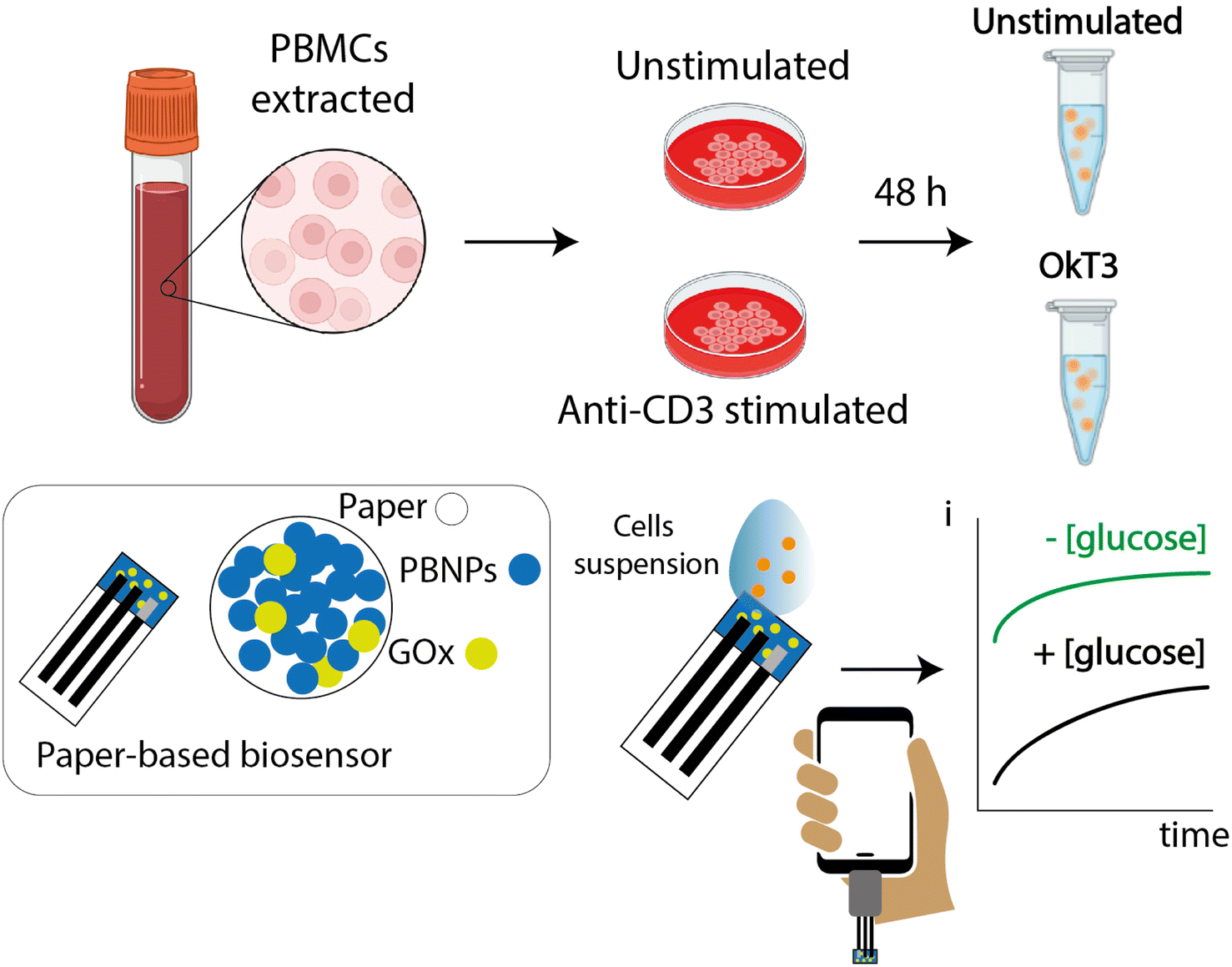 | ||
| Fig. 1 A schematic diagram illustrating the experimental workflow involved in the development and application of a paper-based electrochemical biosensor system for detecting glucose. PBMCs were isolated from peripheral blood samples and stimulated or not with anti-CD3 antibody (OkT3) for 48 h (red-colored Petri dish). Cell lysates obtained after 48 h of culture were resuspended in RIPA buffer, and the glucose was measured using the paper-based biosensor (modified with Prussian blue and glucose oxidase, as shown in the rectangular bar) connected to a smartphone for the measurement of the produced current. | ||
Results and discussion
Electrochemical characterization
The initial characterization of the paper-based devices was performed by CV experiments that were conducted in the presence of potassium ferricyanide to evaluate the electrochemical performance of the screen-printed electrodes, as shown in Fig. 2A.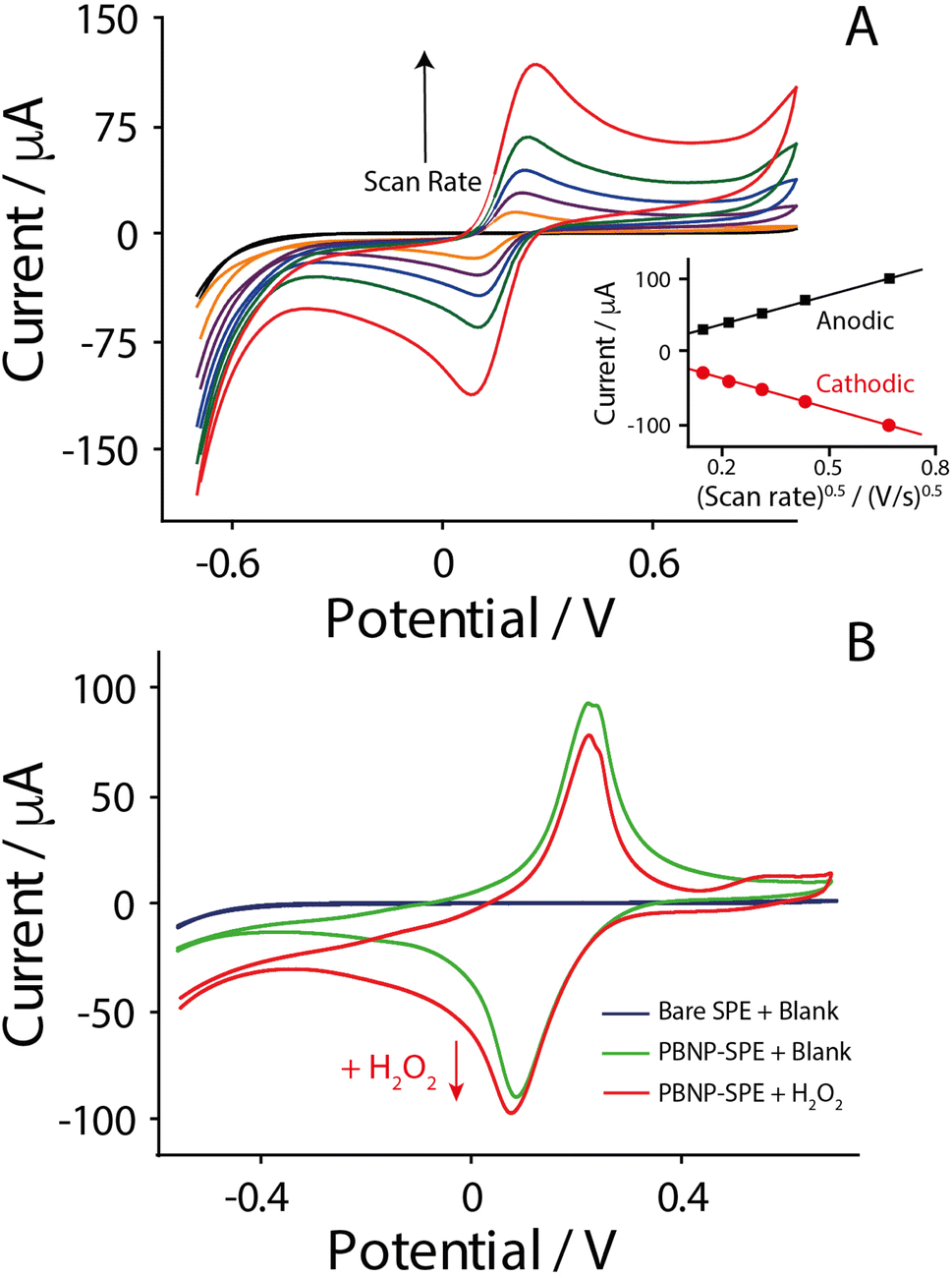 | ||
| Fig. 2 (A) CV curves of screen-printed electrodes at various scan rates ranging from 0.02 to 0.5 V s−1 in a solution containing 5 mM potassium ferricyanide ([K3Fe(CN)6]) in 0.1 M KCl. (Inset) anodic and cathodic peak currents versus the square root of the scan rate of the electrode confirming diffusion-controlled redox behavior, consistent with the Randles–Ševčík equation. (B) Comparative CV curves obtained in 0.1 mol per L KCl solution of bare SPE (blue), SPE modified with 10 mM PB (green), and SPE modified with PB in the presence of 5 mmol per L H2O2 (red). Experimental parameters: starting potential: −0.7 V; first vertex potential (EV1): −0.7 V; second vertex potential (EV2): 0.7 V; potential step (Estep): 0.01 V; scan rate: 0.1 V s−1; no. of scans: 2. | ||
The experiments were conducted in the absence and in the presence of a redox probe, namely potassium ferricyanide dissolved in 0.1 mmol per L potassium chloride, and as observed in Fig. 2A the increase of the scan rate (from 0.02 to 0.5 V s−1) led to an increase of both the anodic and cathodic peaks, respectively, due to the oxidation and reduction of the redox probe at the electrode surface. As reported by the inset of Fig. 2A, a linear relationship between the intensities of the peaks and the square root of scan rate was observed, as explained by the Randles–Ševčìk equation for diffusion-limited processes.33 In addition, the similarity of the slopes of anodic and cathodic curves confirmed the electrochemical reversibility of the redox probe that was used, thus confirming the satisfactory quality of the paper-based printed devices. Subsequently, paper-based strips were modified with PB to be used for the indirect detection of glucose by monitoring the hydrogen peroxide, which is a by-product of the enzymatic reaction catalyzed by the glucose oxidase enzyme.34 In fact, PB is known as an artificial peroxidase for its ability to reduce hydrogen peroxide at overpotentials close to 0 V (vs. Ag/AgCl),35 which has the advantages of limiting the interference of other species existing in complex media. As shown in Fig. 2B, the presence of PB was confirmed by the occurrence of both the anodic and cathodic peaks associated with the presence of Prussian blue and the Prussian white couple that allows for the reduction of hydrogen peroxide. The experiments in the presence of hydrogen peroxide have displayed the highest anodic current close to −0.1 V (vs. Ag/AgCl), and this potential was chosen for further development of the paper-based electrochemical biosensor. It should be noted how this applied potential is capable of yielding high cathodic currents, while more negative potentials did not represent any enhancement of the signal.36
Optimization of the PB concentration
The choice of the chromatographic paper Whatman no. 1 was also confirmed by additional studies that have been carried out using an alternative paper-based substrate, namely filter paper. As reported in the ESI (Fig. S1†), the adoption of Whatman no. 1 paper allowed for better sensitivity and repeatability towards the detection of hydrogen peroxide: it can be a consequence of the fact that the porosity of Whatman paper is finely controlled, and it offers a better substrate for the synthesis and deposition of PB in comparison to the other filter paper tested (Fig. S2†). Subsequently, the concentration of PB was evaluated between 3 and 25 mmol L−1. However, with respect to 3 and 25 mmol L−1, although they were tested in the presence of various concentrations of hydrogen peroxide, the obtained results were not suitable for further development of the device. Specifically, 3 mM PB gave a very low current signal, while 25 mM PB did not allow the testing area to be wetted by the working solution. For this experimental reason, 6.25 and 12.5 mmol per L PB were assessed in the presence of hydrogen peroxide by using chronoamperometry, as reported in Fig. 3.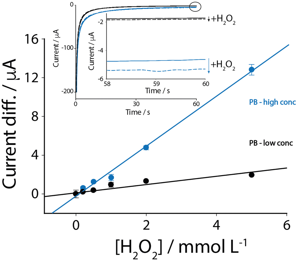 | ||
| Fig. 3 Comparison of calibration curves for H2O2 detection obtained via chronoamperometry using SPEs modified with PB at two concentrations: 12.5 mmol L−1 (blue line) and 6.25 mmol L−1 (black line). The response increases linearly with the concentration of H2O2 (0 to 6 mmol L−1), demonstrating the higher sensitivity of electrodes modified with 12.5 mmol per L PB. Experimental parameters: Edc: −0.1 V; Tinterval: 0.1 s; Trun: 60 s. | ||
As shown in Fig. 3, the paper-based strips modified with 12.5 mmol per L PB showed superior performance, as described by a calibration equation of y = 2.6x − 0.2 (R2 of 0.99), where y indicates the current difference between a chosen concentration of hydrogen peroxide and the current recorded using a blank solution expressed in μA and x indicates the concentration of hydrogen peroxide expressed in mM. The detection limit was calculated to be ca. 500 μmol L−1 (calculated as S/N = 3) with a satisfactory repeatability of 5%, calculated as the relative standard deviation (RSD). In contrast, the 6.25 mmol L−1 concentration yielded a lower sensitivity, as described by the equation y = 0.4x + 0.2 (R2 = 0.87), with a detection limit of ca. 1 mM and a repeatability of 7%.
Glucose detection in cell lysates
Following the optimization of the experimental parameters, i.e., paper-based substrate, PB concentration, and applied potential, the portable device was applied towards the determination of glucose, as shown in Fig. 4. | ||
| Fig. 4 Calibration curve of glucose obtained by measuring signal changes at different concentrations of glucose solutions up to 2 mmol L−1. Bottom inset: current measurements recorded over time for glucose by chronoamperometric technique. Top inset: comparison of glucose levels in the biological sample by chronoamperometric technique, shown as a bar diagram: control in RIPA buffer/KCl, lysates from unstimulated PBMCs and lysates from PBMCs stimulated with OKT3. Experimental parameters: Edc: −0.1 V; Tinterval: 0.1 s; Trun: 60 s. | ||
In order to produce the final platform, the paper-based device was modified with a 1 mg per mL glucose oxidase solution. After the samples were drop-cast onto the strips, the currents were recorded after a waiting time of 5 min, which represented the optimal compromise to have satisfactory sensitivity but avoiding time-consuming procedures. As shown in Fig. 4, the biosensor was tested in the presence of glucose solutions up to 2 mmol L−1, and as it can be observed from the plot, a linear response was observed up to 0.25 mM, while the response reached a plateau at 0.5 mmol L−1, which aligns with enzyme saturation as described by Michaelis–Menten kinetics. The linear response obtained from chronoamperometric measurements was described using the following equation: y = 3x + 0.1 with a R2 of 0.94. A detection limit of 40 μmol L−1, a quantification limit of 120 μmol L−1 and a repeatability of ca. 5% were calculated. Additionally, according to the commercial glucose assay kit for colorimetric detection, purchased from Merck, a correlation described by R2 = 0.968 was obtained. These results confirmed satisfactory sensitivity, repeatability and reliability towards the detection of glucose in the printed paper-based strip. In addition, the specificity was evaluated by measuring samples containing 1 mM each of ascorbic acid, lactic acid, uric acid, and paracetamol: the recorded signals were always lower than 5% of variation with respect to the same concentration of glucose tested. Furthermore, we evaluated the capacity of the biosensor to detect the glucose level in biological samples. We used cell lysates from PBMCs stimulated or not with OkT3. All the cell lysates were resuspended in a mixture composed of RIPA buffer and potassium chloride. In addition, the biosensor was also tested in the presence of a blank solution represented by RIPA buffer in the absence of cells, which represented the control experiment. All the measurements were carried out as follows: 40 μL of sample (both stimulated and unstimulated cells) was added to 60 μL of 0.1 mol per L KCl, while a blank solution was prepared by mixing 40 μL of RIPA buffer with 60 μL of 0.1 mol per L KCl. As shown in the inset of Fig. 4, the results revealed a slight difference between the current recorded for the unstimulated and stimulated cells. The error bars of the histograms are represented by the standard deviation that has been obtained on 5 replicates. However, with respect to the mean values of the recorded currents for both the cell lysates, the slight difference observed was consistent with the expectations: in fact, the use of OKT3 is responsible for higher cellular proliferation, and it should be translated into a higher amount of glucose with respect to the unstimulated cells. However, due to the fact that activation was not selective for the cells tested, we also expected a slight difference in the current responses. However, the intensity of the anodic current reported in the histograms reflects this behaviour: in fact, the higher the glucose, the higher the cathodic current is as a consequence of a higher by-production of hydrogen peroxide following the enzymatic catalysis of glucose oxidase towards glucose.
Conclusion
For the first time, an electrochemical paper-based biosensor has been applied towards the monitoring of glucose in stimulated and unstimulated peripheral blood mononuclear cell lysates. The coupling of paper-based substrates, Prussian blue, glucose oxidase and smartphone-enabled reader has allowed us to evaluate the effect of cellular stimulation with the use of quick, decentralized and low-cost technology. The reported biosensor was sensitive towards glucose detection down to the micromolar range, and its reliability was demonstrated by the satisfactory correlation with the commercial kit with colorimetric detection. This study represents a starting point towards the translation of biosensors into the field of immunology, providing an example of an easy-to-use platform for the rapid screening of personalized therapies. As highlighted, a slight difference was detected in the presence of anti-CD3 stimulated and unstimulated peripheral blood mononuclear cells, according to the recorded currents. However, even if the stimulation was not specific for the treated cells, the slight difference observed in terms of produced glucose represents a good starting point to be generalized for other applications in the fields of immunology and drug discovery.Data availability
The data supporting this article have been included as part of the ESI.†Conflicts of interest
There are no conflicts to declare.Acknowledgements
The PRIN project no. 2022WN89PC is acknowledged for its support. The grant CN00000041 “National Center for Gene Therapy and Drugs based on RNA Technology” (CUP E63C22000940007) is acknowledged for supporting this work. The authors extend their appreciation to the Researchers Supporting Project Number (RSPD2025R944), King Saud University, Riyadh, Saudi Arabia, for funding this work. We acknowledge the support of National Recovery and Resilience Plan (NRRP), Mission 4, Component 2 (M4C2), Investment 1.1, funded by the European Union – NextGenerationEU – CUP E53D23013240006 – Grant Assignment Decree No. 0001111 adopted on 20/07/2023 and the Italian Ministry of Health, NextGenerationEU, M6/C2_CALL 2022 cod. PNRR-MAD-2022-12375634. The authors acknowledge the support of MUR/PNRR Extended Partnership (MNESYS no. PE00000006) to GM. We acknowledge NRRP, M4C2, Investment 1.1, Call for tender no. 104 published on 2.2.2022 by the Italian Ministry of University and Research (MUR), funded by the European Union – NextGenerationEU – (grant no. 2022YMJXYT) – CUP B53D23003710006; NRRP, M4C2, Investment 1.1, funded by the European Union – NextGenerationEU – CUP B53D23031570001; NRRP, M4C2, Investment 1.1, Call for tender no. 104 published on 2.2.2022 by the Italian Ministry of University and Research (MUR), funded by the European Union – NextGenerationEU – (grant no. 20225KH7BZ) – CUP B53D23012500006 for their support.References
- P. Li, T. Lei and L. Ding, Polymer-based bioelectronics go self-powered, Sci. Bull., 2020, 65, 176–180 Search PubMed.
- D. Ohayon, G. Nikiforidis, A. Savva, A. Giugni, S. Wustoni, T. Palanisamy, X. Chen, I. P. Maria, E. Di Fabrizio, P. M. F. J. Costa, I. McCulloch and S. Inal, Biofuel powered glucose detection in bodily fluids with an n-type conjugated polymer, Nat. Mater., 2020, 19, 223–231 CrossRef PubMed.
- K. El Mamoune, L. Barantin, H. Adriaensen and Y. Tillet, Application of Chemical Exchange Saturation Transfer (CEST) in neuroimaging, J. Chem. Neuroanat., 2021, 114, 101944 CrossRef PubMed.
- S. Zhang, B. B. Lachance, M. P. Mattson and X. Jia, Glucose metabolic crosstalk and regulation in brain function and diseases, Prog. Neurobiol., 2021, 204, 102089 CrossRef CAS PubMed.
- P. V. Röder, B. Wu, Y. Liu and W. Han, Pancreatic regulation of glucose homeostasis, Exp. Mol. Med., 2016, 48, e219 CrossRef PubMed.
- K. Sharabi, C. D. J. Tavares, A. K. Rines and P. Puigserver, Molecular pathophysiology of hepatic glucose production, Mol. Aspects Med., 2015, 46, 21–34 CrossRef CAS PubMed.
- A. S. Bolla and R. Priefer, Blood glucose monitoring—an overview of current and future non-invasive devices, Diabetes Metab. Syndr., 2020, 14, 731–741 CrossRef PubMed.
- N. Alyamni, J. L. Abot and A. G. Zestos, Perspective—Advances in Voltammetric Methods for the Measurement of Biomolecules, ECS Sens. Plus, 2024, 3, 27001 CrossRef CAS PubMed.
- B. J. Van Enter and E. Von Hauff, Challenges and perspectives in continuous glucose monitoring, Chem. Commun., 2018, 54, 6576–6586 RSC.
- Z. Karim, M. J. Khan, A. Hussain, F. Ahmed and Z. H. Khan, Multilayer patch functionalized microfibrillated cellulosic paper sensor for sweat glucose monitoring, Sci. Rep., 2024, 14, 1–11 CrossRef PubMed.
- D. Calabria, E. Lazzarini, A. Pace, I. Trozzi, M. Zangheri, S. Cinti, M. Difonzo, G. Valenti, M. Guardigli, F. Paolucci and M. Mirasoli, Smartphone-based 3D-printed electrochemiluminescence enzyme biosensor for reagentless glucose quantification in real matrices, Biosens. Bioelectron., 2023, 227, 115146 CrossRef CAS PubMed.
- E. Lindholm, G. B. Bergmann, H. Haugaa, K. J. Labori, S. Yaqub, B. A. Bjørnbeth, P. D. Line, G. Grindheim, G. Kjøsen, S. E. Pischke and T. I. Tønnessen, Early detection of anastomotic leakage after pancreatoduodenectomy with microdialysis catheters: an observational study, HPB, 2022, 24, 81–90 CrossRef PubMed.
- D. C. Caixeta, C. Lima, Y. Xu, M. Guevara-Vega, F. S. Espindola, R. Goodacre, D. M. Zezell and R. Sabino-Silva, Monitoring glucose levels in urine using FTIR spectroscopy combined with univariate and multivariate statistical methods, Spectrochim. Acta, Part A, 2023, 290, 122259 CrossRef CAS PubMed.
- Y. Cheng, X. Gong, J. Yang, G. Zheng, Y. Zheng, Y. Li, Y. Xu, G. Nie, X. Xie, M. Chen, C. Yi and L. Jiang, A touch-actuated glucose sensor fully integrated with microneedle array and reverse iontophoresis for diabetes monitoring, Biosens. Bioelectron., 2022, 203, 114026 CrossRef CAS PubMed.
- S. Jiang, Y. Zhang, Y. Yang, Y. Huang, G. Ma, Y. Luo, P. Huang and J. Lin, Glucose Oxidase-Instructed Fluorescence Amplification Strategy for Intracellular Glucose Detection, ACS Appl. Mater. Interfaces, 2019, 11, 23628–23636 Search PubMed.
- E. A. Moschou, B. V. Sharma, S. K. Deo and S. Daunert, Fluorescence glucose detection: advances toward the ideal in vivo biosensor, J. Fluoresc., 2004, 14, 535–547 CrossRef CAS PubMed.
- S. Cinti, B. De Lellis, D. Moscone and F. Arduini, Sustainable monitoring of Zn(II) in biological fluids using office paper, Sens. Actuators, B, 2017, 253, 1195–1202 CrossRef.
- A. Miglione, M. Spinelli, A. Amoresano and S. Cinti, Sustainable Copper Electrochemical Stripping onto a Paper-Based Substrate for Clinical Application, ACS Meas. Sci. Au, 2022, 2, 177–184 CrossRef CAS PubMed.
- S. Cinti, Novel paper-based electroanalytical tools for food surveillance, Anal. Bioanal. Chem., 2019, 411, 4303–4311 CrossRef CAS PubMed.
- T. You, S. Xiao, P. Huang, C. Wang, Q. Deng, P. Jiang and D. He, Localized photothermal effect of Co3O4 nanowires boosts catalytic performance in glucose electrochemical detection, Inorg. Chem. Front., 2024, 11, 6527–6535 RSC.
- S. Singh, J. Wang and S. Cinti, Review—An Overview on Recent Progress in Screen-Printed Electroanalytical (Bio)Sensors, ECS Sens. Plus, 2022, 1, 27001 Search PubMed.
- S. Singh, A. Raucci, W. Cimmino and S. Cinti, Paper-Based Analytical Devices for Cancer Liquid Biopsy, Anal. Chem., 2024, 96, 3698–3706 CrossRef CAS PubMed.
- W. Cimmino, A. Raucci, S. P. Grosso, N. Normanno and S. Cinti, Enhancing sensitivity towards electrochemical miRNA detection using an affordable paper-based strategy, Anal. Bioanal. Chem., 2024, 416, 4227–4236 CrossRef CAS PubMed.
- P. M. Kalligosfyri and S. Cinti, 3D Paper-Based Origami Device for Programmable Multifold Analyte Preconcentration, Anal. Chem., 2024, 96, 9773–9779 CrossRef CAS PubMed.
- A. Miglione, A. Raucci, F. Cristiano, M. Mancini, V. Gioia, A. Frugis and S. Cinti, Paper-based 2D configuration for the electrochemical and facile detection of paracetamol in wastewaters, Electrochim. Acta, 2024, 488, 144255 CrossRef CAS.
- S. Cinti, S. Tomassi, C. Ciardiello, R. Migliorino, M. Pirozzi, A. Leone, E. Di Gennaro, V. Campani, G. De Rosa, V. M. D’Amore, S. Di Maro, G. Donati, S. Singh, A. Raucci, F. S. Di Leva, H. Kessler, A. Budillon and L. Marinelli, Paper-based electrochemical device for early detection of integrin αvβ6 expressing tumors, Commun. Chem., 2024, 7, 60 CrossRef CAS PubMed.
- A. Glovi, A. Miglione, D. C. Maresca, F. Somma, B. Romano, A. Ianaro, A. Giordano, M. De Laurentiis, G. Ercolano and S. Cinti, Paper-based electrochemical device for the determination of H2S in murine lysates for liquid biopsy application, Anal. Chim. Acta, 2025, 1342, 343669 CrossRef CAS PubMed.
- M. L. Salva, M. Rocca, C. M. Niemeyer and E. Delamarche, Methods for immobilizing receptors in microfluidic devices: a review, Micro Nano Eng., 2021, 11, 100085 CrossRef CAS.
- S. Khumngern and I. Jeerapan, Synergistic convergence of materials and enzymes for biosensing and self-sustaining energy devices towards on-body health monitoring, Commun. Mater., 2024, 5, 135 CrossRef CAS.
- H. Huang, T. Li, M. Jiang, C. Wei, S. Ma, D. Chen, W. Tong and X. Huang, Construction of flexible enzymatic electrode based on gradient hollow fiber membrane and multi-wall carbon tubes meshes, Biosens. Bioelectron., 2020, 152, 112001 CrossRef CAS PubMed.
- H. A. Silva-Neto, L. F. de Lima, D. S. Rocha, V. N. Ataide, G. N. Meloni, G. Moro, A. Raucci, S. Cinti, T. R. L. C. Paixão, W. R. de Araujo and W. K. T. Coltro, Recent achievements of greenness metrics on paper-based electrochemical (bio) sensors for environmental and clinical analysis, Trac. Trends Anal. Chem., 2024, 174, 117675 CrossRef CAS.
- S. Cinti, R. Cusenza, D. Moscone and F. Arduini, Paper-based synthesis of Prussian Blue Nanoparticles for the development of whole blood glucose electrochemical biosensor, Talanta, 2018, 190, 96–102 Search PubMed.
- S. Cassano, V. Pucino, C. La Rocca, C. Procaccini, V. De Rosa, G. Marone and G. Matarese, Leptin modulates autophagy in human CD4+ CD25− conventional T cells, Metabolism, 2014, 63, 1272–1279, DOI:10.1016/j.metabol.2014.06.010.
- A. Raucci, A. Miglione, L. Lenzi, P. Fabbri, J. Di Tocco, C. Massaroni, D. Lo Presti, E. Schena, V. Pifferi, L. Falciola, W. Aidli, C. Di Natale, P. A. Netti, S. L. Woo, D. Morselli and S. Cinti, Characterization and application of porous PHBV-based bacterial polymers to realize novel bio-based electroanalytical (bio)sensors, Sens. Actuators, B, 2023, 379, 133178 CrossRef CAS.
- F. Ricci and G. Palleschi, Sensor and biosensor preparation, optimisation and applications of Prussian Blue modified electrodes, Biosens. Bioelectron., 2005, 21, 389–407 CrossRef CAS PubMed.
- R. Baretta, A. Raucci, S. Cinti and M. Frasconi, Porous hydrogel scaffolds integrating Prussian blue nanoparticles: A aersatile strategy for electrochemical (bio)sensing, Sens. Actuators, B, 2023, 376, 132985 CrossRef CAS.
Footnote |
| † Electronic supplementary information (ESI) available. See DOI: https://doi.org/10.1039/d5ay00057b |
| This journal is © The Royal Society of Chemistry 2025 |