
 Open Access Article
 This Open Access Article is licensed under a
 Creative Commons Attribution 3.0 Unported Licence
The role of DNA nanotechnology in medical sensing
Darell
Lien

Troy High School, 2200 Dorothy Ln, Fullerton, CA 92831, USA
First published on 6th December 2024
Abstract
This paper explores how DNA nanotechnology enhances biosensors in medicine and pharmacology by taking advantage of the unique characteristics of DNA and the unique advantages of DNA origami technology. DNA origami allows the establishment of complex nanoobjects with precise size and complete molecular writability as well as the possibility of seamless integration and biocompatibility with biological systems. Utilizing this, the chemical denaturation of DNA chains allows for the combination of various functions, including organic fluorescence groups and photoreaction elements, etc. This has allowed DNA origami to become a transformative tool in biotechnology and other fields because of its versatility, use in innovative applications improving the design and function of biosensors, and potential to provide greater possibilities for early disease diagnosis and personalized medicine.
Introduction
Nucleic acids are large biomolecules that define the DNA of every living organism. The unique sequences and combinations of nucleic acids within DNA allow for the vast diversity of life that can be seen today. Aside from their intrinsic value in life, nucleic acids play another important role in aiding scientific innovations and discoveries. In modern society, they have already revolutionized the fields of medicine and technology and are the basis of DNA nanotechnology. DNA nanotechnology focuses on the interactions between nucleic acids to create biosensors, nanoarchitecture, drug delivery systems, and computational tools.Before the emergence of DNA nanotechnology, DNA was primarily studied in life sciences and medicine. However, with DNA nanotech, DNA's exceptional properties in stability, flexibility, and specific binding were emphasized. These properties are crucial for applications such as biosensors and drug development. This can be seen through researchers who have explored DNA-based technologies like DNA origami structures, DNA-based biosensors, vaccines, and therapeutic agents such as aptamers and DNAzymes. These biomolecules mimic antibodies and enzymes, offering advantages like high binding affinity and versatility in production through methods like SELEX. The aptamers can be applied to fields including medicine, pharmacology, and molecular biology, while offering benefits over traditional antibodies in terms of cost and production scalability. Similarly, DNAzymes, acting as catalytic DNA, exhibit diverse functionalities useful in biosensor development and other biochemical applications.
Furthermore, the study of DNA nanotechnology has introduced novel structural designs like DNA 3-way and 4-way junctions, allowing for the enhanced functionality of biosensors through unique design.
In 1982, Ned Seeman set the groundwork for a groundbreaking concept: using DNA as a construction material to assemble geometrically defined structures with nanoscale characteristics.1 This suggestion resulted in what is now known as “structural DNA nanotechnology”. Researchers used DNA's natural self-recognition capabilities, specifically the complementary Watson–Crick base pairing, to create rigid branched DNA patterns between segments of certain oligonucleotides. The resulting superstructures, also known as DNA tiles, serve as basic building blocks for further assembly into discrete finite objects or infinite periodic lattices, which is accomplished through the cohesive force of sticky ends.2 However, this “multi-strand” technique is not without its challenges. First, the building of complex structures necessitates precise stoichiometric control and frequently entails the purification of constituent oligonucleotides and/or tiles, resulting in error-prone and time-consuming synthetic methods. Next, the complexity achieved by this technique is limited to simple geometric shapes and the repeated use of fundamental building components. In 2006, Rothemund introduced the scaffold-based DNA origami approach, which appeared to be an answer to these issues.3 DNA origami, inspired by the precision of Japanese paper folding, includes meticulously folding a long single-stranded DNA strand (scaffold) into a predefined shape using hundreds of small oligonucleotides known as staple strands.4 Rothemund's groundbreaking research demonstrated the folding of a 7.25-kilobase-long circular M13mp18 genome into an antiparallel array of helices via periodic crossings. Unlike previous approaches, DNA origami has a rather efficient self-assembly process that includes thermal annealing with a rapid increase in temperature and a moderate fall, as well as time spent at specific temperatures. However, this efficiency is due to the entropy advantage acquired by folding a single long scaffold strand. Instead of binding with each other, staple strands hybridize with a shared scaffold, removing the need for precise control over their relative stoichiometric ratio and considerably lowering experimental errors and synthesis timeframes.
The DNA origami technology is unique not only in its efficiency, but also in its capacity to create nano-objects with complicated geometries of set dimensions and full molecular addressability. This review will look at the various applications of DNA origami in sensing. These DNA nanostructures have received a lot of interest for their many biological and medicinal uses, which are being driven by a number of fundamental aspects. For starters, their DNA-based makeup makes them intrinsically biocompatible, allowing them to integrate effortlessly with biological systems. Second, DNA structures, particularly origami, provide unmatched addressability at the nanoscale, similar to small breadboards. Staple strands are versatile substrates for grafting and arranging biomolecules such as nucleic acids, proteins, and nanoparticles, providing fine control over functionalization and organization. Thirdly, these nanostructures' usefulness is further increased by the individual DNA strands' chemical modifiability. This versatility makes it possible to add a wider range of functionalities to DNA strands by incorporating different organic fluorophores and photoresponsive moieties like azobenzene. DNA's chemical flexibility and adaptability make it a prime choice for a wide range of uses in various fields, highlighting its potential to spur innovation in biotechnology and other fields.
Detection
DNA nanotechnology offers significant advantages in disease detection due to its adaptability and efficiency. It is particularly adept at pinpointing genetic markers and mutations that are linked to various diseases, which enables the early identification of genetic conditions, hereditary illnesses, and cancers. Since DNA nanotechnology can detect diseases before they become life threatening, it can expedite the treatment process and decrease risk. In addition, because of the nature of DNA sensing nanotechnology, its core being DNA, the development of personalized treatment is made possible. This benefit is especially valuable when it comes to fighting against cancer, a mutative disease that is unique in every occurrence. Having the ability to identify specific genetic mutations or changes allows doctors to identify the best way to combat a patient's specific version of cancer. This can not only speed up the treatment, in that it becomes more effective, but also decrease the risk of adverse reactions to the treatment. Furthermore, existing treatments, such as liquid biopsies, are non invasive, which makes treatment more attractive to patients and less intimidating.In an attempt to prove detecting cancer is possible, Liu et al.5 proposed that targeting specific microRNAs associated with lung cancer could lead to its early detection. As a result, they devised a 3D DNA origami structure tailored for electrochemical detection of lung cancer-related microRNAs, the electrochemical genosensor. This structure comprises a stem-loop DNA with ferrocene labeling on its upper part, coupled with a thiolated tetrahedron DNA nanostructure at the base. While the upper section binds to lung cancer-related microRNAs, the lower portion is affixed onto a gold disk electrode surface, which is modified with gold nanoparticles (Au NPs) and treated with mercaptoethanol (MCH) (Fig. 1). All of the tests regarding the performance of the electrochemical genosensor were conducted through the use of scanning electron microscopy (SEM), atomic force microscopy (AFM), cyclic voltammetry (CV), and differential pulse voltammetry (DPV). Through these tests they found that under optimized conditions, the genosensor demonstrated a detection limit of 10 pM and exhibited a linear response over microRNA concentrations ranging from 100 pM to 1 μM, highlighting its potential for highly sensitive clinical cancer diagnosis applications.
 | ||
| Fig. 1 Scheme5 of the proposed electrochemical genosensor to detect target microRNA. (A) Au NPs were electrochemically deposited on a bare gold disk electrode. (B) Constructing 3D DNA probes with four oligonucleotides (Tetra-A, Tetra-B, Tetra-C and Tetra-D). (C) The 3D DNA probes were self-assembled on the electrode surface. (D) The Au NPs-coated gold electrode was blocked with MCH. (E) With the presence of microRNA, the stem-loop structure of the 3D DNA probe was opened and the ferrocene groups had access to the gold electrode surface after the hybridization with the target microRNA. | ||
Static vs. dynamic
It can be said that biomolecular sensors consist of two main components: target recognition elements and signal transducers, which are integrated into DNA nanostructures. Dynamic DNA structures change their shapes upon recognizing analytes, serving as signal readouts, often detected with atomic force microscopy (AFM). Static structures serve as platforms where target binding events are recorded directly with AFM or indirectly using techniques like fluorescence resonance energy transfer (FRET)-based optical detection. These elements primarily rely on interactions between targets and ligands; ligands commonly being nucleic acids, aptamers, and antibody proteins.Fig. 2 demonstrates an example of how static DNA nanostructures were used in biomolecular sensing. In the figure, Endo & Sugiyama6 engineered origami rectangles with square holes at the center, incorporating DNA cantilevers bridged across the holes (Fig. 2a). This was then utilized to study the dynamic motions and kinetics of biomolecular analytes at the single-molecule level using high-speed AFM. Afterword, DNA binding events, such as DNA base pairing, guanine quadruplex formation, and DNA-enzyme binding, induced association or dissociation of the cantilever bridges, were detected by the AFM. Similar methodologies were employed to detect B–Z transitions of DNA strands in response to varying Mg2+ concentrations and to demonstrate photo regulated DNA base pairing using photoisomerizable azobenzene moieties.
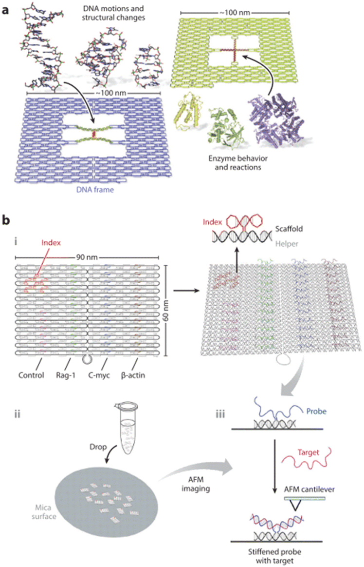 | ||
| Fig. 2 (a) Single-molecule biosensing and biophysics platform.6 (b) DNA origami-based label-free RNA hybridization assay.7 Abbreviation: AFM, atomic force microscopy. | ||
Next, the programmability and addressability of DNA origami structures were showcased in a label-free hybridization assay (Fig. 2b). Yan and colleagues7 designed an origami rectangle containing three different probes arranged spatially. The capture of target RNA strands by the probes stiffened the probes, enabling detection by AFM. This study is important because it shows the straightforward yet effective approach of DNA nanostructures, and paved the way for the development of other DNA assembly-based biosensors.
In dynamic biosensor schemes, structural reconfigurations are linked to analyte binding events. A prevalent sensor design involves a scissor/plier-type structure, demonstrated independently by multiple research groups. A project run by Kuzuya et al.,8 started by crafting single-molecule beacons from DNA origami that would close upon binding to a target molecule (Fig. 3a). Their design enabled the examination of various analytes, including streptavidin, immunoglobulin G, telomere strands, microRNA (miRNA), and ATP. Structural changes could also be detected by AFM and fluorescence measurements. A similar structural design was adopted by Niemeyer and colleagues9 to investigate analyte binding kinetics (Fig. 3b).
 | ||
| Fig. 3 (a and b) Nanopliers as a single-molecule sensing platform.8,9 The DNA plier pinches closed or open upon target binding. (c) Enzymatic nanoreactor enabled by reconfigurable DNA assemblies.10 (d) Biosensing via allosteric activation.11 Abbreviations: AFM, atomic force microscopy; BamHI, a type II restriction endonuclease from Bacillus amyloliquefaciens; G6pDH, glucose-6-phosphate dehydrogenase; miR-210, a short microRNA; NAD+, an oxidized form of nicotinamide adenine dinucleotide (NAD). | ||
Expanding on this concept, the Liu group10 devised a DNA tweezer that pinches near a fuel strand, initiating a NAD+/NADH reaction by glucose-6-phosphate dehydrogenase (Fig. 3c). The reconfiguration dynamics of the structure were analyzed by reading and recording the fluorescence signals resulting from a by-product molecule and a FRET pair. Additionally, Fig. 3d presents a nanomechanical origami device capable of long-range allosteric activation, proposed by Ke et al.11 This device, which consists of four rigid rods connected by flexible single-stranded hinges, allows for the reconfiguration of the entire structure via allosteric changes upon recognition of analyte molecules. Dynamic nanodevices such as these demonstrate how applications in biosensing and single-molecule biophysics studies are viable.
Biosensors for virus detection
DNA nanotechnology is a strong “bottom-up” way to design and create nucleic acid nanostructures. By employing DNA molecules as structural materials, this method enables precise control at the nanoscale, resulting in the development of standardized and modular tools suitable for novel virus-detection systems. Researchers can employ DNA nanotechnology to create personalized, highly sensitive biosensors that detect viruses more accurately and efficiently. The self-assembly of DNA tiles generates DNA nanostructures with addressable properties, high sensitivity, and affinity. These distinctive features have paved the way for DNA nanotechnology-based biosensors. Rapid progress in this discipline has accelerated the development of highly sensitive and precise viral detection technologies. DNA nanobiosensors,12,13 which use DNA nanotechnology, may detect the presence of target DNA via electrical, optical, or audio signals. These devices typically consist of two key components: a molecular recognition element and a transducer. The molecular recognition element detects the target DNA in the sample, and the transducer turns the signal into viewable and recordable data. In DNA nanobiosensors, DNA molecules form programmable supramolecular nanostructures. These nanostructures serve as templates for accurately assembling sensing elements, hence improving sensing performance and enabling the construction of advanced biosensors.14–16 Supramolecular DNA structures, including DNA tetrahedron-based biosensors,17 function as programmable anchor points. Recently, many DNA nanobiosensors have been produced, with optical and electrochemical DNA nanobiosensors being the most investigated due to their great sensitivity, inexpensive cost, and short detection times.Surface-enhanced Raman scattering (SERS)
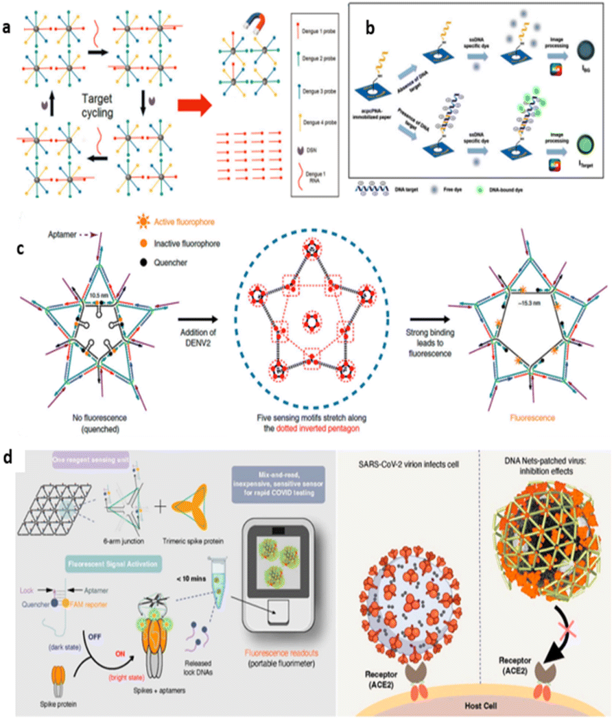
When nearby noble metal nanoparticles are close together, the gap between them generates an amplified electromagnetic field, greatly increasing the signal strength of Raman-active molecules.18,19 Surface-Enhanced Raman Scattering (SERS) aims to amplify Raman-scattering signals by adhering the analyte to the surfaces of metal nanoparticles or nanostructures, hence boosting the signal and making it easier to detect. SERS-based sensing techniques have grown in popularity over the last few decades due to numerous advantages: (i) exceptional sensitivity; (ii) capability for multiplex sensing; (iii) suitability for point-of-care (POC) devices; and (iv) ease of sample preparation.20–23 The main advantage of DNA nanobiosensors based on SERS technology is their capacity to detect specific analytes at extremely low concentrations.24 However, a key difficulty for SERS-based DNA nanobiosensors is the need for tight contact between the analyte and the surface, which might degrade with prolonged usage, reducing signal intensity.25 SERS-sensing technologies have demonstrated significant potential for a variety of immunoassays,26 meeting the growing demand for precise and quick virus detection.To identify the DNA sequence of the dengue virus (DENV),27 Song et al. developed a SERS sensor based on a locally catalyzed hairpin assembly (LCHA) and hybridization chain reaction (HCR) from DNA tiles (Fig. 4a). The LCHA system, which consists of L1 and L2 strands and Hairpin C1, self-assembles with the ROX dye-labeled Hairpin C2, allowing for continuous recycling of the DENV DNA sequence. ROX-labeled hairpins (H1 and H2) were also added to the SERS-AgNRS array, triggering HCR and increasing the ROX signal. This combined LCHA and HCR biosensor had a SERS intensity that was approximately 2.8 times larger than the single LCHA method and more than 4.5 times that of the ordinary CHA strategy. The signal-to-noise ratio (S/N, IDENV/Iblank) of LCHA-HCR was 5.4 times greater than that of individual CHA, with a limit of detection (LOD) for DENV as low as 0.49 fM, which was much better than earlier SERS sensors.29
 | ||
| Fig. 4 SERS DNA nanobiosensors for virus detection. (a) Schematic diagram of SERS biosensor for DENV gene by cascade enzyme-free signal amplification strategy of local catalytic hairpin assembly (LCHA) and hybridization chain reaction (HCR).27 Copyright 2020, Elsevier. (b) SERS-based design strategy for the SARS-CoV-2 label-free sensitive-sensor platform.28 Copyright 2023, Elsevier. | ||
Park et al. created a sensitive, label-free SERS aptamer sensor to identify SARS-CoV-2 variations of interest (Fig. 4b).28 High-throughput screening revealed two DNA aptamers with affinities of 1.47 ± 0.30 nM and 1.81 ± 0.39 nM for the SARS-CoV-2 spike protein. The combination of aptamers and silver nanoforests resulted in an ultrasensitive SERS platform with a detection limit of 10−18 M for recombinant trimeric spike proteins. They also developed a label-free aptamer sensor that does not require Raman labels and relies on the aptamer's intrinsic signals. This label-free SERS sensor performed admirably in clinical samples, detecting wild-type, Delta, and Omicron variations.
Surface plasmon resonance (SPR)
Surface plasmon resonance (SPR)-based DNA nanobiosensors work by sensing changes in the refractive index at the interface of the dielectric and metal layers.30–32 The SPR response is highly dependent on the characteristics of the metal layer, with gold being the most desired material.33 Traditional SPR sensors are made up of a stationary recognition element, an optical prism, and an analyte.34 In an SPR-based DNA nanobiosensor, manufactured DNA nanostructures are initially immobilized on the surface of a metal film. Following surface functionalization, a sample solution containing analytes is applied to the surface. When incident light strikes the medium from various angles, photons are absorbed by the plasma wave at the critical angle, which is affected by the medium's refractive index. When a virus sample's nucleic acid interacts with DNA nanoparticles, the refractive index near the metal film's surface changes, resulting in a resonant angular shift of the plasma wave and virus detection.35 SPR-based DNA nanobiosensors have shown tremendous potential for rapid point-of-care (POC) virus detection due to their sensitive and label-free detection mechanism.36 These sensors provide high accuracy, label-free monitoring, speed, and sensitivity. However, additional advancements in specificity and sensitivity are required to properly employ SPR technology for early viral detection.37Diao et al. created an SPR DNA biosensor to detect HIV-related DNA using an entropy-driven strand displacement reaction (ESDR) and dsDNA tetrahedron (DDT) (Fig. 5a).38 This biosensor detects target DNA within a linear range of 150 nM to 1 pM, with a limit of detection (LOD) of 48 fM. Lee et al. developed a label-free biosensor for detecting avian influenza (AIV H5N1) utilizing localized surface plasmon resonance (LSPR) technology (Fig. 5b).39 This biosensor, which consists of a multifunctional DNA three-way junction (3WJ) on a hollow gold spike-shaped nanomaterial, can detect AIV and other viruses. SPR biosensors have several advantages, including label-free detection, simpler molecular hybridization, and rapid detection. They do not require labels, such as fluorescence or radioisotopes, which simplifies sample processing and minimizes the influence on biological molecules.
 | ||
| Fig. 5 SPR DNA nanobiosensors for virus detection. (a) Schematic diagram of SPR biosensors based on ESDRs and DDTs nanostructures for the detection of HIV-related DNA.38 Copyright 2017, Elsevier. (b) Diagram of the AIV detection biosensor fabricated based on the LSPR method.39 Copyright 2019, Elsevier. (c) Schematic of the mechanism of DENV detection by AuNPs and hairpin ssDNA-CdSeTeS QDs.40 | ||
Chowdhury et al. created a DNA nanobiosensor for the quick and quantitative detection of all four dengue virus serotypes using the distance-based LSPR effect between cadmium telluride selenide fluorescent quantum dots (CdSeTeS QDs) and gold nanoparticles (AuNPs) (Fig. 5c).40 They created four nanoprobes covalently coupled to serotype-specific hairpin ssDNA primers at various places on the CdSeTeS QDs. The hairpin structure contained a self-complementary anchoring area composed of six polyguanines (poly-G) and polycytosines (poly-C) coupled to CdSeTeS QDs. Thiolated poly-C functionalized AuNPs were also produced. We employed both synthetic ssDNA and actual RNA samples to detect DENV serotypes. DENV's target ssDNA/RNA sequences hybridized with the hairpins' complementary ssDNA loop sequence, resulting in the formation of a linear ssDNA probe strand conjugated to QDs. The target DNA/RNA sequences are subsequently precisely coupled to the nanoprobe using complementary binding. The distance effect based on LSPR allowed for rapid and quantitative detection of DENV serotypes. This approach showed excellent sensitivity, with LODs of 24.6 fM for DENV1, 11.4 fM for DENV2, 39.8 fM for DENV3, and 39.7 fM for DENV4. This DNA nanobiosensor has enormous promise for practical applications in identifying dengue virus serotypes with high accuracy and efficiency.
Fluorescence-based (FB)
The fluorescence-based DNA nanobiosensor detects changes in fluorescence signals as a fluorescent dye-labeled DNA probe attaches to the target molecule.41–43 Typically, the fluorescent dye on the DNA probe is not activated until it attaches to the target molecule. The spatial position of the fluorescent dye varies when it is specifically paired with the target, resulting in changed fluorescence signals and allowing for target identification. This type of biosensor has good spatial and temporal resolution, sensitivity, and a quick reaction time, making it perfect for multiplexed assays.44 These properties make fluorescence-based DNA nanobiosensors a useful instrument for virus detection and biomolecule analysis.45–47 However, fluorescence-based DNA nanobiosensors encounter problems such as fluorophore scintillation and photobleaching, which may limit their uses. Furthermore, non-specific binding of fluorescent markers to ambient components can hamper their application.48–50Shen et al. created a fluorescent DNA biosensor that detects four strains of dengue virus (DENV) using a quantum dot-capped DNA capture probe (QD-CPs) (Fig. 6a).51 In this biosensor, DNA capture probes are bonded to the surfaces of quantum dots and magnetic beads. During detection, DENV ssRNA creates heterologous double strands with the DNA capture strand, which are then broken by double-strand-specific nuclease (DSN) to release quantum dots from the magnetic beads. This approach allows for continued hybridization with the leftover DNA capture probe, resulting in ultrasensitive DENV detection at a limit of detection (LOD) of 0.5 fM, which is substantially more sensitive than earlier methods.
 | ||
| Fig. 6 Fluorescence-based DNA nanobiosensors for virus detection. (a) Schematic representation of the working principle of the fluorescence DENV assay based on QD-CPs.51 Copyright 2015, Elsevier. (b) The working principle of the fluorescence-based detection of HCV using the acpcPNA-immobilized PAD and ssDNA-specific fluorescence dye.52 Copyright 2021, Elsevier. (c) Dimensional analysis and design concept of the DNA tile structure corresponding to the ED3 cluster on the DENV surface.53 Copyright 2019, Springer Nature. (d) Schematic design concept and detection of DNA mesh structures binding to SARS-CoV-2.54 Copyright 2019, Springer Nature. | ||
Teengam et al. developed a paper-based fluorescent DNA biosensor that uses acpcDNA to selectively detect the Hepatitis C virus (HCV) (Fig. 6b).52 This biosensor demonstrated a linear relationship between fluorescence changes and HCV DNA concentration, with an LOD of 5 pM, and successfully detected HCV complementary DNA (cDNA) in clinical samples. Jiao et al. developed a DNA nanoscaffold hybridization chain reaction (DNHCR)-based nucleic acid assay for quick SARS-CoV-2 detection.55 When the target SARS-CoV-2 RNA is present, a cascade reaction occurs within the DNA nanoscaffold, resulting in fluorescence recovery and instantaneous, highly amplified signal generation. This assay can be done in serum and saliva samples in 10 minutes at temperatures ranging from 15 to 35 °C.
Kwon et al. created fluorescent DNA biosensors that target the DENV surface antigen and paired them with nucleic acid aptamers to detect fluorescence and suppress DENV activity (Fig. 6c).53 They revealed that 2D spatial pattern recognition is critical for binding DNA nanostructures to targets, offering suggestions for enhancing ligand-DNA nanostructure binding. Ochmann et al. improved fluorescence signals for target nucleic acids by combining DNA folding-based optical antennas with metal nanoparticles and utilizing plasma phenomena to generate particular signals.56 This approach successfully detected Zika-specific artificial DNA and RNA in buffer solutions and heat-inactivated human serum, demonstrating sensitivity to minor nucleotide differences. Chowdhury et al. created a fluorescent DNA biosensor to measure four DENV serotypes.57 The researchers employed AuNP-graphene quantum dots nanocomposites (GQD-AuNP) with dye-labeled DNA probes to detect DENV at concentrations ranging from 10−14 to 10−6 M, with a LOD of 9.4 fM. This sensor also worked admirably in clinical applications for DENV detection.
Chauhan et al. demonstrated a unique mesh DNA nanostructure known as “DNA Net” for specifically identifying and trapping intact SARS-CoV-2 viruses (Fig. 6d).54 This DNA Net, which includes aptamers that target viral surface spike glycoproteins, emits a fluorescent signal when bound to the virus, allowing for rapid and sensitive COVID-19 detection. The sensor, which operates at room temperature and does not require complicated equipment, has a LOD comparable to clinical SARS-CoV-2 viral loads (1 × 10⁴ to 1 × 10¹⁰ copies per mL). It can identify intact virus in a sample.
Gogianu et al. created an innovative microarray platform to improve DNA detection.58 They created three-dimensional microarray chips on silicon nanowire substrates using metal-assisted chemical etching, then coated them with SU-8 polymer and customized them with carbon quantum dots (CDs). When high-quality tagged HPV 16-targeted ssDNA was hybridized to the SiNWs/SU-8/CDs platform, it produced a robust fluorescence signal, which was increased by the addition of CDs. Coimmobilizing the HPV 16 probe with PDDA resulted in optimal signal intensity and coefficient of variation.
Electrochemical
Electrochemical DNA biosensors use single-stranded DNA (ssDNA) probes with recognition capabilities that are mounted on an electrode. These probes can hybridize with target ssDNA, forming bgymdouble-stranded DNA (dsDNA) on the electrode surface. This hybridization alters electrical signals like current, potential, and impedance, which can subsequently be utilized to calculate the amount of target DNA. Electrochemical biosensors can recognize complementary target sequences by immobilizing DNA probes on a substrate surface and converting sequence-recognition hybridization signals into electrical signals using a signal transduction device.The resulting electrical signals can be classed as amperometric or resistive sensors based on the electrical quantity they measure. These biosensors, with their real-time monitoring and quick on-site detection capabilities, are a potential point-of-care (POC) diagnostic method for viral detection.59 They provide great sensitivity, simplicity, low cost, and ease of downsizing, making them an appealing choice for constructing portable diagnostic equipment.
Voltammetry
Voltammetry uses voltage ramps to monitor current changes, yielding different oxidation and reduction peaks for each analyte. Recently, researchers have concentrated on cyclic voltammetry (CV), differential pulse voltammetry (DPV),60,61 and square-wave voltammetry (SWV)62 for virus identification. DPV involves administering a differential potential pulse with a constant amplitude while constantly increasing the scanning potential. The current in each pulse is separated into two phases: before the potential application and after the pulse. The differential between these currents is shown against the potential. SWV, a differential voltammetry technique distinguished by large-amplitude pulses, entails applying a constant-amplitude pulse as the scanning potential gradually increases. In SWV, the potential–time plot is created by subtracting the current at the end of the pulse from the current at the start of the pulse, and then plotting this difference against the growing scanning potential.63,64 SWV's advantage over DPV is its rapid speed.Lee et al. created a DNA-based biosensor using label-free porous gold nanoparticles (pAuNPs) to detect the H5N1 virus (Fig. 7a).65 The electrodes, coated with pAuNPs to improve surface roughness and electron transfer, used DNA three-way junctions (3WJs) that included HA protein detection (recognition fragment), electrochemical signal generation (DNAzyme), and immobilization parts (thiol group) introduced on the Au electrode. The biosensor had a limit of detection (LOD) of 9.43 pM in HEPES buffer and 1 pM in diluted chicken serum. Ju et al. created a very sensitive and adaptable electrochemical biosensing approach for detecting DENV nucleic acids (Fig. 7b).66 This approach uses a dendritic hybridization chain reaction to immobilize trapped DNA on the electrode. The target DENV DNA sequence recognizes the dsDNA in the block state, releases the initiator strand, and a gold electrode modified with capture DNA captures the initiator strand, forming a triplet-state nanostructure. Following the introduction of supplementary Strand 2, a tree-like HCR is triggered, resulting in a branching DNA nanostructure and an enhanced current signal. This approach has a linear detection range for DENV of 1.6 to 1000 pM, a limit of detection of 188 fM, and can discriminate single-base alterations.
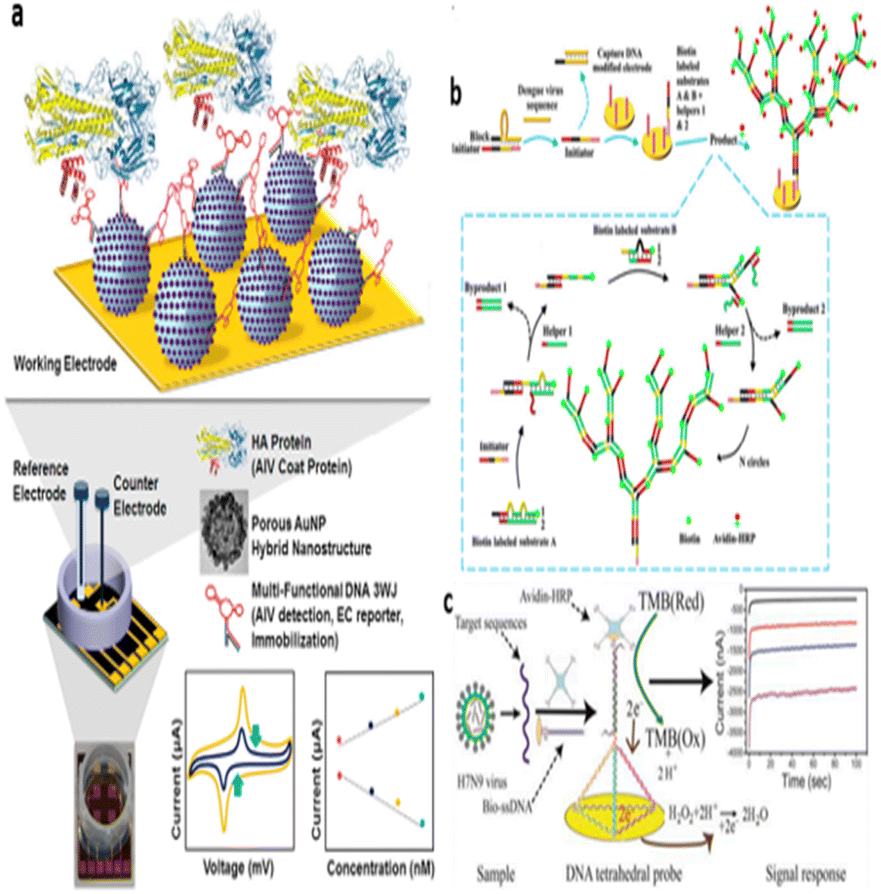 | ||
| Fig. 7 Electrochemical DNA nanobiosensors for virus detection. (a) Schematic image of the fabricated AIV detection biosensor.65 Copyright 2019, Elsevier. (b) Schematic illustration of triplet nanostructure-mediated dendritic HCR for electrochemical detection of DENV.66 Copyright 2021, Elsevier. (c) Schematic diagram of an electrochemical biosensor based on DNA tetrahedral nanostructures for the detection of H7N9 virus.67 Copyright 2015, American Chemical Society. | ||
Dong et al. created an electrochemical biosensor using DNA tetrahedral nanostructures to detect the H7N9 virus by detecting portions of the hemagglutinin gene sequence (Fig. 7c).67 A DNA tetrahedral probe was mounted on a gold electrode surface, and hybridization to the target ssDNA was accomplished through the self-assembly of three thiol-modified nucleotide sequences and longer nucleotide sequences containing complementary DNA. The collected target DNA strands hybridize to a biotinylated ssDNA probe, which is then activated by the affinity hormone horseradish peroxidase, resulting in an amperometric signal when reacting with a 3,3′,5,5′-tetramethylaniline substrate. This biosensor identifies the H7N9 virus DNA uniquely, distinguishing it from other influenza viruses (e.g., H1N1 and H3N2) and single-base mismatched oligonucleotides, with a limit of detection (LOD) of 100 fM. The study also shows that DNA tetrahedral probe-based electrochemical biosensors can detect target DNA in clinical samples.
The tetrahedral DNA maximizes surface area, allowing for greater binding capacity and improving sensitivity, especially critical for detecting low viral loads. Furthermore, the high specificity of the tetrahedral DNA probe reduces the likelihood of false positives, thus ensuring reliable results essential for clinical applications. The study also underscores the practical potential of this technology, demonstrating how tetrahedral DNA nanostructures can be used in real-world virus detection scenarios. This work opens up possibilities for rapid diagnostic tools that could play a vital role in managing viral outbreaks effectively.
Mahmoodi et al. described a DNA-based, selective, and sensitive electrochemical biosensor for HPV-18 detection.68 They deposited reduced graphene oxide (rGO) and multi-walled carbon nanotubes (MWCNTs) on printed carbon electrodes, then added gold nanoparticles (AuNPs) dropwise to the modified electrodes and immobilized ssDNA probes. DPV identified HPV-18 by measuring variations in the oxidation signal of anthraquinone sulfonate (AQMS) before and after probe hybridization with target DNA. The biosensor demonstrated linear detection throughout a concentration range of 0.01 fM to 0.01 nM, with an LOD of 0.05 fM, and performed well with clinical samples.
Impedance-based
Impedance-based sensors detect target molecules via impedance changes, as opposed to direct current (DC) measurements employed in voltammetry.69,70 Impedance-based DNA nanobiosensors produce DNA duplexes when biomolecules interact with DNA probes on the sensor surface. When an alternating electric field is provided, the process of duplex construction and breakdown influences the current through the sensor, resulting in a change in impedance. By monitoring this change, the target molecule's existence and concentration can be determined.71 These biosensors typically have low limits of detection (LOD) in the picomolar (pM) range and are highly stable, making them excellent for viral detection.72Shariati et al. created a label-free electrochemical impedance DNA biosensor for HBV detection that employs gold nanocrystals (AuNCs).73 This biosensor was created by immobilizing the HBV DNA probe (ssDNA) on the surface of AuNCs. The electrochemical impedance spectroscopy (EIS) investigation demonstrated that a LOD of 0.1 fM could be reached for HBV DNA, as well as the ability to differentiate between complementary and non-complementary DNA targets, including those with one, two, or three mismatches. The biosensor also showed the ability to detect HBV in serum samples. Wang et al. created an EIS-based DNA sensor to detect HBV and HPV.71 This sensor uses ssDNA probes that self-assemble on AuNP-coated single-walled carbon nanotubes (SWCNT/Au). The limit of detection (LOD) for this biosensor is 0.1 pM for HBV and 1 aM for HPV. Furthermore, this biosensor is extremely stable, delivering dependable and consistent results throughout time. Steinmetz et al. created a DNA biosensor for the Zika virus (ZIKV) in human serum samples by modifying an oxidized glassy carbon electrode (ox-GCE) with silsesquioxane-functionalized gold nanoparticles (AuNPs-SiPy) (Fig. 8a).74 The biosensor detects ZIKV by analyzing charge transfer resistance (ΔRct) and electrode surface roughness (Rq). The detection limit was 0.82 pM, with a linear range of 1.0 × 10⁻¹² to 1.0 × 10⁻⁶ M.
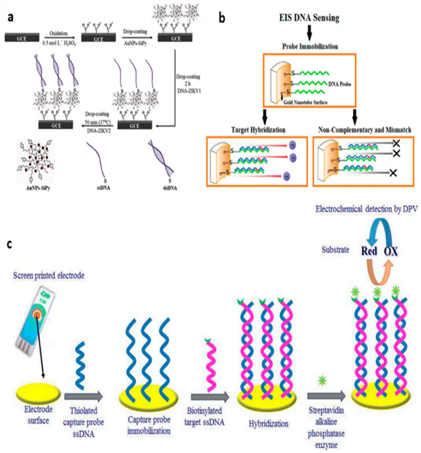 | ||
| Fig. 8 Impedance-based DNA nanobiosensors for virus detection. (a) Schematic representation of the construction and DNA hybridization stages of the oxidized-GCE-[AuNP-SiPy]/ZIKV1 biosensor.74 Copyright 2019, Elsevier. (b) Schematic representation of different steps for the fabrication of electrochemical DNA biosensors.75 Copyright 2018, Elsevier. (c) AuNT PC surface probe immobilization and hybridization of HPV-DNA target sequences with AuNT surface modifications and schematic diagrams.76 Copyright 2019, Elsevier. | ||
Ilkhani et al. created a unique electrochemical DNA nanobiosensor for detecting Ebola virus DNA, which uses an enzymatic amplification assay to improve sensitivity and selectivity (Fig. 8b).75 This nanobiosensor was created by immobilizing a sulfhydrylated DNA capture probe on a printed electrode, which then hybridized with a biotinylated target strand DNA. The biotinylated hybridization product was tagged with streptavidin–alkaline phosphatase conjugate on the working electrode's surface, allowing the enzymatic product to be detected using DPV. EIS was used to optimize the experimental technique, resulting in suitable circumstances for biosensor development. This nanobiosensor had a LOD of 4.7 nM for Ebola virus DNA, indicating its potential as a sensitive detection tool.
Zucolotto et al. demonstrated an electrochemical DNA nanobiosensor that detects the Zika virus label-free using an impedance method.77 This biosensor uses disposable electrodes made by thermal evaporation on a polyethylene terephthalate substrate covered with a nanoscale gold layer. A three-contact electrode arrangement can identify DNA sequences in a tiny sample volume, eliminating the necessity for labeling in Zika virus sequence identification. The nanobiosensor has a reaction time of 1.5 hours and a limit of detection (LOD) of 25.0 ± 1.7 nM using impedance measurements, indicating selectivity for Zika virus synthetic DNA.
Shariati et al. developed a label-free DNA nanobiosensor for HPV detection using gold nanotubes (AuNTs) (Fig. 8c).76 AuNT-modified nanoporous polycarbonate (AuNT-PC) templates were created by electrodeposition, and ssDNA probes were covalently anchored on the AuNT-PC electrodes. The hybridization between the target DNA sequence and the ssDNA probe was evaluated using the EIS approach. This nanobiosensor detects HPV DNA with a LOD of 1 fM and a linear response range of 0.01 pM to 1 μM. It is highly selective for complementary, mismatched, and non-complementary DNA sequences.
Advanced DNA-based biosensors
CRISPR-Cas12a
DNA origami have led to innovative detection tools in biological testing. One notable application is using DNA origami with CRISPR-Cas12a to achieve high-sensitivity detection of genetic mutations in cancer diagnostics. Researchers have engineered DNA origami structures that, when combined with CRISPR, can discriminate between single-base differences in DNA sequences, making it possible to identify specific cancer mutations at extremely low concentrations, even at the zeptomolar level (10−21 mol L−1). This level of sensitivity far surpasses conventional methods like PCR, allowing for more accurate detection of mutations crucial for personalized cancer treatment. The method has shown promise in detecting mutations in non-small cell lung cancer samples, even in cases where PCR failed to identify mutations, highlighting its potential to reduce false negatives in clinical diagnostics.78SPECTRA tool
Developed specifically for the detection of aggressive cancer cells, SPECTRA use DNA origami scaffolds combined with Raman spectroscopy to improve cancer imaging accuracy. It utilizes folded DNA structures to maintain Raman reporter genes and cancer-targeting DNA sequences in controlled arrangements, thereby enhancing signal strength. SPECTRA probes differentiate metastatic cancer cells by generating strong Raman signals in the presence of molecular markers specific to these cells. This allows clinicians to locate metastatic cells earlier and more accurately than traditional imaging methods such as MRI.79Challenges facing DNA nanotech
Although DNA nanotechnology has the potential to significantly advance therapeutic applications through sensing, stability concerns may arise about DNA structures. DNA molecules are intrinsically subject to deterioration by nucleases, which are enzymes that break down DNA. Furthermore, DNA structures can become unstable in settings that depart from physiological standards, such as high temperatures, severe pH levels, or the presence of specific chemicals. Ensuring the stability of DNA nanostructures in various conditions is critical for their practical uses, particularly in medicine and biotechnology. Strategies for improving stability, such as chemical changes to DNA or protective coatings, are being investigated, but obtaining consistent and long-term stability remains a challenge.80,81 Another key challenge is producing DNA nanostructures on a large scale suitable for clinical applications. While traditional pharmaceuticals and nanomedicines have well-established manufacturing procedures, DNA nanotechnology remains mostly in the research stage. It is difficult to increase the production of DNA nanostructures while maintaining their quality, usefulness, and safety. Furthermore, the cost of creating these nanostructures remains considerable, thereby limiting their accessibility and widespread use in the medical area. The use of DNA nanotechnology in medicine also raises regulatory and ethical concerns. The FDA and other regulatory organizations demand substantial testing and validation to assure the safety and efficacy of new medical innovations. Because DNA nanotechnology is innovative, existing regulatory frameworks may be inadequate to analyze it, thereby delaying approval and deployment. Furthermore, ethical issues are raised about the use of synthetic DNA, particularly in terms of genetic privacy, the possibility of unintentional genetic changes, and the long-term effects on human health.Conclusions
DNA origami not only simplifies the assembly process, but also allows for the production of nanostructures with complicated shapes and precise functionality. In terms of sensing, DNA nanostructures are extremely versatile in illness detection, providing early detection of genetic problems, infectious diseases, and many malignancies. These structures are used as platforms to detect specific biomolecular interactions with great sensitivity and precision. DNA nanotechnology represents a revolutionary approach to medicine, providing unique options for illness detection, therapy, and imaging. The amazing progress made in this discipline demonstrates its potential to change healthcare and pave the path for personalized therapy in the future.Data availability
No new data were created or analysed during this study. Data sharing is not applicable to this article.Author contributions
Darell Lien provided analysis on the use of DNA nanotechnology structures in research on medical sensing applications and technologies. He prepared the writing – original draft and writing – review and editing.Conflicts of interest
There are no conflicts to declare.Notes and references
- N. C. Seeman, Nucleic Acid Junctions and Lattices, J. Theor. Biol., 1982, 237–247, DOI:10.1016/0022-5193(82)90002-9.
- N. C. Seeman, DNA in a material world, Nature, 2003, 421, 427–431, DOI:10.1038/nature01406.
- P. W. Rothemund, Folding DNA to create nanoscale shapes and patterns, Nature, 2006, 440(7082), 297–302, DOI:10.1038/nature04586.
- S. M. Douglas, H. Dietz, T. Liedl, B. Högberg, F. Graf and W. M. Shih, Self-assembly of DNA into nanoscale three-dimensional shapes, Nature, 2009, 459(7245), 414–418, DOI:10.1038/nature08016.
- S. Liu, W. Su, Z. Li and X. Ding, Electrochemical detection of lung cancer specific microRNAs using 3D DNA origami nanostructures, Biosens. Bioelectron., 2015, 71, 57–61, DOI:10.1016/j.bios.2015.04.006.
- M. Endo and H. Sugiyama, Single-molecule imaging of dynamic motions of biomolecules in DNA origami nanostructures using high-speed atomic force microscopy, Acc. Chem. Res., 2014, 47(6), 1645–1653, DOI:10.1021/ar400299m.
- Y. Ke, S. Lindsay, Y. Chang, Y. Liu and H. Yan, Self-assembled water-soluble nucleic acid probe tiles for label-free RNA hybridization assays, Science, 2008, 319(5860), 180–183, DOI:10.1126/science.1150082.
- A. Kuzuya, Y. Sakai, T. Yamazaki, Y. Xu and M. Komiyama, Nanomechanical DNA origami ‘single-molecule beacons’ directly imaged by atomic force microscopy, Nat. Commun., 2011, 2(1), 449, DOI:10.1038/ncomms1452.
- H. K. Walter, J. Bauer, J. Steinmeyer, A. Kuzuya, C. M. Niemeyer and H. A. Wagenknecht, DNA Origami Traffic Lights with a Split Aptamer Sensor for a Bicolor Fluorescence Readout, Nano Lett., 2017, 17(4), 2467–2472, DOI:10.1021/acs.nanolett.7b00159.
- M. Liu, et al., A DNA tweezer-actuated enzyme nanoreactor, Nat. Commun., 2013, 4, 2127, DOI:10.1038/ncomms3127.
- Y. Ke, T. Meyer, W. M. Shih and G. Bellot, Regulation at a distance of biomolecular interactions using a DNA origami nanoactuator, Nat. Commun., 2016, 7, 10935, DOI:10.1038/ncomms10935.
- Y. Hua, J. Ma, D. Li and R. Wang, DNA-Based Biosensors for the Biochemical Analysis: A Review, Biosensors, 2022, 12(3), 183, DOI:10.3390/bios12030183.
- J. Zhai, H. Cui and R. Yang, DNA based biosensors, Biotechnol. Adv., 1997, 15(1), 43–58, DOI:10.1016/s0734-9750(97)00003-7.
- E. Sameiyan, E. Bagheri, M. Ramezani, M. Alibolandi, K. Abnous and S. M. Taghdisi, DNA origami-based aptasensors, Biosens. Bioelectron., 2019, 143, 111662, DOI:10.1016/j.bios.2019.111662.
- Y. Liu, S. Kumar and R. E. Taylor, Mix-and-match nanobiosensor design: Logical and spatial programming of biosensors using self-assembled DNA nanostructures, Wiley Interdiscip. Rev.:Nanomed. Nanobiotechnol., 2018, 10(6), e1518, DOI:10.1002/wnan.1518.
- S. Fan, J. Cheng, Y. Liu, D. Wang, T. Luo, B. Dai, C. Zhang, D. Cui, Y. Ke and J. Song, Proximity-Induced Pattern Operations in Reconfigurable DNA Origami Domino Array, J. Am. Chem. Soc., 2020, 142(34), 14566–14573, DOI:10.1021/jacs.0c06061.
- N. Xie, S. Liu, X. Yang, X. He, J. Huang and K. Wang, DNA tetrahedron nanostructures for biological applications: biosensors and drug delivery, Analyst, 2017, 142(18), 3322–3332, 10.1039/c7an01154g.
- L. Tong, H. Xu and M. Käll, Nanogaps for SERS applications, MRS Bull., 2014, 39(2), 163–168, DOI:10.1557/mrs.2014.2.
- C. Song, et al., High-Sensitive Assay of Nucleic Acid Using Tetrahedral DNA Probes and DNA Concatamers with a Surface-Enhanced Raman Scattering/Surface Plasmon Resonance Dual-Mode Biosensor Based on a Silver Nanorod-Covered Silver Nanohole Array, ACS Appl. Mater. Interfaces, 2020, 12(28), 31242–31254, DOI:10.1021/acsami.0c08453.
- I. Khalil, et al., Graphene oxide and gold nanoparticle based dual platform with short DNA probe for the PCR free DNA biosensing using surface-enhanced Raman scattering, Biosens. Bioelectron., 2019, 131, 214–223, DOI:10.1016/j.bios.2019.02.028.
- G. M. Das, S. Managò, M. Mangini and A. C. De Luca, Biosensing Using SERS Active Gold Nanostructures, Nanomaterials, 2021, 11, 2679, DOI:10.3390/nano11102679.
- Q. Li, H. Huo, Y. Wu, L. Chen, L. Su, X. Zhang, J. Song and H. Yang, Design and Synthesis of SERS Materials for In Vivo Molecular Imaging and Biosensing, Adv. Sci., 2023, 10, 2202051, DOI:10.1002/advs.202202051.
- E. Seymour, F. Ekiz Kanik, S. Diken Gür, M. Bakhshpour-Yucel, A. Araz, N. Lortlar Ünlü and M. S. Ünlü, Solid-Phase Optical Sensing Techniques for Sensitive Virus Detection, Sensors, 2023, 23, 5018, DOI:10.3390/s23115018.
- J. Xia, W. Li, M. Sun and H. Wang, Application of SERS in the Detection of Fungi, Bacteria and Viruses, Nanomaterials, 2022, 12, 3572, DOI:10.3390/nano12203572.
- S. M. Mousavi, S. A. Hashemi, V. Rahmanian, M. Y. Kalashgrani, A. Gholami, N. Omidifar and W.-H. Chiang, Highly Sensitive Flexible SERS-Based Sensing Platform for Detection of COVID-19, Biosensors, 2022, 12, 466, DOI:10.3390/bios12070466.
- F. Saviñon-Flores, E. Méndez, M. López-Castaños, A. Carabarin-Lima, K. A. López-Castaños, M. A. González-Fuentes and A. Méndez-Albores, A Review on SERS-Based Detection of Human Virus Infections: Influenza and Coronavirus, Biosensors, 2021, 11, 66, DOI:10.3390/bios11030066.
- C. Song, J. Zhang, Y. Liu, X. Guo, Y. Guo, X. Jiang and L. Wang, Highly sensitive SERS assay of DENV gene via a cascade signal amplification strategy of localized catalytic hairpin assembly and hybridization chain reaction, Sens. Actuators, B, 2020, 325, 128970, DOI:10.1016/j.snb.2020.128970.
- K. S. Park, A. Choi, H. J. Kim, I. Park, M.-S. Eom, S.-G. Yeo, R. G. Son, T.-I. Park, G. Lee, H. Tom Soh, Y. Hong and S. Pil Pack, Ultra-sensitive label-free SERS biosensor with high-throughput screened DNA aptamer for universal detection of SARS-CoV-2 variants from clinical samples, Biosens. Bioelectron., 2023, 228, 115202, DOI:10.1016/j.bios.2023.115202.
- Y. S. Huh, A. J. Chung, B. Cordovez and D. Erickson, Enhanced on-chip SERS based biomolecular detection using electrokinetically active microwells, Lab Chip, 2009, 9(3), 433–439, 10.1039/b809702j.
- Y. W. Fen, W. M. M. Yunus, N. A. Yusof, N. S. Ishak, N. A. S. Omar and A. A. Zainudin, Preparation, characterization and optical properties of ionophore doped chitosan biopolymer thin film and its potential application for sensing metal ion, Optik, 2015, 126(23), 4688–4692, DOI:10.1016/j.ijleo.2015.08.098.
- A. A. Zainudin, Y. W. Fen, N. A. Yusof and N. A. S. Omar, Structural, optical and sensing properties of ionophore doped graphene based bionanocomposite thin film, Optik, 2017, 144, 308–315, DOI:10.1016/j.ijleo.2017.07.001.
- M. Dass, F. N. Gür, K. Kołątaj, M. J. Urban and T. Liedl, DNA Origami-Enabled Plasmonic Sensing, J. Phys. Chem. C, 2021, 125(11), 5969–5981, DOI:10.1021/acs.jpcc.0c11238.
- N. A. S. Omar and Y. W. Fen, Recent development of SPR spectroscopy as potential method for diagnosis of dengue virus E-protein, Sens. Rev., 2018, 38(1), 106–116, DOI:10.1108/SR-07-2017-0130.
- S. Murali, R. R. Rustandi, X. Zheng, A. Payne and L. Shang, Applications of Surface Plasmon Resonance and Biolayer Interferometry for Virus-Ligand Binding, Viruses, 2022, 14(4), 717, DOI:10.3390/v14040717.
- K. Takemura, Surface Plasmon Resonance (SPR)- and Localized SPR (LSPR)-Based Virus Sensing Systems: Optical Vibration of Nano- and Micro-Metallic Materials for the Development of Next-Generation Virus Detection Technology, Biosensors, 2021, 11, 250, DOI:10.3390/bios11080250.
- M. M. Hassan, F. S. Sium, F. Islam and S. M. Choudhury, A review on plasmonic and metamaterial based biosensing platforms for virus detection, Sens. Bio-Sens. Res., 2021, 33, 100429, DOI:10.1016/j.sbsr.2021.100429.
- P. S. Pandey, S. K. Raghuwanshi, A. Shadab, M. T. I. Ansari, U. K. Tiwari and S. Kumar, SPR Based Biosensing Chip for COVID-19 Diagnosis - A Review, IEEE Sens. J., 2022, 22(14), 13800–13810, DOI:10.1109/JSEN.2022.3181423.
- W. Diao, et al., Highly sensitive surface plasmon resonance biosensor for the detection of HIV-related DNA based on dynamic and structural DNA nanodevices, Biosens. Bioelectron., 2018, 100, 228–234, DOI:10.1016/j.bios.2017.08.042.
- T. Lee, G. H. Kim, S. M. Kim, K. Hong, Y. Kim, C. Park, H. Sohn and J. Min, Label-free localized surface plasmon resonance biosensor composed of multi-functional DNA 3 way junction on hollow Au spike-like nanoparticles (HAuSN) for avian influenza virus detection, Colloids Surf., B, 2019, 182, 110341, DOI:10.1016/j.colsurfb.2019.06.070.
- A. D. Chowdhury, et al., The detection and identification of dengue virus serotypes with quantum dot and AuNP regulated localized surface plasmon resonance, Nanoscale Adv., 2020, 2(2), 699–709, 10.1039/c9na00763f.
- N. Oliveira, et al., A sensitive and selective label-free electrochemical DNA biosensor for the detection of specific dengue virus serotype 3 sequences, Sensors, 2015, 15(7), 15562–15577, DOI:10.3390/s150715562.
- P. Damborský, J. Švitel and J. Katrlík, Optical biosensors, Essays Biochem., 2016, 60(1), 91–100, DOI:10.1042/EBC20150010.
- T. Celiker, et al., Fluorescent bioassay for SARS-CoV-2 detection using polypyrene-g-poly(ε-caprolactone) prepared by simultaneous photoinduced step-growth and ring-opening polymerizations, Mikrochim. Acta, 2022, 189(5), 202, DOI:10.1007/s00604-022-05244-2.
- A. Sharma, et al., Optical Biosensors for Diagnostics of Infectious Viral Disease: A Recent Update, Multidisciplinary Digital Publishing Institute (MDPI), 2021, DOI:10.3390/diagnostics11112083.
- F. Kulzer and M. Orrit, Single-molecule optics, Annu. Rev. Phys. Chem., 2004, 55, 585–611, DOI:10.1146/annurev.physchem.54.011002.103816.
- M. Shirani, H. Kalantari, M. J. Khodayar, M. Kouchak and N. Rahbar, A novel strategy for detection of small molecules based on aptamer/gold nanoparticles/graphitic carbon nitride nanosheets as fluorescent biosensor, Talanta, 2020, 219, 121235, DOI:10.1016/j.talanta.2020.121235.
- A. M. Salama, G. Yasin, M. Zourob and J. Lu, Fluorescent Biosensors for the Detection of Viruses Using Graphene and Two-Dimensional Carbon Nanomaterials, Biosensors, 2022, 12(7), 460, DOI:10.3390/bios12070460.
- F. Ekiz-Kanik, D. D. Sevenler, N. L. Ünlü, M. Chiari and M. S. Ünlü, Surface chemistry and morphology in single particle optical imaging, Nanophotonics, 2017, 6(4), 713–730, DOI:10.1515/nanoph-2016-0184.
- A. Sharma, R. Khan, G. Catanante, T. A. Sherazi, S. Bhand, A. Hayat and J. L. Marty, Designed Strategies for Fluorescence-Based Biosensors for the Detection of Mycotoxins, Toxins, 2018, 10(5), 197, DOI:10.3390/toxins10050197.
- H. Maddali, C. E. Miles, J. Kohn and D. M. O'Carroll, Optical Biosensors for Virus Detection: Prospects for SARS-CoV-2/COVID-19, ChemBioChem, 2021, 22, 1176, DOI:10.1002/cbic.202000744.
- W. Shen and Z. Gao, Quantum dots and duplex-specific nuclease enabled ultrasensitive detection and serotyping of Dengue viruses in one step in a single tube, Biosens. Bioelectron., 2015, 65, 327–332, DOI:10.1016/j.bios.2014.10.060.
- P. Teengam, N. Nisab, N. Chuaypen, P. Tangkijvanich, T. Vilaivan and O. Chailapakul, Fluorescent paper-based DNA sensor using pyrrolidinyl peptide nucleic acids for hepatitis C virus detection, Biosens. Bioelectron., 2021, 189, 113381, DOI:10.1016/j.bios.2021.113381.
- P. S. Kwon, et al., Designer DNA architecture offers precise and multivalent spatial pattern-recognition for viral sensing and inhibition, Nat. Chem., 2020, 12(1), 26–35, DOI:10.1038/s41557-019-0369-8.
- N. Chauhan, et al., Net-Shaped DNA Nanostructures Designed for Rapid/Sensitive Detection and Potential Inhibition of the SARS-CoV-2 Virus, J. Am. Chem. Soc., 2023, 145(37), 20214–20228, DOI:10.1021/jacs.2c04835.
- J. Jiao, C. Duan, L. Xue, Y. Liu, W. Sun and Y. Xiang, DNA nanoscaffold-based SARS-CoV-2 detection for COVID-19 diagnosis, Biosens. Bioelectron., 2020, 167, 112479, DOI:10.1016/j.bios.2020.112479.
- S. E. Ochmann, C. Vietz, K. Trofymchuk, G. P. Acuna, B. Lalkens and P. Tinnefeld, Optical Nanoantenna for Single Molecule-Based Detection of Zika Virus Nucleic Acids without Molecular Multiplication, Anal. Chem., 2017, 89(23), 13000–13007, DOI:10.1021/acs.analchem.7b04082.
- A. D. Chowdhury, et al., Femtomolar Detection of Dengue Virus DNA with Serotype Identification Ability, Anal. Chem., 2018, 90(21), 12464–12474, DOI:10.1021/acs.analchem.8b01802.
- L. Gogianu, et al., Microarray biochip fabricated on silicon nanowires/carbon dots heterostructures for enhanced viral DNA detection, Appl. Surf. Sci., 2023, 636, 157878, DOI:10.1016/j.apsusc.2023.157878.
- S. Li, C. Li, Y. Wang, H. Li and F. Xia, Re-engineering Electrochemical Aptamer-Based Biosensors to Tune Their Useful Dynamic Range via Distal-Site Mutation and Allosteric Inhibition, Anal. Chem., 2020, 92(19), 13427–13433, DOI:10.1021/acs.analchem.0c02782.
- H. Huang, W. Bai, C. Dong, R. Guo and Z. Liu, An ultrasensitive electrochemical DNA biosensor based on graphene/Au nanorod/polythionine for human papillomavirus DNA detection, Biosens. Bioelectron., 2015, 68, 442–446, DOI:10.1016/j.bios.2015.01.039.
- E. Vermisoglou, D. Panáček, K. Jayaramulu, M. Pykal, I. Frébort, M. Kolář, M. Hajdúch, R. Zbořil and M. Otyepka, Human virus detection with graphene-based materials, Biosens. Bioelectron., 2020, 166, 112436, DOI:10.1016/j.bios.2020.112436.
- K. Kurzatkowska, A. Sirko, W. Zagórski-Ostoja, W. Dehaen, H. Radecka and J. Radecki, Electrochemical Label-free and Reagentless Genosensor Based on an Ion Barrier Switch-off System for DNA Sequence-Specific Detection of the Avian Influenza Virus, Anal. Chem., 2015, 87(19), 9702–9709, DOI:10.1021/acs.analchem.5b01988.
- E. Kianfar, M. Salimi and B. Koohestani, Methanol to Gasoline Conversion over CuO/ZSM-5 Catalyst Synthesized and Influence of Water on Conversion, Fine Chem. Eng., 2020, 75–82, DOI:10.37256/fce.122020499.
- E. Kianfar, An Experimental Study PVDF and PSF Hollow Fiber Membranes for Chemical Absorption Carbon Dioxide, Fine Chem. Eng., 2020, 92–103, DOI:10.37256/fce.122020552.
- T. Lee, et al., Fabrication of electrochemical biosensor consisted of multi-functional DNA structure/porous au nanoparticle for avian influenza virus (H5N1) in chicken serum, Mater. Sci. Eng., C, 2019, 99, 511–519, DOI:10.1016/j.msec.2019.02.001.
- J. Fu, J. Wu, R. Zhang, Q. Wu and H. Ju, Electrochemical biosensing of DENV nucleic acid amplified with triplet nanostructure-mediated dendritic hybridization chain reaction, Sens. Actuators, B, 2021, 345, 130436, DOI:10.1016/j.snb.2021.130436.
- S. Dong, et al., Electrochemical DNA biosensor based on a tetrahedral nanostructure probe for the detection of avian influenza A (H7N9) virus, ACS Appl. Mater. Interfaces, 2015, 7(16), 8834–8842, DOI:10.1021/acsami.5b01438.
- P. Mahmoodi, M. Rezayi, E. Rasouli, A. Avan, M. Gholami, M. Ghayour Mobarhan, E. Karimi and Y. Alias, Early-stage cervical cancer diagnosis based on an ultra-sensitive electrochemical DNA nanobiosensor for HPV-18 detection in real samples, J. Nanobiotechnol., 2020, 18(1), 11, DOI:10.1186/s12951-020-0577-9.
- X. Weng, C. Li, C. Chen, G. Wang, C. Xia and L. Zheng, A Microfluidic Device for Tobacco Ringspot Virus Detection by Electrochemical Impedance Spectroscopy, Micromachines, 2023, 14, 1118, DOI:10.3390/mi14061118.
- Y.-S. Chen, C.-H. Huang, P.-C. Pai, J. Seo and K. F. Lei, A Review on Microfluidics-Based Impedance Biosensors, Biosensors, 2023, 13, 83, DOI:10.3390/bios13010083.
- S. Wang, et al., Electrochemical detection of hepatitis B and papilloma virus DNAs using SWCNT array coated with gold nanoparticles, Biosens. Bioelectron., 2013, 41(1), 205–210, DOI:10.1016/j.bios.2012.08.021.
- Z. Štukovnik and U. Bren, Recent Developments in Electrochemical-Impedimetric Biosensors for Virus Detection, Int. J. Mol. Sci., 2022, 23, 15922, DOI:10.3390/ijms232415922.
- M. Shariati, Impedimetric Biosensor for Monitoring Complementary DNA from Hepatitis B Virus Based on Gold Nanocrystals, J. Electrochem. Soc., 2021, 168(1), 016512, DOI:10.1149/1945-7111/abdc72.
- M. Steinmetz, et al., A sensitive label-free impedimetric DNA biosensor based on silsesquioxane-functionalized gold nanoparticles for Zika Virus detection, Biosens. Bioelectron., 2019, 141, 111351, DOI:10.1016/j.bios.2019.111351.
- H. Ilkhani and S. Farhad, A novel electrochemical DNA biosensor for Ebola virus detection, Anal. Biochem., 2018, 557, 151–155, DOI:10.1016/j.ab.2018.06.010.
- M. Shariati, M. Ghorbani, P. Sasanpour and A. Karimizefreh, An ultrasensitive label free human papilloma virus DNA biosensor using gold nanotubes based on nanoporous polycarbonate in electrical alignment, Anal. Chim. Acta, 2019, 1048, 31–41, DOI:10.1016/j.aca.2018.09.062.
- H. A. M. Faria and V. Zucolotto, Label-free electrochemical DNA biosensor for zika virus identification, Biosens. Bioelectron., 2019, 131, 149–155, DOI:10.1016/j.bios.2019.02.018.
- J. S. Chen, et al., CRISPR-Cas12a target binding unleashes indiscriminate single-stranded DNase activity, Science, 2018, 360(6387), 436–439, DOI:10.1126/science.aar6245.
- L. Wu, S. Tanwar, G. Kaur, S. Date, L. Goel, A. Chatterjee, P. McGuiggan and I. Barman, DNA Origami-Engineered Plasmonic Nanoprobes for Targeted Cancer Imaging, Adv. Funct. Mater., 2024, 34(30), 2309929, DOI:10.1002/adfm.202309929.
- N. P. Agarwal, M. Matthies, F. N. Gür, K. Osada and T. L. Schmidt, Block Copolymer Micellization as a Protection Strategy for DNA Origami, Angew. Chem., Int. Ed., 2017, 56(20), 5460–5464, DOI:10.1002/anie.201608873.
- N. Ponnuswamy, M. M. C. Bastings, B. Nathwani, J. H. Ryu, L. Y. T. Chou, M. Vinther, W. A. Li, F. M. Anastassacos, D. J. Mooney and W. M. Shih, Oligolysine-based coating protects DNA nanostructures from low-salt denaturation and nuclease degradation, Nat. Commun., 2017, 8, 15654, DOI:10.1038/ncomms15654.
| This journal is © The Royal Society of Chemistry 2025 |