
 Open Access Article
 This Open Access Article is licensed under a Creative Commons Attribution-Non Commercial 3.0 Unported Licence
Effect of the Sr–Fe layered double hydroxide coating based on the microenvironment response on implant osseointegration in osteoporotic rats†
Chenyu
Liao
a,
Dongcai
He
b,
Kaiwen
Yin
a,
Yuhung
Lin
 a,
Yihan
Chen
fg,
Ziqiang
Zhang
b,
Jing
Zhang
e,
Hongrong
Luo
d,
Xianchun
Chen
*c and
Yunfeng
Li
*a
a,
Yihan
Chen
fg,
Ziqiang
Zhang
b,
Jing
Zhang
e,
Hongrong
Luo
d,
Xianchun
Chen
*c and
Yunfeng
Li
*a
aState Key Laboratory of Oral Diseases & National Center for Stomatology & National Clinical Research Center for Oral Diseases & Department of Oral & Maxillofacial Surgery, West China Hospital of Stomatology, Sichuan University, Chengdu, 610041, China. E-mail: liyunfeng@scu.edu.cn
bCollege of Materials Science and Engineering, Sichuan University, No. 24 South Section 1, Yihuan Road, Chengdu, 610065, China
cCollege of Polymer Science and Engineering, Sichuan University, No. 24 South Section 1, Yihuan Road, Chengdu, 610065, China. E-mail: chenxianchun@scu.edu.cn
dCollege of Biomedical Engineering, Sichuan University, No. 24 South Section 1, Yihuan Road, Chengdu, 610065, China
eCollege of Architecture and Environment, Sichuan University, Chengdu, Sichuan 610065, China
fShanghai Institute of Ceramics, Chinese Academy of Science, Research Unit of Nanocatalytic Medicine iSpecific Therapy for Serious Disease, Chinese Academy of Medical Sciences, Shanghai 200050, P.R. China
gCenter of Materials Science and Optoelectronics Engineering, University of Chinese Academy of Sciences, Beijing 100049, P.R. China
First published on 12th January 2024
Abstract
Osteoporosis is a disease that manifests itself as an abnormality of bone metabolism and is characterized by low bone mass and destruction of the bone microstructure. Since bone resorption occurs more rapidly than new bone formation, osteoporosis leads to reduced orthopedic implant stability. From a microenvironmental point of view, the rationale for this outcome is that osteoclasts are overactive in the bone tissue of patients with osteoporosis, and the large amount of H+ they produce leads to local chronic acidosis, which promotes bone mineral loss. Therefore, we designed a weakly alkaline layered double hydroxide (LDH) coating to modulate the pathologically acidic microenvironment and the osteogenic–osteoclastic coupling by releasing Sr2+. We prepared Sr–Fe LDH coatings on pure titanium implants using a hydrothermal method in this study and characterized the material using SEM, AFM, XRD, XPS, EDS, ICP, pH acidimeter, etc. We found that the coatings had good nanomorphology and were able to efficiently neutralize H+ as well as steadily release Sr2+ for up to 21 days. In vitro, the coating not only significantly promoted the adhesion, proliferation, and differentiation of osteoblasts, but also inhibited the differentiation of osteoclasts at the same time. In addition, in animal experiments, the coating significantly improved the mechanical stability of the implant in osteoporotic rats, increasing Sr–Fe LDH@Ti maximal push-out force by 72.2% compared to Ti. At the same time, the coating was effective in reversing the osteoporotic state, resulting in a 58.5% increase in BV/TV (%), and a 12.4% increase in Tb. N (1 mm−1), a 31.6% increase in Tb. Th (μm), and a 30.9% increase in BA (%). Our results suggest that this Sr–Fe LDH nanocoating material with acid-neutralizing, as well as long-term Sr2+-releasing capabilities, is a novel and effective orthopedic implant coating material under osteoporotic conditions.
1. Introduction
Today, osteoporosis affects more than 1.02 billion people worldwide,1 and the vast majority of these people are postmenopausal women and middle-aged and older men.2 There is a very high demand for bone implants as these people are at high risk of tooth loss and bone joint diseases.3It is well known that in osteoporotic patients, it is difficult to form enough bone to support the implant because of the osteogenic–osteoclastic uncoupling, which leads to loosening and detachment of the implant.4–6
In osteoporosis, the bone tissue microenvironment is characterized by reduced osteoblast activity and impaired bone tissue regeneration. In addition, hyperactive osteoclasts produce a slightly acidic environment by secreting large quantities of hydrogen ions, resulting in bone mineral loss and destruction of the bone microstructure. In addition, a high concentration of H+ will reduce osteoblast activity and inhibit bone tissue regeneration.7–9 All of these factors affect bone metabolism and inhibit osseointegration, thereby increasing the risk of implant failure.10 Therefore, in order to improve the osseointegration of implants, the coating material should not only focus on the modulation of cellular function but also on the modulation of the pathological acidic microenvironment.11–13
In order to modulate cellular function, many studies have been conducted to release growth factors such as bone morphogenetic proteins (BMPs), transforming growth factor-β (TGF-β), vascular endothelial growth factor (VEGF), interleukin-4 (IL-4), and interleukin-10 (IL-10) by constructing coatings on the implant surface that can be loaded with drugs.14–18 However, these drugs, cytokines, or growth factors have disadvantages such as short half-life, susceptibility to inactivation, lack of long-term stability, tissue selectivity, potential toxicity, and dose-dependence,19,20 making it difficult to achieve better results. The strategy of introducing strontium elements into coating materials and modifying pure titanium and titanium alloy implants to modulate cellular function is a strategy that is simple to prepare, inexpensive, and no less effective than strategies based on the release of drugs, cytokines, and growth factors and it has already produced impressive results. Coatings containing strontium can emit Sr2+ relatively steadily during in vivo degradation. Sr2+ is a known biologically active ion that, at appropriate concentrations,21–23 increases pro-osteoblast activity and inhibits osteoclast overactivity, thereby regulating bone metabolism and promoting osseointegration.24–28
Layered double hydroxide (LDH), a biomaterial composed of a body laminate of divalent and trivalent cations and a guest laminate of anions, has been investigated extensively in the field of bone repair.29,30 The formula for this substance is [M1−x2+Nx3+(OH)2]x+[Anx/nn−]x−·mH2O. LDH's chemical composition is modifiable, and various biological effects can be conferred on LDH by introducing different bioactive elements.31,32 Notably, LDH is an alkaline substance, and its first biomedical application was as a pH-raising anti-acid medication. LDH has an acidic environmental response, and LDH coatings containing bioactive elements can undergo an acid-neutralization reaction with H+ in acidic environments and degrade swiftly, releasing bioactive ions. In contrast, it self-degrades slowly in neutral and alkaline environments.33,34
Therefore, in order to regulate the abnormal acidic microenvironment of osteoporosis, as well as to improve the dysfunctional state of osteoblasts and osteoclasts, and ultimately to enhance osteointegration, we combined the excellent performance of Sr2+ in improving bone metabolism in this work with the ability of alkaline LDH to neutralize acid and utilized the biologically-safe Fe3+ to assist in the construction of the Sr–Fe LDH coating. To verify that this was achieved, we used ICP to measure the ability of Sr2+ to be released in PBS and a pH acidimeter to detect the ability of Sr–Fe LDH to neutralize H+. The effects of Sr–Fe LDH coating on the function of osteoblasts and osteoclasts were investigated using mouse embryonic osteoblasts (3T3-E1) and mouse monocyte macrophage leukemia cells (RAW264.7) as cell models. The effect of Sr–Fe LDH coating on osseointegration was investigated using ovariectomized rats as a model. Our results show that the Sr–Fe LDH coating can release Sr2+ for a long time and stably, and has a good acid-neutralising ability. In vitro, it can significantly promote the proliferation and differentiation of osteoblasts while inhibiting the differentiation of osteoclasts. In addition, the Sr–Fe LDH coating was able to improve the mechanical stability, peripheral bone formation, and osseointegration of implants in vivo.
2. Methods
2.1 Preparation of materials
The pure titanium size used for in vitro experiments in this study was 10 mm × 1 mm and the bar implant size used for in vivo experiments was 10 mm × 1 mm. The surface of pure titanium was ground, polished, and then acid etched for 90 s using 0.2 vol% hydrofluoric acid. We used the most widely used and simple hydrothermal method for the preparation of Sr–Fe LDH: firstly, 4.008 g of sodium hydroxide was added to the PTFE liner with 60 ml of deionised water, and mechanically stirred completely and allowed to dissolve, then pure titanium flakes or rods were added, followed by the addition of 1.5899 g of sodium carbonate, 6.2172 g of strontium oxide, and 6.4122 g of ferric hydroxide, in that order. Stirring was continued for 30 min. Finally, the PTFE liner was placed in a hydrothermal kettle and the hydrothermal reaction was carried out at a temperature of 120 °C for 14 h. To remove the residual impurities, the prepared material was required to be ultrasonically cleaned with 50% ethanol more than 5 times. In this work, we refer to pure titanium sheets and pure titanium implants as Ti, and to sheets and implants prepared with Sr–Fe LDH coatings as Sr–Fe LDH@Ti.2.2 Characterization of materials
A field emission scanning electron microscopy (FE-SEM, Apreo 2 S Lovac, Czech Republic) and an atomic force microscope (AFM, SPM-9600, Japan) were used to investigate the surface morphology and the roughness of the samples. The crystal structure of the samples was examined via X-ray diffraction (XRD). X-ray photoelectron spectroscopy (XPS) was used to analyze the elemental composition and chemical state of the samples. The thickness, elemental species, and elemental distribution of the samples were detected using X-ray energy spectrometry (EDS). The hydrophilicity of the materials was analyzed using an optical contact angle meter (DSA25E, DEU). Sr2+, which has a bone repair regulatory function, was quantified using inductively coupled plasma emission spectroscopy (ICP-OES). Analysis of the acid-neutralizing capacity of samples using a pH acidimeter.2.3 In vitro study
![[thin space (1/6-em)]](https://www.rsc.org/images/entities/char_2009.gif) :3 or 1:4.
:3 or 1:4.
Method for laser confocal microscopy observation: 500 ml of MC3T3-E1 and RAW264.7 cell suspensions were prepared at a density of 1 × 10⁴ cells per ml and 2 × 10⁴ cells per ml, respectively. The cells were then inoculated onto the surface of the material and placed in 48-well plates, which were then placed in a cell-specific culture incubator at 37 °C for 24 hours. After twenty-four hours, the cells were rinsed twice with PBS and then incubated at room temperature. After two washes, the cells were sequentially fixed with 4% paraformaldehyde, permeabilized with 0.2% Triton X-100, and stained at room temperature with YF®/rhodamine-labelled ghost pen cyclic peptide (Uelandy, CHN) and DAPI (Biosharp, CHN) for the cell membrane and nucleus, respectively. Observations were carried out using a laser confocal microscope (Olympus SpinSR10, Japan).
The CCK8 method was used to determine the effect of the materials on the cell viability of MC3T3-E1 and RAW264. 7: 500 μl of the cell suspension was configured at a density of 1 × 10⁴ cells per ml and 2 × 10⁴ cells per ml, respectively, and then the cells were inoculated on the surface of the materials placed in a 48-well plate and co-cultured with the materials for 1, 3, and 5 days. Every two days, the medium was substituted with the new medium. After 2 thorough washes with 500 ml of PBS (biosharp, CHN) on 1, 3, and 5 days, 200 μl of 10% CCK8 solution (Biosharp, CHN) freshly configured with the culture medium was added to each well. The absorbance was measured at 450 nm using an enzyme marker (Synergy H1, USA) following a 2 hour incubation at 37 °C in a cell-specific incubator.
Method for ALP staining: 500 μl of the MC3T3-E1 cell suspension with a density of 4 × 10⁴ cells per ml was inoculated onto the surface of both materials in 48-well plates, respectively, and the new induction solution was replaced every 2 days. Following a 7 day incubation at 37 °C in a cell-specific incubator, the cells were rinsed twice with PBS and fixed with 4% paraformaldehyde. The cells were stained with a BCIP/NBT alkaline phosphatase chromogenic reagent (Beyotime, CHN) for 24 hours at room temperature. After adding 200 μl of DMSO to each well and stirring for 30 minutes to dissolve the blue alkaline phosphatase crystals, 100 μl of the solution was aspirated, and the absorbance was measured at 520 nm using an enzyme marker.
ARS staining method: 500 μl of the MC3T3-E1 cell suspension with a density of 4 × 10⁴ cells per ml was inoculated onto the surface of both materials placed in 48-well plates, respectively, and the new induction solution was replaced every 2 days. After 21 days of incubation at 37 °C in a cell-specific incubator, the cells were twice rinsed with PBS and then fixed with 4% paraformaldehyde. The cells were then stained for 24 hours at room temperature with 1% alizarin red solution (Solarbio, CHN). Finally, 200 μl of 10% cetylpyridinium chloride solution was added to each well, agitated for 30 minutes, and 100 μl of the solution was aspirated and analyzed using an enzyme marker for absorbance at 562 nm.
TRAP staining procedure: RAW264.7 cells were seeded at a density of 6000 cells per cm2 in 48-well plates, and the induction solution was replaced every two days. After 6 days of induction, the cells were rinsed twice with PBS and successively permeabilized with 4% paraformaldehyde fixation and 0.2% Triton-X 100. The cells were then stained for 1 hour with a TRAP staining reagent (Servicebio, CHN) and observed under a light microscope.
RAW264.7 cells were seeded onto the surface of the material in 48-well plates at a density of 6000 cells per cm2, and the induction solution was replaced every two days. After 6 days of induction, the cells were washed twice with PBS, fixed with 4% paraformalde, permeabilized with 0.2% Triton-X 100, and stained with YF®/rhodamine-labelled phantom pen cyclopeptide and DAPI, respectively, for cell membrane and nucleus. Lastly, laser confocal microscopy was utilized for observation purposes.
6000 cells per cm2 RAW264.7 were seeded onto the surface of the material in 48-well plates for RT-qPCR, and the induction solution was replaced every two days. After incubation for six days, the plate was rinsed twice with PBS. RNA was extracted using the RNAiso Plus kit. The relative expression of osteoblast-associated genes NFATc1, c-FOS, CTSK, and TRAP was determined using a real-time fluorescence quantitative PCR instrument (Bio-rad CFX96 Touch, USA) and -actin as an internal reference gene.
2.4 In vivo study
Regarding the method of anesthesia: we anesthetized the animals three times in our animal experiments: the osteoporosis model establishment experiments, the implant placement experiments, and at the time of animal execution. We anesthetized the rats by intraperitoneal injection of 3% sodium pentobarbital solution (45 mg kg−1). At the same time, 2% lidocaine was used for local infiltration anesthesia within the incision area to achieve the best anesthesia effect.
Regarding the method of execution: we euthanized the animals by injecting an overdose of 3% pentobarbital sodium solution (150 mg kg−1) into the peritoneal cavity of SD rats.
Ti and Sr–Fe LDH@Ti were implanted into the bone marrow compartments of the bilateral tibiae of rats in both groups parallel to the long axis of the tibiae and slightly below the articular surface twelve weeks after bilateral ovariectomy. All rodents received intramuscular antibiotics and analgesics for three days following surgery. After 8 W of implant placement, all rodents were euthanized, and specimens were collected for further study.
000 μm). The VOI region was evaluated for bone volume fraction (BV/TV), trabecular thickness (Tb. Th), trabecular number (Tb. N), trabecular separation (Tb. Sp), and bone-implant contact (BIC).
2.5 Statistical analysis
All data were expressed as mean ± standard deviation and statistically analyzed using IBM SPSS Statistics 26 software (SPSS, 14hicago, IL, USA). Comparisons between groups were made using the independent samples t-test. p < 0.05 was considered statistically significant.3. Result and discussion
3.1 Synthesis and characterization of the Sr–Fe LDH coating
Scanning electron microscopy (Fig. 1A) and atomic force microscopy (Fig. 1B and Fig. S1A, ESI†) were utilized to observe the morphology of the samples, and uniform nanospherical Sr–Fe LDHs were found on the surface of pure titanium but there is no significant difference in roughness between Ti and Sr–Fe LDH@Ti (Fig. S1B, ESI†). We then conducted XRD and XPS analyses. In the XRD mapping characterization of Ti and Sr–Fe LDH@Ti (Fig. 1C), it can be seen that Sr–Fe LDH@Ti, in addition to the diffraction peaks of Ti, diffraction peaks are also detected at around 10° and 20°, corresponding to the (0 0 3) and (0 0 6) crystal planes, which are consistent with the typical LDH physical structure (PDF# 50-0652). From the XRD patterns (Fig. S2, ESI†), we can observe that the Sr–Fe LDH powders are mainly composed of two main phases, namely Sr3Fe2OH12 and FeOOH. The diffraction peaks of these phases exhibit sharp and symmetric features, which is a typical sign of the integrity and crystallinity of the crystal structure. This means that during the synthesis of Sr–Fe LDH, both phases formed a structure with excellent crystallinity, and it can also prove that the synthesis of Sr–Fe LDH was successful. XPS spectroscopy analysis was performed to further ascertain the surface elemental states of the coatings, the Ti, Fe, Sr, C, and O elements were observed in the complete XPS spectra of the sample surface (Fig. 1D). A split-peak fitting procedure was performed on the Fe 2p binding energy peaks (Fig. 1E), and it can be seen that the peaks at 714.5 eV and 724.8 eV represent the peaks of Fe 2p3/2 and Fe 2p1/2, whereas the binding energy peaks at 718.3 eV and 732.4 eV are satellite peaks of Fe 2p3/2 and Fe 2p1/2. This indicates the presence of both Fe2+ and Fe3+ in Sr–Fe LDH. It indicates that Sr–Fe LDH contains both Fe2+ and Fe3+. The results of the O 1s split peak fitting (Fig. 1F) reveal that the two peaks appearing at 529.1 and 531.2 eV correspond to metal oxides and metal hydroxides, respectively, and the ratio of the half-peak widths of the two peaks is close to 1:6, indicating that the majority of the elemental oxygen exists in the form of metal hydroxides. EDS (Fig. 1G) revealed that Sr, Fe, O, and C are uniformly distributed on the surface of pure titanium and Sr2+ of 6.72% weight percent (wt%) and 2.5% atomic percent (at%). Combining SEM and EDS analyses, we found that the thickness of the coating was 2.1 μm (Fig. S3, ESI†). Hydrophilicity is an important factor affecting the surface activity of implants, and the majority of studies35,36 have concluded that hydrophilic surfaces are preferable to hydrophobic surfaces for promoting early cell proliferation and osteogenic differentiation. Consequently, we employed the seated drop method for contact angle analysis of Ti and Sr–Fe LDH@Ti (Fig. 1H), and the result demonstrates that Sr–Fe LDH@Ti is more hydrophilic than Ti. Sr2+ is believed to promote osteoblast differentiation while inhibiting osteoclast differentiation,37,38 so to verify the degradation ability of the Sr–Fe LDH coating, we used inductively coupled plasma emission spectrometry (ICP) to quantify the Sr2+ released from Sr–Fe LDH@Ti in PBS buffer at a specific time. Quantitative analysis of Sr2+ in the medium at specific times by inductively plasma emission spectroscopy (ICP) (Fig. 1I) showed that Sr2+ was released at a high rate during 2 days, and then gradually reached the ion release equilibrium, which allowed Sr2+ to be released slowly. Among them, the Sr2+ release concentration of Sr–Fe LDH@Ti in pH = 4.5 PBS within 2 days was 38.3 mg L−1, while that at pH = 7.4 PBS was 8.05 mg L−1. Analyses by EDS showed that the content of Sr on the surface of the material after 7 days of immersion in pH = 4.5 solution was significantly lower than that in pH = 7.4 solution (Fig. S4, ESI†). The higher degradation rate in acidic PBS compared with that in neutral PBS illustrated the sensitive and efficient response of Sr–Fe LDH@Ti to the acidic environment. More importantly, Sr2+ could be slowly released in PBS at pH = 7.4 for up to 21 days (Fig. S5, ESI†), indicating that Sr–Fe LDH@Ti could slowly release Sr2+ for a long period, which is important for the long-term regulation of the local pathological microenvironment in osteoporosis. In the pathological microenvironment of osteoporosis, mature and functionally active osteoclasts secrete large amounts of H+ on the bone surface, leading to the dissolution of hydroxyapatite, the main inorganic component of bone tissue, and can promote the degradation of organic components by histaminase K (CTSK) and anti-tartaric acid phosphatase (TRAP).39 At the same time, large amounts of H+ negatively affect calcium salt deposition and mineralization of the bone-like material. From the point of view of the cell itself, excess H+ can have a great impact on osteoclast function, mainly in the form of increased expression of genes that promote bone resorption function, including carbonic anhydrase II,40 calcitonin receptor, tissue protease K (CTSK), and tartrate-resistant acid phosphatase (TRAP). Notably, since H+ is supplied by carbonic anhydrase II from osteoblasts, the low pH microenvironment prompts osteoblasts to secrete more H+, thus creating a vicious cycle that promotes bone resorption.41 For osteoblasts, excess H+ down-regulates the expression of osteogenesis-related genes, so it is important for materials to have good acid-neutralizing properties to inhibit bone resorption.7,34 Since Sr–Fe LDH is an alkaline material, we speculate that it can be degraded while neutralizing H+. To verify our conjecture, we formulated 50 mg ml−1 pulverized Sr–Fe LDH with purified water and compared it to gradients of NaOH concentration (Fig. 1J). It can be observed that its acid neutralization ability is between 0.1 M and 1 M NaOH, which is a strong indication of its excellent acid neutralization ability.
 | ||
| Fig. 1 (A) SEM images of Ti and Sr–Fe LDH@Ti. (B) AFM images and quantitative analysis of Sr–Fe LDH@Ti; (C) XRD images of Ti and Sr–Fe LDH@Ti. (D) Complete XPS spectrum images of Sr–Fe LDH@Ti. (E) Split-peak fitting of the Fe 2p binding energy peaks of Sr–Fe LDH@Ti. (F) O 1s split peak fitting images of Sr–Fe LDH@Ti. (G) Elemental analyses of Sr–Fe LDH@Ti by EDS. (H) Analyses of contact angle test of Ti and Sr–Fe LDH@Ti; n = 6. (I) The release curves of Sr2+ ions from Sr–Fe LDH@Ti. (J) pH-value monitoring during the titration of Sr–Fe LDHs and NaOH into acidic PBS (pH = 4.5). All values are presented as mean ± s.d. *p < 0.05, **p < 0.01, and ***p < 0.001. | ||
3.2 In vitro study
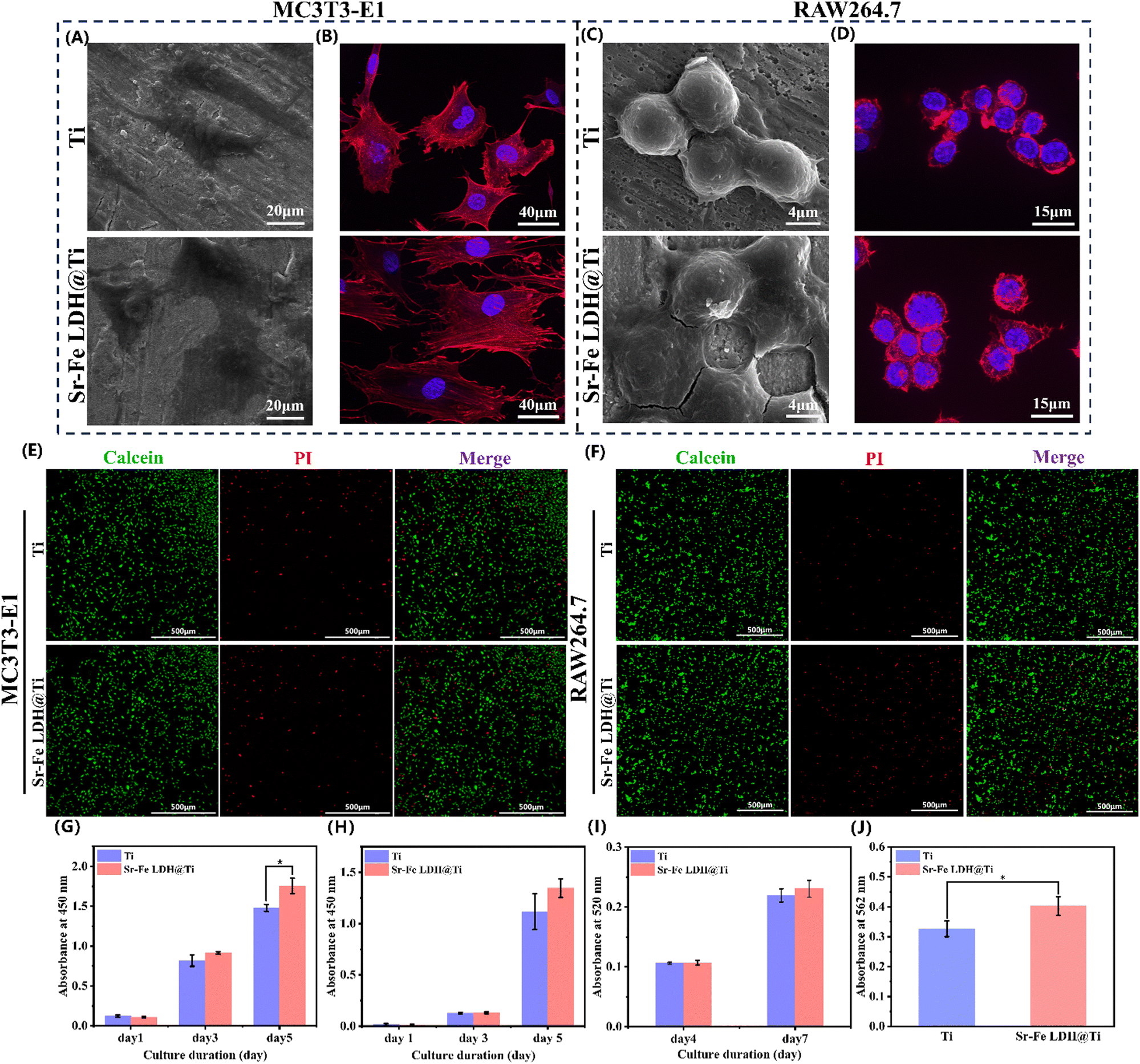 | ||
| Fig. 2 (A) SEM images of MC3T3-E1 cultured with Ti and Sr–Fe LDH@Ti for 24 h. (B) Confocal laser microscopy images of MC3T3-E1 costained with rhodamine–phalloidin (red) and DAPI (blue) on the surface of Ti and Sr–Fe LDH@Ti. (C) SEM images of RAW264.7 cultured with Ti and Sr–Fe LDH@Ti for 24 h. (D) Confocal laser microscopy images of RAW265.7 are combined with rhodamine–phalloidin (red) and DAPI (blue) on the surface of Ti and Sr–Fe LDH@Ti. (E) and (F) Fluorescence staining images of MC3T3-E1 and RAW264.7 with Calcein-AM/PI after coculture on the surface of different materials for 24 h. CCK8 test of MC3T3-E1 (G) and RAW264.7 (H) Cultured with Ti and Sr–Fe LDH@Ti; n = 3. Analysis of (I) ALP staining and (J) alizarin red staining of the MC3T3-E1 treated with Ti and Sr–Fe LDH@Ti scaffolds at days 21 and 7, respectively; n = 3. All values are presented as mean ± s.d. *p < 0.05, **p < 0.01, and ***p < 0.001. | ||
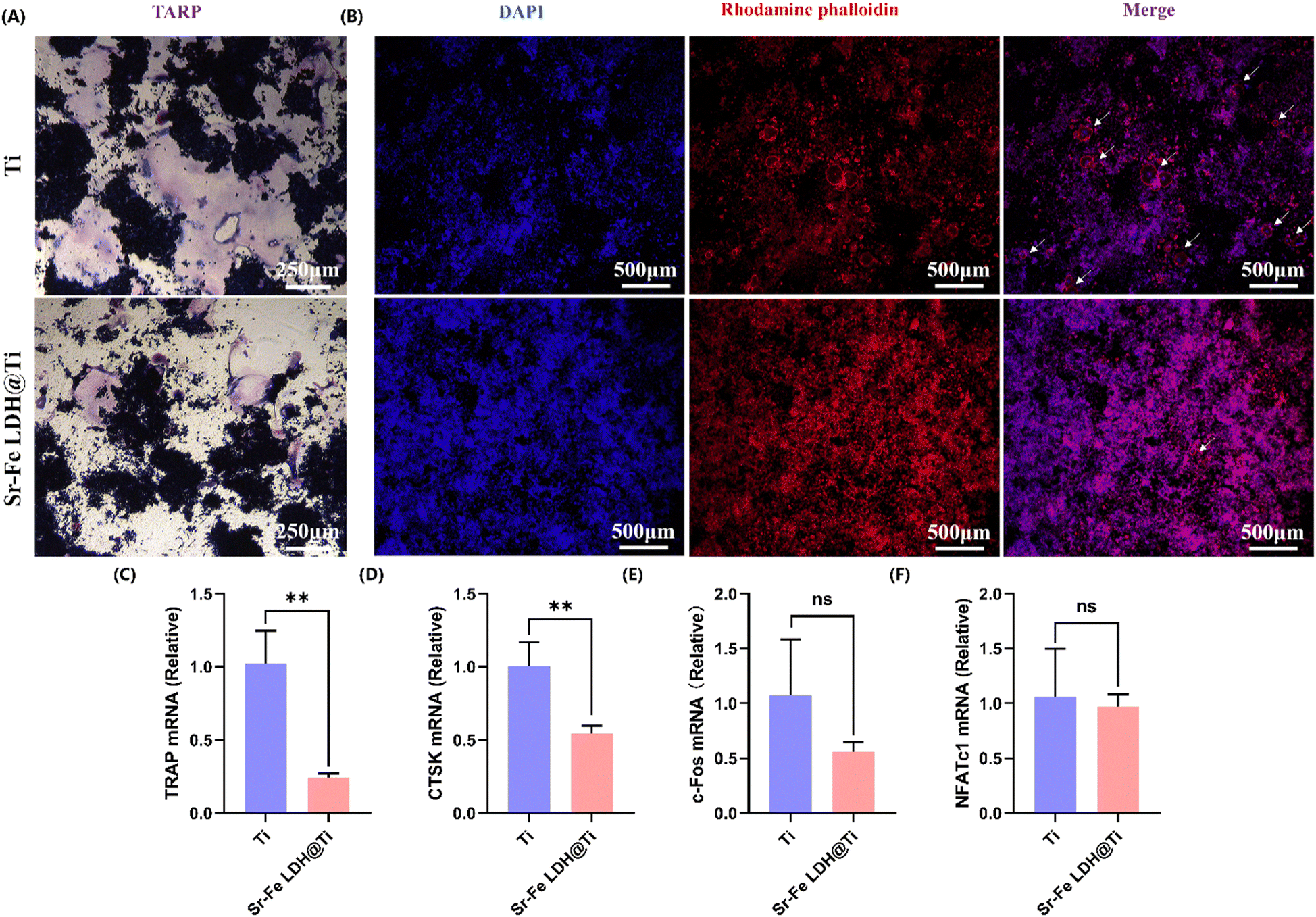 | ||
| Fig. 3 (A) TRAP staining images of RAW264.7 cultured with Ti and Sr–Fe LDH@Ti extract in the presence of RANKL (50 ng ml−1) for 6 days. (B) Confocal laser microscopy images of osteoclasts costained with anti-vinculin (green), rhodamine phalloidin (red), and DAPI (blue). (C)–(F) The qRT-PCR results of the expression of osteoclast marker genes including TRAP, CTSK, c-Fos, and NFATc1. All values are presented as mean ± s.d. *p < 0.05, **p < 0.01, and ***p < 0.001; n = 3. | ||
3.3 In vivo study
 | ||
| Fig. 4 (A) Quantitative results of the biomechanical test of implant fixation 8w after implant insertion, presented as the maximal push-out force; n = 9. (B) X-ray illustration of implant implantation and VOI. (C) Three-dimensional micro-CT images of the bone tissue around Ti and Sr–Fe LDH@Ti implants in the volume of interest (VOI). (D)–(G) Quantitative results of the micro-CT evaluation of implant osseointegration and trabecular microstructure around implants within the volume of interest; n = 11. All values are presented as mean ± s.d. *p < 0.05, **p < 0.01, and ***p < 0.001. | ||
 | ||
| Fig. 5 (A) Hematoxylin eosin staining of the heart, liver, spleen, lungs, and kidneys in rats. (B) Histological images of the proximal tibiae with implants below the epiphyseal plate 8w after implantation; n = 11 and (C) result of the Bone area ratio analysis; n = 11. All values are presented as mean ± s.d. *p < 0.05, **p < 0.01, and ***p < 0.001. | ||
4. Conclusions
To regulate the abnormal acidic microenvironment caused by osteoporosis, as well as to improve the dysfunctional state of osteoblasts and osteoclasts, the ultimate goal is to improve osseointegration. In this work, we combined the excellent performance of Sr2+ in improving bone metabolism and the ability of alkaline LDH to neutralize acids and prepared Sr–Fe LDH coatings on the surface of pure titanium implants by the hydrothermal method using Fe3+, which has good biosafety. The alkaline Sr–Fe LDH coating has good acid neutralization capacity as well as the ability to release Sr2+ for a long and stable period, is biocompatible with osteoblasts and monocyte macrophages, and can inhibit osteoclast differentiation and maturation while promoting osteoblast proliferation and differentiation. Mechanical stability, peripheral bone formation, and osseointegration of pure titanium implants in OVX rats were promoted by the modulation of the acidic microenvironment, as well as osteoblast and osteoclast functions. Our results demonstrate that the Sr–Fe LDH coating is a promising implant coating for patients with osteoporosis and has the potential to reduce instability, loosening, and dislodgement of implants in osteoporosis patients and to improve the success rate of implant placement surgery. In the next study, we will focus on the angiogenic and immunomodulatory ability of Sr–Fe LDH coating on osteoporotic rats.Conflicts of interest
The authors declare that they have no known competing financial interests or personal relationships that could have appeared to influence the work reported in this paper.Acknowledgements
This work was supported by the National Key R&D Program of China (no. 2021YFE0205000), the Sichuan Science and Technology Program (no. 2023NSFSC0570), the National Key R&D Program of China (no. 2022YFB3804500), and the Chengdu Science and Technology Program (no. 22022-YF05-01838-SN). We appreciate the platform and technical guidance provided by Associate Professor Liu Yan (the College of Life Sciences, Sichuan University) and we appreciate Li-Ying Hao (the State Key Laboratory of Oral Diseases, West China Stomatological Hospital, Sichuan University, China) for their help in AFM characterization.References
- I. R. Reid, Nat. Rev. Endocrinol., 2020, 16, 333–339 CrossRef PubMed.
- W. D. Leslie and S. N. Morin, Curr. Osteoporos. Rep., 2020, 18, 115–129 CrossRef PubMed.
- M. Fini, G. Giavaresi, T. Greggi, L. Martini, N. N. Aldini, P. Parisini and R. Giardino, J. Biomed. Mater. Res., Part A, 2003, 66A, 176–183 CrossRef CAS PubMed.
- Y. Li, F. Li, C. C. Zhang, B. A. Gao, P. Tan, B. G. Mi, Y. Zhang, H. Cheng, H. Liao, K. F. Huo and W. Xiong, J. Nanosci. Nanotechnol., 2015, 15, 4136–4142 CrossRef CAS PubMed.
- Y. D. He, Y. X. Gao, Q. L. Ma, X. G. Zhang, Y. M. Zhang and W. Song, J. Nanobiotechnol., 2022, 20, 22 CrossRef PubMed.
- Y. D. He, Z. Li, X. Ding, B. Y. Xu, J. J. Wang, Y. Li, F. H. Chen, F. H. Meng, W. Song and Y. M. Zhang, Bioact. Mater., 2022, 8, 109–123 CAS.
- X. F. Lin, Q. Q. Wang, C. H. Gu, M. B. Li, K. Chen, P. F. Chen, Z. B. Tang, X. Liu, H. H. Pan, Z. M. Liu, R. K. Tang and S. W. Fan, J. Am. Chem. Soc., 2020, 142, 17543–17556 CrossRef CAS PubMed.
- J. E. Compston, M. R. McClung and W. D. Leslie, Lancet, 2019, 393, 364–376 CrossRef CAS PubMed.
- Y. H. Shen, W. C. Liu, C. Y. Wen, H. B. Pan, T. Wang, B. W. Darvell, W. W. Lu and W. H. Huang, J. Mater. Chem., 2012, 22, 8662–8670 RSC.
- L. Kyllonen, M. D'Este, M. Alini and D. Eglin, Acta Biomater., 2015, 11, 412–434 CrossRef CAS PubMed.
- J. K. Zhang, Y. Zhuang, R. L. Sheng, H. Tomás, J. Rodrigues, G. Y. Yuan, X. D. Wang and K. L. Lin, Mater. Horiz., 2024, 11, 12–36 RSC.
- H. P. Wei, J. J. Cui, K. L. Lin, J. Xie and X. D. Wang, Bone Res., 2022, 10, 17 CrossRef CAS PubMed.
- M. U. Joshi, S. P. Kulkarni, M. Choppadandi, M. Keerthana and G. Kapusetti, Smart Mater. Med., 2023, 4, 661–679 CrossRef.
- R. Agarwal and A. J. Garcia, Adv. Drug Delivery Rev., 2015, 94, 53–62 CrossRef CAS PubMed.
- T. Wang, J. X. Bai, M. Lu, C. L. Huang, D. C. Geng, G. Chen, L. Wang, J. Qi, W. G. Cui and L. F. Deng, Nat. Commun., 2022, 13, 17 CrossRef PubMed.
- S. Song, G. H. Zhang, X. T. Chen, J. Zheng, X. D. Liu, Y. Q. Wang, Z. J. Chen, Y. X. Wang, Y. L. Song and Q. Zhou, J. Nanobiotechnol., 2023, 21, 257 CrossRef CAS PubMed.
- R. Smeets, B. Stadlinger, F. Schwarz, B. Beck-Broichsitter, O. Jung, C. Precht, F. Kloss, A. Grobe, M. Heiland and T. Ebker, Biomed Res. Int., 2016, 2016, 16 Search PubMed.
- E. Campodoni, M. Montanari, C. Artusi, G. Bassi, F. Furlani, M. Montesi, S. Panseri, M. Sandri and A. Tampieri, J. Compos. Sci., 2021, 5, 278 CrossRef CAS.
- M. Dermience, G. Lognay, F. Mathieu and P. Goyens, J. Trace Elem. Med. Biol., 2015, 32, 86–106 CrossRef CAS PubMed.
- Z. Ciosek, K. Kot, D. Kosik-Bogacka, N. Lanocha-Arendarczyk and I. Rotter, Biomolecules, 2021, 11, 26 CrossRef PubMed.
- E. O'Neill, G. Awale, L. Daneshmandi, O. Umerah and K. W. H. Lo, Drug Discovery Today, 2018, 23, 879–890 CrossRef PubMed.
- Z. S. Patel, S. Young, Y. Tabata, J. A. Jansen, M. E. K. Wong and A. G. Mikos, Bone, 2008, 43, 931–940 CrossRef CAS PubMed.
- U. Bilati, E. Allemann and E. Doelker, Eur. J. Pharm. Biopharm., 2005, 59, 375–388 CrossRef CAS PubMed.
- P. Marquis, C. Roux, C. de la Loge, M. Diaz-Curiel, C. Cormier, G. Isaia, J. Badurski, J. Wark and P. J. Meunier, Osteoporosis Int., 2008, 19, 503–510 CrossRef CAS PubMed.
- R. Yan, J. H. Li, Q. J. Wu, X. K. Zhang, L. W. Hu, Y. W. Deng, R. X. Jiang, J. Wen and X. Q. Jiang, Front. Chem., 2022, 10, 16 Search PubMed.
- T. Y. Geng, Y. R. Wang, K. L. Lin, C. Zhang, J. Wang, Y. Liu, C. Y. Yuan and P. L. Wang, Front. Bioeng. Biotechnol., 2022, 10, 12 Search PubMed.
- C. Zhou, Z. Y. Ge, L. Song, J. H. Yan, X. R. Lang, Y. Z. Zhang and F. M. He, Clin. Oral Implant. Res., 2023, 34, 297–311 CrossRef CAS PubMed.
- M. R. Katunar, J. I. Pastore, A. Cisilino, J. Merlo, L. S. Alonso, M. Baca, K. Haddad, S. Cere and J. Ballarre, Surf. Coat. Technol., 2022, 433, 9 CrossRef.
- Y. W. Ge, Z. H. Fan, Q. F. Ke, Y. P. Guo, C. Q. Zhang and W. T. Jia, Mater. Today Bio, 2022, 16, 13 Search PubMed.
- D. D. Zhang, S. Cheng, J. Tan, J. N. Xie, Y. Zhang, S. H. Chen, H. H. Du, S. Qian, Y. Q. Qiao, F. Peng and X. Y. Liu, Bioact. Mater., 2022, 17, 394–405 CAS.
- D. D. Cao, Z. L. Xu, Y. X. Chen, Q. F. Ke, C. Q. Zhang and Y. P. Guo, J. Biomed. Mater. Res., Part B, 2018, 106, 863–873 CrossRef CAS PubMed.
- Q. W. Li, D. H. Wang, J. J. Qiu, F. Peng and X. Y. Liu, Biomater. Sci., 2018, 6, 1227–1237 RSC.
- T. T. Hu, Z. Gu, G. R. Williams, M. Strimaite, J. J. Zha, Z. Zhou, X. C. Zhang, C. L. Tan and R. Z. Liang, Chem. Soc. Rev., 2022, 51, 6126–6176 RSC.
- H. Fu, L. T. Wang, Q. Q. Bao, D. L. Ni, P. Hu and J. L. Shi, J. Am. Chem. Soc., 2022, 144, 8987–8999 CrossRef CAS PubMed.
- M. M. Bornstein, P. Valderrama, A. A. Jones, T. G. Wilson, R. Seibl and D. L. Cochran, Clin. Oral Implant. Res., 2008, 19, 233–241 CrossRef PubMed.
- C. Eriksson, H. Nygren and K. Ohlson, Biomaterials, 2004, 25, 4759–4766 CrossRef CAS PubMed.
- Z. Y. Zhong, X. D. Wu, Y. F. Wang, M. D. Li, Y. Li, X. L. Liu, X. Zhang, Z. Y. Lan, J. L. Wang, Y. Y. Du and S. M. Zhang, Bioact. Mater., 2022, 10, 195–206 CAS.
- C. B. Tovani, T. M. Oliveira, M. P. R. Soares, N. Nassif, S. Y. Fukada, P. Ciancaglini, A. Gloter and A. P. Ramos, ACS Appl. Mater. Interfaces, 2020, 12, 43422–43434 CrossRef CAS PubMed.
- D. L. Kendler, F. Marin, C. A. F. Zerbini, L. A. Russo, S. L. Greenspan, V. Zikan, A. Bagur, J. Malouf-Sierra, P. Lakatos, A. Fahrleitner-Pammer, E. Lespessailles, S. Minisola, J. J. Body, P. Geusens, R. Möricke and P. López-Romero, Lancet, 2018, 391, 230–240 CrossRef CAS PubMed.
- D. M. Biskobing and D. Fan, Calcif. Tissue Int., 2000, 67, 178–183 CrossRef CAS PubMed.
- N. Udagawa, J. Takito and T. Suda, Nihon rinsho, 1992, 50, 2133–2138 CAS.
- S. J. Jenkins, D. Ruckerl, P. C. Cook, L. H. Jones, F. D. Finkelman, N. van Rooijen, A. S. MacDonald and J. E. Allen, Science, 2011, 332, 1284–1288 CrossRef CAS PubMed.
- M. A. Kafi, K. Aktar, Y. Phanny and M. Todo, J. Mater. Sci.-Mater. Med., 2019, 30, 131 CrossRef CAS PubMed.
- M. Kingsak, P. Maturavongsadit, H. Jiang and Q. Wang, Biomater. Transl., 2022, 3, 221–233 Search PubMed.
- M. Chu, Z. Y. Sun, Z. H. Fan, D. G. Yu, Y. Q. Mao and Y. P. Guo, Theranostics, 2021, 11, 6717–6734 CrossRef CAS PubMed.
- Y. J. Wang, X. Mei, Y. Y. Bian, T. T. Hu, X. S. Weng, R. Z. Liang and M. Wei, Nanoscale, 2020, 12, 19075–19082 RSC.
- J. F. Li, L. Yang, X. D. Guo, W. Cui, S. Y. Yang, J. P. Wang, Y. Z. Qu, Z. W. Shao and S. Y. Xu, Biomed. Mater., 2018, 13, 13 Search PubMed.
- Y. Z. Zhu, H. Liang, X. M. Liu, J. Wu, C. Yang, T. M. Wong, K. Y. H. Kwan, K. M. C. Cheung, S. L. Wu and K. W. K. Yeung, Sci. Adv., 2021, 7, 13 Search PubMed.
- Z. C. Cao, H. M. Wang, J. L. Chen, Y. A. Zhang, Q. Y. Mo, P. Zhang, M. Y. Wang, H. Y. Liu, X. Y. Bao, Y. Z. Sun, W. Zhang and Q. Q. Yao, Bioact. Mater., 2023, 20, 221–242 CAS.
- J. H. Hwang, Y. S. Park, H. S. Kim, K. Dong-ha, S. H. Lee, C. H. Lee, S. H. Lee, J. E. Kim, S. Lee, H. M. Kim, H. W. Kim, J. Kim, W. Seo, H. J. Kwon, B. J. Song, D. K. Kim, M. C. Baek and Y. E. Cho, J. Controlled Release, 2023, 355, 184–198 CrossRef CAS PubMed.
- Y. H. Shen, W. C. Liu, K. L. Lin, H. B. Pan, B. W. Darvell, S. L. Peng, C. Y. Wen, L. F. Deng, W. W. Lu and J. A. Chang, Langmuir, 2011, 27, 2701–2708 CrossRef CAS PubMed.
- K. Chen, P. C. Qiu, Y. Yuan, L. Zheng, J. B. He, C. Wang, Q. Guo, J. Kenny, Q. Liu, J. M. Zhao, J. H. Chen, J. Tickner, S. W. Fan, X. F. Lin and J. K. Xu, Theranostics, 2019, 9, 1634–1650 CrossRef CAS PubMed.
- C. L. Gregson, D. J. Armstrong, J. Bowden, C. Cooper, J. Edwards, N. J. L. Gittoes, N. Harvey, J. Kanis, S. Leyland, R. Low, E. McCloskey, K. Moss, J. Parker, Z. Paskins, K. Poole, D. M. Reid, M. Stone, J. Thomson, N. Vine and J. Compston, Arch. Osteoporos., 2022, 17, 46 CrossRef PubMed.
- F. Cosman, S. J. de Beur, M. S. LeBoff, E. M. Lewiecki, B. Tanner, S. Randall and R. Lindsay, Osteoporosis Int., 2014, 25, 2359–2381 CrossRef CAS PubMed.
- C. B. Johnston and M. Dagar, Med. Clin. N. Am., 2020, 104, 873–884 CrossRef PubMed.
- N. Yousefzadeh, K. Kashfi, S. Jeddi and A. Ghasemi, Excli J., 2020, 19, 89–107 Search PubMed.
- O. Barou, D. Valentin, L. Vico, C. Tirode, A. Barbier, C. Alexandre and M. H. Lafage-Proust, Invest. Radiol., 2002, 37, 40–46 CrossRef PubMed.
- L. Maïmoun, T. C. Brennan-Speranza, R. Rizzoli and P. Ammann, Bone, 2012, 51, 586–591 CrossRef PubMed.
- Y. F. Li, Q. Li, S. S. Zhu, E. Luo, J. H. Li, G. Feng, Y. M. Liao and J. Hu, Biomaterials, 2010, 31, 9006–9014 CrossRef CAS PubMed.
Footnote |
| † Electronic supplementary information (ESI) available. See DOI: https://doi.org/10.1039/d3tb02410e |
| This journal is © The Royal Society of Chemistry 2024 |