
Flexible resistive tactile pressure sensors
Qianhe
Shu
a,
Yuncong
Pang
b,
Qiqi
Li
a,
Yuzhe
Gu
a,
Zhiwei
Liu
 *c,
Baoguang
Liu
a,
Jianmin
Li
*a and
Yang
Li
*ab
*c,
Baoguang
Liu
a,
Jianmin
Li
*a and
Yang
Li
*ab
aCollege of Electronic and Optical Engineering, College of Flexible Electronics (Future Technology), Nanjing University of Posts & Telecommunications (NJUPT), Nanjing, 210023, China. E-mail: lijm@njupt.edu.cn; yli@njupt.edu.cn
bState Key Laboratory of Organic Electronics and Information Displays, Jiangsu Key Laboratory for Biosensors, Institute of Advanced Materials (IAM), Nanjing University of Posts and Telecommunications (NJUPT), Nanjing, 210023, China
cSchool of Energy and Environmental Engineering, University of Science and Technology Beijing, Beijing 100083, China. E-mail: liuzhiwei@ustb.edu.cn
First published on 26th March 2024
Abstract
The widespread integration of sensors into our everyday existence has paved the path for groundbreaking progress across various domains, including healthcare, robotics, and human–computer interaction. In this context, flexible resistive tactile pressure sensors have emerged as vital instruments due to their outstanding electrical and mechanical properties, cost-effectiveness, and ease of manufacturing. They have become pivotal in driving innovation, from wearable devices to human–machine interfaces. This comprehensive review article delves into recent advancements in this rapidly growing field, focusing on operational principles, performance metrics, material choices, structural design, and the applications of flexible resistive tactile pressure sensors. The challenges and opportunities in the field, such as enhancing sensitivity, durability, and reproducibility, and emerging trends, such as the integration of machine learning algorithms for real-time data analysis are also addressed, providing insights into the future direction of this rapidly evolving technology. By consolidating the current state-of-the-art in flexible resistive tactile pressure sensors, this article aims to inspire further innovation and collaboration in the pursuit of more sophisticated and versatile tactile pressure sensing technologies.
1. Introduction
Sensors, serving as tools for collecting, detecting, converting, and transmitting information, possess the capability to convert target signals that are not directly measurable into electrical or other output signals following specific patterns.1–5 The need for precise and versatile tactile pressure sensing is evident in numerous applications, spanning from the delicate touch needed in medical diagnostics to the responsive interaction vital in modern robotics. Serving as essential components in smart devices to comprehend the external surroundings, tactile sensors are primarily responsible for empowering devices to identify and perceive various physical attributes during the device's functioning while engaging with target objects and the surrounding milieu.6–8 Nonetheless, traditional tactile sensors predominantly employ a variety of rigid materials as their sensitive components. Due to the substantial advancements in rigid electronic components and materials over time, sensor systems of various types have become inherently inflexible, leading to a standardization of their operational principles and environmental adaptability. In previous sensor applications, the key requirements were stability, cost-effectiveness, and durability, which rigid sensors could effectively address and fulfill for the majority of cases. However, as robotics continues to advance and the demand for high processing performance in sensors persists, sensors based on rigid materials have begun to exhibit numerous issues.9 Despite their technological maturity, the constraints of form and material inherent to these sensors result in drawbacks such as bulkiness and fragility, which hinder their applicability in flexible human–machine interaction and portable wearable smart devices.In contrast to “rigidity,” flexibility in tactile sensors refers to human skin-like properties, including adaptability to diverse shapes, which ensures effective functionality across various technological domains.10 Flexible sensors, which enhance surface contact for increased sensitivity, play a crucial role in applications like robotics, where precise pressure measurements are vital for safe and efficient interactions.11 Furthermore, the relationship between flexibility and durability empowers sensors to withstand mechanical stress, proving advantageous in applications involving repetitive strains.12
In practical tactile perception, the forces acting on the sensor are highly complex, leading to intricate mechanical stimuli. Tactile sensors need to precisely perceive forces of various magnitudes and directions, generating electrical signals in real-time based on different operating principles to be transmitted to the system for analysis. Based on different ways of generating electrical signals, flexible tactile sensors can be classified into resistive,13 capacitive,14 inductive,15 piezoelectric,16 and triboelectric types.17 Among them, researchers have focused extensively on flexible resistive tactile sensors due to their notable advantages, including high precision, sensitivity, a broad sensing range, uncomplicated structure, stability, reliability, ease of miniaturization, and robust overload capacity. In 2013, Canavese et al. proposed a piezoflexible resistive composite material that can be used to make flexible tactile sensors that achieve real-time 3D response to pressure and can be used for tactile sensing in robotics.18 As more experiments were conducted, flexible tactile piezoresistive sensors were found to have good integration with humans in addition to robots. In 2015, flexible tactile sensors were made into electronic skin by Gerratt et al. and applied to the surface of human skin.19 In pursuit of better portability and usage experience, after material optimization and structure design, Gao et al. developed a wearable microfluidic diaphragm pressure sensor in 2017.20 This sensor is thinner, lighter, more sensitive, and has more comprehensive and detailed haptic feedback. In 2019, Wu built on this foundation by iterating on a sensor that can be self-powered.21 Two years later, Wang et al. designed a self-powered tactile sensor with an even lower limit of pressure detection, opening up the possibility of ultra-small signal detection.22 In 2022 and 2023, researchers' dedicated efforts and the integration of flexible tactile piezoresistive sensors into human–machine interaction, coupled with machine learning and material and structural optimization, significantly enhanced sensor performance, reaching an unprecedented level of sensitivity and practicality.23,24
Presently, there have been noteworthy and substantial advancements in the field of flexible resistive tactile sensors. The progression extends from the initial resistive rigid tactile sensors to the inception and evolution of the flexible tactile sensing concept. In aspects related to substrate materials, sensitive material choices, unit structures, and the design of integrated arrays, the manufacturing technology for flexible resistive tactile sensors has progressively matured, allowing for a wider range of functionalities (Fig. 1). In addition to detecting pressure and tension, numerous flexible resistive tactile sensors can also detect friction, torsion, bending force, temperature, humidity, proximity, and other physical stimuli. Some even possess features such as transparency,25 magnetism sensitivity,26 and self-healing.27 The refinement of performance and diversification of functionality have led them from perceiving force on objects to monitoring posture movements,28 facial expressions,29 physiological health,30 and further into establishing tactile sensing systems, protecting against external environmental interference, and assisting in medical health monitoring.
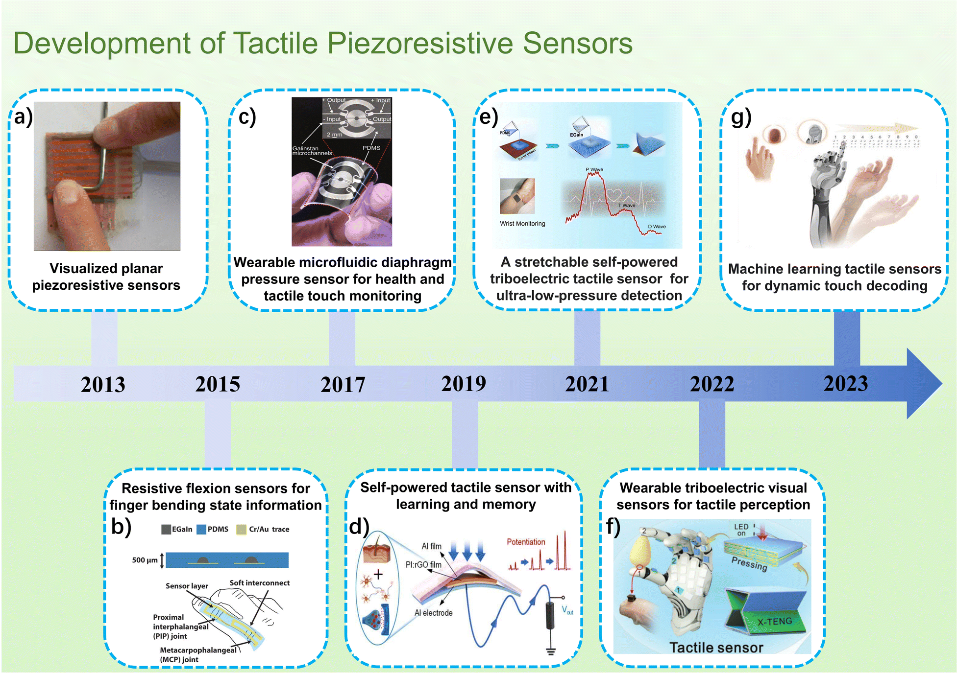 | ||
| Fig. 1 The development of piezoresistive tactile sensors in recent years. (a) Polymeric composite with nanostructured spiky particles as filler.18 Reproduced with permission. Copyright at Sensors and Actuators A: Physical, 2014 (b) schematic of the cross-section and graphical representation on a finger of the resistive flexion sensors for finger bending state information.19 Reproduced with permission. Copyright at Advanced Functional Materials, 2015 (c) Wearable Microfluidic Diaphragm Pressure Sensor for Health and Tactile Touch Monitoring.20 Reproduced with permission. Copyright at Advanced Materials, 2017 (d) Self-Powered Tactile Sensor with Learning and Memory.21 Reproduced with permission. Copyright at ACS Nano, 2019 (e) A stretchable self-powered triboelectric tactile sensor with EGaIn alloy electrode for ultra-low-pressure detection.22 Reproduced with permission. Copyright at Nano Energy, 2021 (f) Wearable Triboelectric Visual Sensors for Tactile Perception.23 Reproduced with permission. Copyright at Advanced Materials, 2022 (g) Machine Learning-Enabled Tactile Sensor Design for Dynamic Touch Decoding.24 Reproduced with permission. Copyright at Advance Science, 2023. | ||
In recent years, there has been a swift evolution in flexible resistive tactile sensors, characterized by the continuous emergence of new materials and structures, often accompanied by the development of high-performance sensors. To consolidate and synthesize this wealth of information, this review initiates with an exploration of the two fundamental detection principles underpinning flexible resistive tactile sensors. Additionally, it serves as a reference for researchers aiming to create novel materials and structures. Furthermore, the sensors' performance can be summarized using six key performance parameters based on empirical data. Subsequently, in the subsection of flexible substrates and conductive materials that most researchers are committed to innovating and optimizing, the vast variety of materials and common structural designs used in flexible resistive tactile sensors up to the present time are described in detail, and different types of flexible resistive tactile sensors are compared and analyzed. Finally, the three main application areas of the sensor are summarized: tactile sensing and human–computer interaction, healthcare, and electronic skin. While pointing out the advantages of flexible resistive tactile sensors, this subsection also identifies the technical challenges faced by current research and provides an outlook on their future development.
2. Principle of flexible resistive tactile sensors
In tactile sensing, pressure, strain and friction may exist. According to different tactile sensing methods, tactile sensors are classified into various types, including resistive, piezoelectric, capacitive, and friction-based. However, in practical applications, the signal output is actually multiple effects from mechanical stimuli. In order to enhance the signal output quality of tactile sensors, the combination of multiple sensing mechanisms has become an innovative improvement approach.31–33 Among the various tactile sensors, resistive tactile sensors have garnered significant attention from scholars due to their relatively low cost, ease of manufacturing, and large sensitivity range, making them suitable for larger contact areas. Here, we provide a detailed description and explanation of the sensing principles of flexible resistive tactile pressure sensors. The unique qualities of flexible resistive tactile sensors are a direct result of their distinct sensing mechanism and operational concepts. By capitalizing on these two principles, researchers are committed to improving essential performance metrics that serve as critical indicators of overall functionality. This next section will provide a detailed exploration of the underlying rationale for optimizing materials and structures, offering insights into the two sensing principles and the six primary performance parameters of flexible resistive tactile sensors.2.1. Detection principle of flexible resistive tactile sensors
In the realm of detection principles, flexible resistive tactile sensors can be broadly classified into two mechanisms: pressure-sensitive and resistive strain-sensitive mechanisms. While both mechanisms involve generating resistance responses to external stimuli and quantifying input signal magnitudes by monitoring variations in resistance values, it's important to note that their underlying principles differ.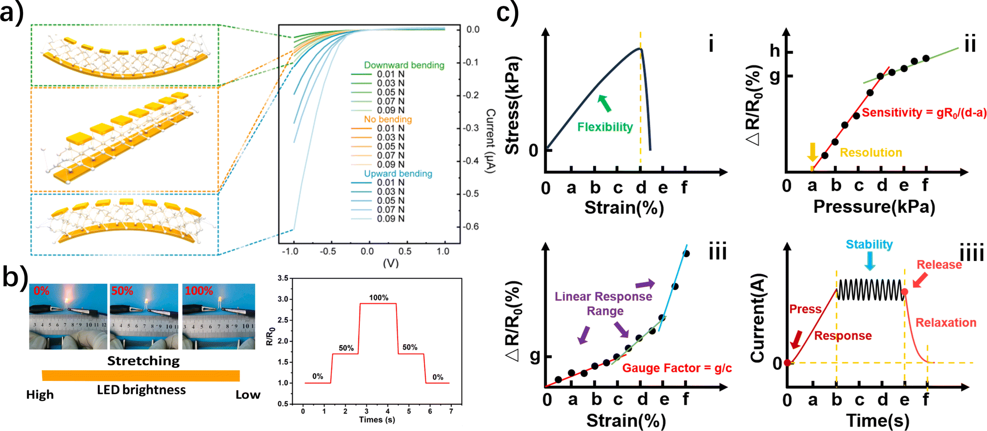 | ||
| Fig. 2 Principles of flexible resistive tactile sensors. (a) Principle of flexible piezoresistive sensors. The lattice changes under different degrees of bending, resulting in a change in resistance.35 Reproduced with permission. Copyright at Nano Energy, 2022 (b) principle of resistance-strain flexible sensors. Different deformations after applying different external forces lead to changes in resistance, which is externally manifested as a change in the brightness of LEDs.37 Reproduced with permission. Copyright at Composites Part A: Applied Science and Manufacturing, 2022 (c) six key performance parameters of flexible resistive tactile sensors. | ||
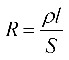
Unlike traditional pressure-sensitive sensors, pressure-sensitive flexible resistive sensors use single-crystal silicon as the sensitive material, but they utilize substrates that are more flexible to enhance their deformation capabilities. When the single-crystal silicon material in a pressure-sensitive flexible resistive sensor experiences force, its resistivity undergoes a corresponding change.
 | (1) |
Compared to traditional resistive strain sensors, resistive strain-sensitive flexible sensors offer increased flexibility due to the choice of flexible materials. This allows the attached sensitive unit to undergo greater deformation under smaller strain forces when the elastic element is deformed by applied force. Consequently, these sensors have higher transmission capabilities. The sensitive unit then converts this deformation into changes in resistance values, making it possible to measure various physical quantities such as pressure, stress, acceleration, and temperature.
The difference between pressure-sensitive flexible sensors and resistive strain-sensitive flexible sensors lies in how resistance changes with pressure. In the former, the change in resistance primarily depends on resistivity variations, while in the latter, resistance changes mainly depend on geometric dimensional changes (strain).
2.2. Performance parameters of flexible resistive tactile sensors
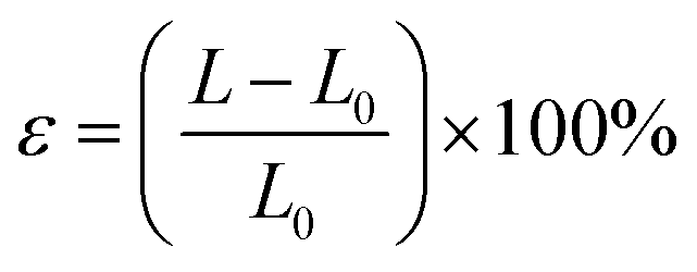
The performance parameters of flexible resistive tactile sensors, such as flexibility, sensitivity, resolution, stability, linear response range, response and relaxation time, etc., can reflect the performance and excellence of the sensor intuitively (Fig. 2c). These indicators are extremely important, and will affect the sensor's operating environment and limit state to varying degrees. Flexible resistive tactile sensors with excellent performance tend to strike a balance between these indicators.Strain ε is usually expressed using the following formula:
 | (2) |
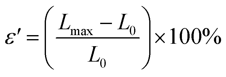
The strain range refers to the maximum deformation range of the flexible resistive tactile sensor without fracturing while maintaining its sensing performance. The strain at which the flexible resistive tactile sensor fractures is referred to as the elongation at break ε′, and its formula is as follows:
 | (3) |
In flexible resistive tactile sensors, flexibility primarily depends on the elasticity of the substrate material, such as polydimethylsiloxane (PDMS), polyethylene terephthalate (PET), polyurethane (PU), and copolyesters (Ecoflex). Additionally, methods such as reducing sensor thickness, decreasing the sensor's Young's modulus, and optimizing sensor structural design can all enhance the flexibility of flexible resistive tactile sensors.38,39
 | (4) |
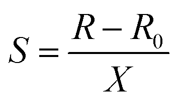
For instance, in Fig. 2c(ii), we can use the pressure change (d–a) in the section from a to d with its corresponding resistance change to calculate the sensitivity of the section. Furthermore, the sensitivity factor (K) can be used to describe the sensor's sensitivity. It reflects the degree of change in the output response of the sensor under a unit input, as shown in eqn (5):
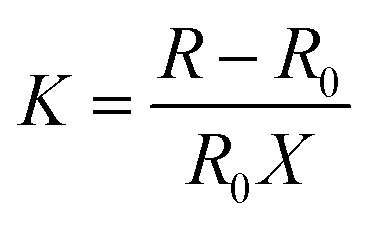 | (5) |
In addition to sensitivity, scholars also use the relationship between strain and resistance changes in flexible resistive tactile sensors to describe their sensitivity, which is referred to as the Gauge Factor (GF). The expression for GF is as follows:
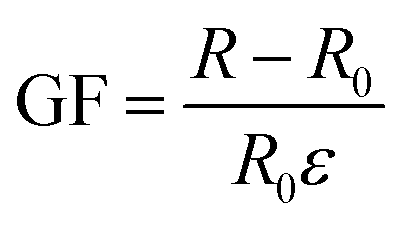 | (6) |
The sensitive material in flexible resistive tactile sensors is the primary factor determining their sensitivity. Typically, the better the conductivity of the sensitive material, the higher the sensitivity and responsiveness to external stimuli. Among various sensitive materials, those with excellent conductivity include silver nanowires,40 graphene (Gr),41 carbon nanotubes (CNTs),42 and single-crystal silicon (Si).43 Additionally, sensitivity in flexible resistive tactile sensors can also be improved through methods such as adjusting the quality ratio of conductive fillers, optimizing the structural design of sensitive units, reducing the initial strength of electrical signals, using novel sensing materials, and introducing non-steady small microcracks.
For flexible resistive tactile sensors, to better simulate the human skin's sensitivity to environmental factors like spatial orientation, pressure, and stress, it's necessary to increase their resolution as much as possible. To enhance spatial resolution, scholars often utilize methods like increasing the number of sensor elements, reducing the size of sensor elements, and developing array-based tactile sensors.44–46 Additionally, in extremely low-pressure and high-pressure environments, the demands for accurately detecting changes in force are also very stringent, necessitating high resolution.
It's important to note that resolution is a more critical parameter than sensitivity. Sensitivity is a relative measure, while resolution directly reflects the sensor's responsiveness to stimuli. Scholars often focus their research on material selection and microstructure design of the sensitive unit to continually improve both the sensor's operating range and its resolution.
Factors influencing a sensor's long-term stability extend beyond the sensor's own structure and include the sensor's operating environment. Therefore, for a sensor to exhibit good stability, it must also possess strong environmental adaptability. The sensor's inherent environmental adaptability can be improved through both material selection (hydrophobic, corrosion-resistant, friction-resistant materials) and structural design, enabling the sensor to be hydrophobic,47 oxidation-resistant,48 and corrosion-resistant,49 among other qualities.
 | (7) |
Additionally, the linearity can also be described using the linear fitting coefficient R2. A higher value of R2 suggests better linearity. Assuming there are n responses yi corresponding to n excitations xi, the formula for R2 is:
 | (8) |
 | (9) |
Often, for flexible resistive tactile sensors with extremely wide response ranges, the linear response range is not continuous. Depending on their responsiveness, they can be divided into three sections: low-pressure, medium-pressure, and high-pressure. Each section has a different sensitivity. This phenomenon is mainly due to the occurrence of varying degrees of microcracks within the sensitive unit as it deforms, leading to the interruption of internal conductive pathways. This is reflected as different degrees of resistance change within different excitation ranges. As shown in Fig. 2c(iii), the sensor's rate of change of resistance varies significantly from 0 to c, from c to e, and after e, which is characterized by an abrupt change in slope. The three linear response ranges mean that the sensor has three operating intervals. To mitigate the influence of internal microcracks, more flexible materials can be chosen for the flexible substrate, or nano-materials with fibrous50 or scale-like51 structures can be selected for the sensitive unit. This helps the sensor maintain its conductive pathways to a greater extent even when microcracks occur.
Relaxation time, on the other hand, is the time it takes for a flexible resistive tactile sensor to return to a steady state after the removal of an external stimulus. It represents the time it takes for the transient response of the sensor's output electrical signal to settle after the removal of the stimulus signal, which is represented in Fig. 2c(iiii) as the time period from e to f. Relaxation time can characterize the extent of the influence of fast variables. A shorter relaxation time indicates that fast variables are more easily eliminated or reduced.
3. Design and construction of flexible resistive tactile sensors
The diverse performance parameters of flexible resistive tactile sensors are intricately linked to the materials chosen and the structural design employed. As previously discussed, the selection of substrate materials plays a critical role in determining the flexibility of flexible resistive tactile sensors. Sensitive materials primarily govern their sensitivity, linear response range, and resolution, while the structural design exerts a significant influence on the sensor's overall performance. Consequently, investigating flexible substrate materials, conductive sensitive materials, structural design, and performance enhancement remains a significant research direction.3.1. Flexible substrate materials
Flexible substrate materials encompass the foundational materials that house the conductive sensitive elements in flexible resistive tactile sensors, making up a significant portion of the sensor's composition. The flexibility of flexible resistive tactile sensors is largely determined by the flexible substrate material. This is mainly reflected in aspects such as the sensor's stretch range, Young's modulus, tear strength, hardness, and density, all of which indicate the sensor's deformability to varying degrees. Furthermore, for the purpose of emulating human skin and considering aesthetic factors, researchers often opt for materials that closely resemble the modulus and high transparency of human skin.52,53Table 1 presents a performance comparison of several key flexible substrate materials.| Type | Material | Young's modulus | Mechanical strength | Flexibility | Reference |
|---|---|---|---|---|---|
| Rubber | PDMS (normally cured high-molecular-weight) | 73 kPa to 2.1 MPa | F S = 1.5 MPa | ε max = 300% | 54 and 73 |
| Ecoflex | 50 kPa to 200 kPa | F S = 0.3–0.9 MPa | ε max = 860% (Ecoflex 00–50) | 55 | |
| EP | 2.5 GPa to 5 GPa | F S = 0.2 MPa | ε max = 54.2% | 56 and 74 | |
| Plastic | PET | 2 GPa to 4 GPa | F S = 62 MPa | ε max = 6% | 61 |
| PVA fiber | 40.02 GPa | F S = 1.73 GPa | ε max = 2.88% | 62 | |
| Fiber | Kevlar fibers | 70.5 GPa (Kevlar 29/Longitudinal) | F S = 3.6 GPa | ε max = 5.5% | 64 |
| Nonwoven fabrics | — | F S = 220 kPa (fiber density: 0.12 g cm−3) | ε max = 70% | 70 | |
| Tissue paper | — | F S = 100 kPa | — | 72 | |
| Others | PU (medical grade) | (8.5 ± 0.3) MPa | — | ε max > 400% | 75 |
There are many substrate materials available for flexible resistive tactile sensors, roughly categorized into rubber, plastic, and fibers. Among them, rubber materials commonly used include PDMS,54 Ecoflex,55 and epoxy resin (EP).56 PDMS is a polymer known for its main chain structure composed of Si–O–Si bonds, providing 100% transparency, along with properties such as heat and cold resistance, hydrophobicity, excellent shear resistance, low stiffness (∼2 MPa) and a high thermal expansion coefficient (α = 310 × 10−6 K−1). Tai utilized the thermal expansion property of PDMS to create programmable thermal capabilities in sensors.57 The value of the real/imaginary part of the impedance of this sensor increased from 104 to 105 ohms (25 °C) to more than 109 ohms (65 °C) at different temperatures, demonstrating its excellent temperature sensing capability (Fig. 3a and b). Ecoflex is a member of the super-elastic family, with a fracture strain exceeding 900%. Additionally, its Young's modulus was only 125 kPa, closely matching the Young's modulus of human skin (25–220 kPa), significantly lower than PDMS's Young's modulus. Wang and others utilized nickel nanowires and Ecoflex to fabricate flexible strain sensors with a GF reaching 200 at 100% strain.58 EP is an organic polymer compound containing two or more epoxy groups. While EP is classified as a type of rubber, it lacks the flexibility seen in materials like PDMS or Ecoflex, demonstrating poor tensile performance, low impact resistance, and a relatively brittle texture. However, EP boasts strong adhesive properties, particularly with metal materials.59 Based on a screen-printing process, Lin et al. prepared a high-performance flexible piezoresistive sensor based on Gr and EP, with response and recovery times of 40.8 ms and 3.7 ms, and a pressure detection range of 2.5–500 kPa.60 The addition of EP increased the bonding force between the substrate and the conductive material, further expanding the application in sensor.
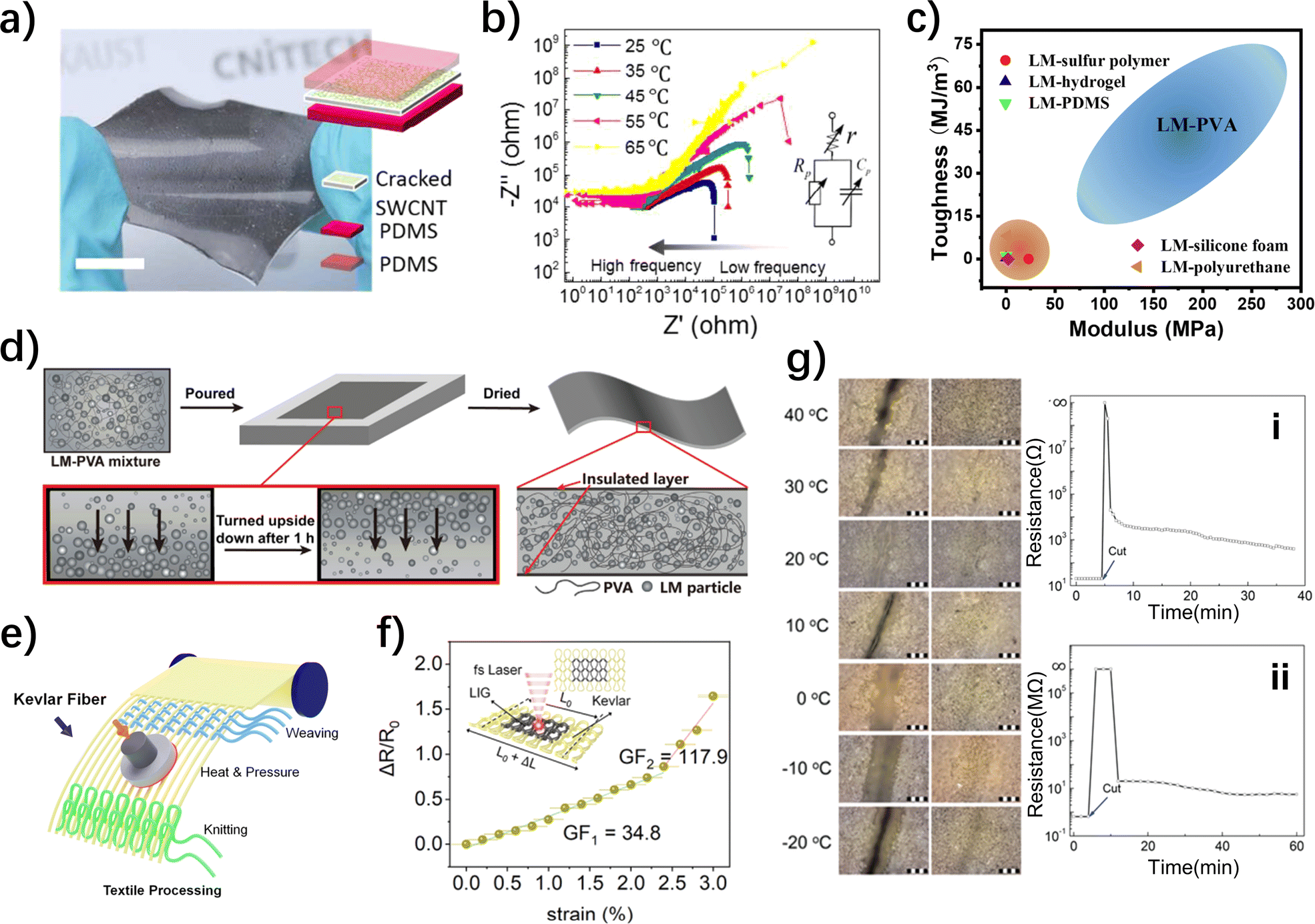 | ||
| Fig. 3 Structure and performance of different substrate materials for flexible resistive tactile sensors. (a) PDMS is used as a temperature-sensitive flexible material in the substrate.57 Reproduced with permission. Copyright at ACS Applied Materials & Interfaces, 2017 (b) electrical impedance spectroscopies of the sensor at different temperatures from 1 kHz to 2 MHz.57 Reproduced with permission. Copyright at ACS Applied Materials & Interfaces, 2017 (c) mechanical performance of LM-PVA and several other LM-polymer composites.63 Reproduced with permission. Copyright at Chemical Engineering Journal, 2020 (d) scheme for the preparation of LM-PVA film via physical blending and drying.63 Reproduced with permission. Copyright at Chemical Engineering Journal, 2020 (e) textile processing with various structures involving Kevlar fibers.69 Reproduced with permission. Copyright at ACS Nano, 2023 (f) sensitivity plots of knit Kevlar/LIG strain sensor at different strains.69 Reproduced with permission. Copyright at ACS Nano, 2023 (g) Images of sh-μAg-PU electrode before cutting and after self-healing at different temperatures: time dependence of logarithmic resistance and healing efficiency of the (i) electrode and (ii) sensing layer.76 Reproduced with permission. Copyright at Advanced Materials, 2016. | ||
In plastic materials, PET is widely used due to its linear polymer structure featuring highly symmetrical aromatic rings, which makes it easily orientable and crystallizable.61 Additionally, PET stands out due to its resistance to folding, creep, fatigue, and friction, coupled with excellent dimensional stability and the highest toughness among thermoplastic plastics. However, its relatively low fracture elongation (150%) and extremely high Young's modulus (4000 MPa) prevented it from being the preferred substrate material. Besides PET, polyvinyl alcohol (PVA) is an excellent water-soluble plastic.62 Lou et al. developed a super-tough force sensor based on liquid metal-polyvinyl alcohol composites (LM-PVA), and the addition of PVA resulted in a dramatic improvement in the toughness and wear resistance of the sensor (Fig. 3d).63 The sensor exhibited a modulus of elasticity in stress–strain tests that is not found in most other LM-polymer composites, with a 12.3-fold increase in toughness, and possesses exceptional mechanical durability, remaining stable over 1000 tensile and compressive cycles (Fig. 3c).63
Fiber materials have lower flexibility and are not the first choice for electronic skin preparation.64 However, Kevlar fibers, known for permanent heat resistance, corrosion resistance, high strength, abrasion and tear resistance, unexpectedly caught the attention of scholars.65–67 Despite its very low fracture elongation (2.8%), Kevlar's advantages in easy processing and material deposition make it a focal point for wearable and protective electronic products. Wang and others deposited shear stiffening polymers (S-ST polymers) and multi-walled carbon nanotubes onto Kevlar to create wearable electronic textiles.68 The addition of S-ST polymers improved the sensor's impact resistance significantly (dynamic impact resistance increased by 190%). Yang et al. transformed pristine Kevlar textiles into conducting laser-induced Gr (LIG) for direct laser writing of e-textiles by using femtosecond laser pulses in ambient air (Fig. 3e). Due to the incorporation of Kevlar fibers, the sensor combined excellent tensile resistance and sensitivity, which changed from 34.8 to 117.9 as the strain increased from 0 to 3.0% (Fig. 3f).69 Furthermore, affordable and readily available nonwoven fabrics and tissue paper composed of cotton fibers are also favored by scholars. Nonwoven fabric arranges short fibers or filaments in an oriented or random manner to form a fibrous network structure, which was then reinforced using mechanical, thermal, or chemical methods.70 Liu and others used nonwoven fabric to produce a flexible tactile sensor with a maximum sensitivity of 81.6 kPa−1, a working range of 0–100 kPa, a rapid response time of 6 ms, and a slow response time of 30 ms.71 Additionally, Pataniya and others coated tissue paper with tungsten diselenide (WSe2) nanosheets to create a pressure sensor with an ultra-wide pressure working range of 1 Pa to 100 kPa.72 This sensor exhibited a high sensitivity of 29.24 kPa−1 within the pressure range of 1–12 Pa and the capability to detect pressure from small liquid droplets.
Not all materials can be strictly distinguished, and one notable example is PU, which stands out within the superelastic materials category.77 PU is a block copolymer formed by the polymerization of long and short chain segments, and the properties of PU, such as softness, hardness, and strength, are influenced by the type of segments used.78 As a result, PU can be produced not only as PU plastics (mainly foam plastics) but also as PU fibers (spandex),79 PU rubber,80 and elastomers. PU exhibits exceptional elasticity, with a fracture elongation of up to 800%. Even at 300% elongation, it maintains a rebound rate of over 95%. Moreover, PU possesses flame retardancy, recyclability, and self-healing properties, making it a preferred material for creating self-healing flexible resistive tactile sensors. Huynh and others utilized PU's self-healing ability to develop a self-healing, fully functional, and multiparametric flexible sensing platform which contained the substrate, electrodes, and sensing layer.76 As shown in Fig. 3g, within a wide temperature range (−20 to 40 °C), the base, electrodes, and chemical resistance of this sensor could fully recover within 16 hours, 30 minutes, and 16 hours, respectively. In addition, both the sensing layer and the electrodes of the platform were able to heal quickly to their original state after being cut.
In addition to the mentioned conventional materials that can serve as flexible substrates, there are also many emerging materials, such as biomimetic materials, showing tremendous potential. In the field of biomimicry material innovation, whether assembling existing materials based on biomimetic structures or directly developing novel biomimetic materials, it can enhance the elasticity and biocompatibility of flexible resistive tactile sensors, achieving more intelligent and biologically analogous sensor responses.81 Wang et al. replicated the anisotropic one-dimensional microstructure of reed leaves, using the multilayer stacking of microgrooved polydimethylsiloxane (m-PDMS) to develop a highly sensitive flexible resistive tactile sensor with a sensitivity of 2.54 kPa−1. This sensor achieves a rapid response within 30 ms in a working range of 107 kPa and is applicable in medical and human–machine interaction.82 Matteo Paolieri and colleagues, inspired by the strong adhesive properties of mussels in the ocean, developed Biomimetic Flexible Electronic Materials using Silk Fibroin-MXene Composites. This material exhibits both high adhesiveness and high stretchability (∼600%) and can be employed in flexible resistive tactile sensors.83 Additionally, following the principles of biomimicry, we can integrate biomimetic sensing molecules or cell-mimicking materials into sensors to create biomimetic sensing materials. This approach allows for selective sensing of specific stimuli.
In summary, PDMS, Ecoflex, PET, PVA, and PU materials offer notable flexibility and elasticity, making them suitable options for producing highly deformable flexible resistive tactile sensors. EP and PET can serve as substrate materials for sensors that prioritize adhesive properties and friction resistance over extreme flexibility. Biomimetic materials, as novel and advanced materials, have the potential to significantly enhance the performance of substrates. Alternatively, when easy processing and excellent tear resistance are essential, Kevlar and nonwoven fabrics are viable candidates for crafting electronic textiles. It's worth emphasizing that multiple alternative flexible substrate materials exist, each with unique properties, and while this paper offers a broad overview, the selection of materials should align with specific application needs.
3.2. Conductive sensitive materials
The performance of a wide range of parameters in flexible resistive tactile sensors is significantly influenced by the choice of conductive sensitive materials. When coupled with substrate materials that are less flexible, the selection of suitable conductive sensitive materials can empower the sensor to fulfill most requirements and excel in its sensing capabilities.Commonly used conductive sensitive materials can be broadly categorized into metal-based materials, carbon-based materials, conductive polymers, etc. In this context, this paper primarily focuses on metal-based materials such as silver, nickel, gold, copper, and metal compounds. It also discusses commonly used carbon nanocomposite materials like Gr, CNTs, and MXene. Conductive polymers are also frequently utilized in the conductive layers of flexible resistive tactile sensors, with examples including ionic hydrogels, polypyrrole (PPy), and polyaniline (PANI).
Silver (Ag), characterized by its exceptional electrical and thermal conductivity, alongside stable chemical properties and remarkable ductility, finds extensive applications and stands out as the most preferred metal-based material. One common approach is to fabricate silver nanowires (Ag NW) or deposit Ag onto other materials to enhance their conductivity.84 In the conductive pathways formed by silver nanowires, there are three connection states: end-point contact,85 cross-contact,86 and non-contact. These different connection states have a significant impact on the formation of conductive pathways and the sensing capabilities of the sensor. Research of Amjadi et al. indicated that the more Ag NW deposited on the substrate, the easier it was to form a conductive pathway (Fig. 4a).87 Consequently, this resulted in a lower initial resistance in the pathway, a broader strain range, and improved linearity (Fig. 4b). However, increasing the thickness of the sensitive layer could lead to a decrease in sensor sensitivity. Therefore, the number and thickness of Ag NW depositions require theoretical consideration and experimental verification. Additionally, despite silver's relatively inert chemical nature, it can react with sulfur-containing substances in the air, which primarily affects silver through sulfidation. To address this issue without compromising the performance of flexible resistive tactile sensors, researchers often embed Ag NW in materials like Gr and reduced Gr oxide to enhance their antioxidation properties.88
 | ||
| Fig. 4 Structure and properties of the metallic and carbon-based materials based flexible resistive tactile sensors. (a) AgNW is embedded in the sensor's sandwich.87 Reproduced with permission. Copyright at ACS Nano, 2014 (b) relative change of resistance versus strain for the sensors with different levels of initial resistance.87 Reproduced with permission. Copyright at ACS Nano, 2014 (c) change in the sheet resistance of pristine Cu NWs network and Al2O3@Cu NWs network under ultraviolet ozone irradiation.91 Reproduced with permission. Copyright at Chemical Engineering Journal, 2023 (d) ΔR/R0versus magnetic flux density.92 Reproduced with permission. Copyright at Sensors and Actuators B: Chemical, 2020 (e) schematic illustration of the magnetic-electric coupling sensing mechanism.92 Reproduced with permission. Copyright at Sensors and Actuators B: Chemical, 2020 (f) structures in sensors filled with FGS conductive layers.93 Reproduced with permission. Copyright at Small, 2017 (g) GF versus strain for FGS/SBS and FGS/SBS/Ag composite.93 Reproduced with permission. Copyright at Small, 2017 (h) circuit drawn using carbon nanotube ink. The LED lights up when the switch is closed and the power is on.94 Reproduced with permission. Copyright at Advanced Functional Materials, 2021 (i) schematic illustration of the MXene/ZIF-67/PAN nanofibers and MXene/ZIF-67/PAN film-based pressure sensor.95 Reproduced with permission. Copyright at ACS Applied Materials & Interfaces, 2022 (j) sensitivity curve of this device at 200 Pa to 100 kPa.95 Reproduced with permission. Copyright at ACS Applied Materials & Interfaces, 2022. | ||
Nickel is a hard, ductile, and ferromagnetic metal with good oxidation resistance, conductivity, magnetism, and plasticity. Initially, researchers used nickel nanoparticles to fill the substrate material for flexible sensors. However, pure nickel nanoparticles alone did not achieve high-performance sensor metrics. Therefore, methods such as embedding nickel in other polymers, growing gold needle shells on silver-plated nickel nanoparticles and electrodepositing nickel nanoparticles on Gr coatings gradually emerged.89 Han et al. achieved an initial microcrack formation on the metal surface through electrodepositing nickel nanoparticles on Gr, which then generated additional microcracks during the tensile strain process.90 This dual microcrack mechanism allowed the sensor to achieve a highly sensitive GF value of up to 3360.09 within strain range from 20% to 60%. Furthermore, similar to silver, nickel can also exist in the form of nickel nanowires (Ni NW) in flexible resistive tactile sensors. Wang et al. proposed a simple and cost-effective magnetic field-assisted chemical reduction method to fabricate Ni NWs with widths as low as 21 nm and a high aspect ratio (L/D) of up to 300.58 Surprisingly, the strain sensor fabricated from these Ni NWs exhibited a GF of approximately 200 within the 0% to 100% strain range, indicating good sensitivity. While the strain sensor model may seem relatively uncomplicated, it effectively showcases the potential of Ni NWs in flexible resistive tactile sensor applications.
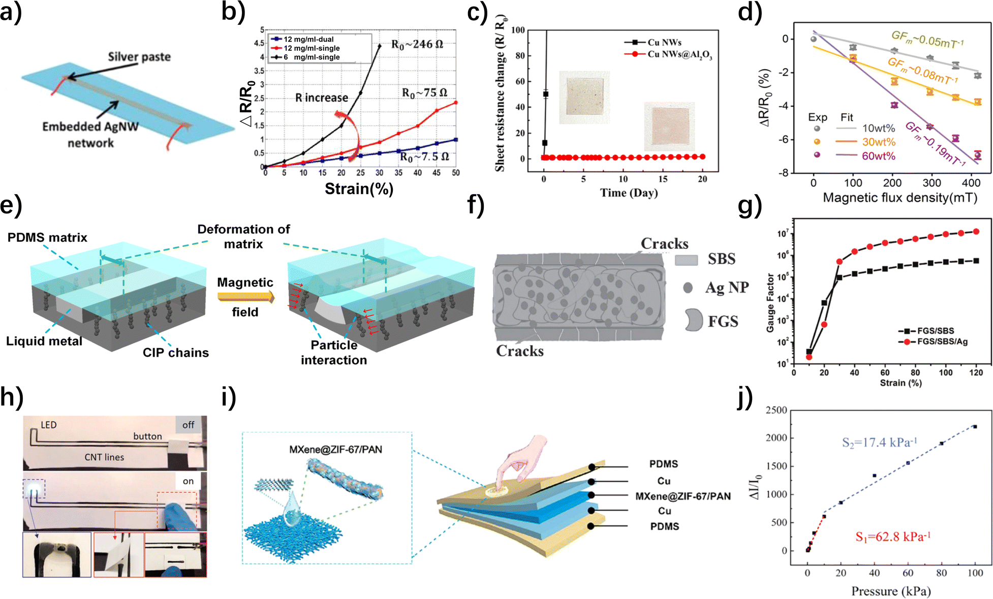
In addition to silver and nickel, gold, which has high density, excellent conductivity, and good ductility, has also attracted the attention of researchers. However, due to its high cost, gold is mostly added in small amounts to flexible resistive tactile sensors in the form of nanoparticles or nanowires.96 For instance, Huynh and Haick introduced self-repairing gold nanoparticles into a chemically resistant crosslinked polyurethane base, sandwiched between two layers of silver micro-particle polyurethane, resulting in the development of a versatile, flexible sensor possessing the ability to self-heal.76 The loaded sensitivity of the chemiresistive sensor after cutting remained stable at (0.09 ± 0.01) g F−1. Furthermore, copper nanowires (Cu NW), known for their good conductivity, have also captured researchers' attention. Although Cu NW is more susceptible to oxidation compared to the previously mentioned metal-based materials, addressing the oxidation issue and utilizing Cu NW to fabricate high-performance flexible resistive tactile sensors is of significant importance, particularly in the context of resource scarcity. Whether using quasi in situ polymerization, using PDMS as a sealing material to isolate air, or encapsulating Cu NW with other conductive materials, these are viable approaches to address the oxidation issue of Cu NWs. Zhao et al. enhanced the thermochemical stability of Cu NWs using solution-grown Al2O3 nanoshells.91 After the covering treatment with Al2O3 nanoshells, the Cu NWs maintained a relatively stable resistive resistance value in the comparison experiment for up to 20 days (Fig. 4c).
Besides elemental metals, certain highly conductive metal compounds and liquid metals can also serve as sensitive materials for flexible resistive tactile sensors, displaying impressive performance.97 Rana et al. utilized polycrystalline MoS2 to fabricate flexible strain sensors that exhibited excellent sensitivity (GF = 80 ± 2) and stability over a wide range of stresses (≥14 MPa).98 Similarly, Pataniya employed adaptable pressure-sensitive paper devices that were enhanced with WSe2 nanosheets. These devices showcased an exceptionally broad pressure operating range spanning from 1 Pa to 100 kPa, and achieved the highest sensitivity at 29.24 kPa−1.72 They demonstrated swift response times of 200 ms and relaxation times of 100 ms, enabling them to detect pressures as minute as 1.4 Pa, such as those generated by water droplets. Additionally, Hu et al. employed the liquid metal GaInSn for electronic circuits, creating a magnetoflexible resistive strain sensor with self-healing capabilities (Fig. 4e).92 When the content of carbonyl iron particles was increased from 10 wt% to 30 wt% and 60 wt%, the absolute value of GFm(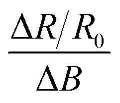 , ΔB was the variation of magnetic flux density) was significantly increased, with a corresponding significant increase in magnetic sensitivity (Fig. 4d).
Gr, a material formed by closely packed carbon atoms in a single-layer two-dimensional honeycomb lattice, exhibits outstanding optical, electrical, and mechanical properties, with a carrier mobility of approximately 15![[thin space (1/6-em)]](https://www.rsc.org/images/entities/char_2009.gif) 000 cm2 (V s)−1 at room temperature. Researchers often blend Gr with other conductive sensitive materials to enhance the sensing capability of the sensitive layer.99 Beyond traditional sheet-like Gr, materials like Gr foams and Gr sponges have also gained traction. However, the fabrication process for Gr foams can be complex and costly. To reduce costs and enhance sensitivity, some researchers have sought process improvements. In addition to conventional methods of synthesizing Gr foams via vapor deposition, Li et al. have developed a simple and scalable self-assembly approach under milder conditions to create high-performance Gr foams.100 The resulting flexible resistive tactile sensors exhibit advantages such as high tensile sensitivity and good reproducibility. Likewise, Gr sponges offer exceptional performance. For instance, Zhao et al. employed fragmentized Gr sponges (FGS) to fabricate high-performance sensors (Fig. 4f).93 Gr sponges are porous three-dimensional networks composed of interconnected graphene sheets, while FGS refers to graphene sponges that have been fragmented into micro-sized structures. Sensors crafted from FGS exhibited remarkable electrical conductivity (1521 S cm⁻1) along with impressive mechanical properties, displaying a fracture elongation of 680% and a tensile strength of 3.5 MPa. Upon doping FGS with polystyrene-butadiene-block styrene (SBS) and silver (Ag) nanoparticles, they displayed extraordinarily high sensitivity. The GF increased from 20.5 at 10% strain to 1.25 × 10⁷ at 120% strain (as shown in Fig. 4g). Additionally, these sensors demonstrated rapid response times of less than 20 ms and maintained excellent stability throughout tests surpassing 2000 cycles.
000 cm2 (V s)−1 at room temperature. Researchers often blend Gr with other conductive sensitive materials to enhance the sensing capability of the sensitive layer.99 Beyond traditional sheet-like Gr, materials like Gr foams and Gr sponges have also gained traction. However, the fabrication process for Gr foams can be complex and costly. To reduce costs and enhance sensitivity, some researchers have sought process improvements. In addition to conventional methods of synthesizing Gr foams via vapor deposition, Li et al. have developed a simple and scalable self-assembly approach under milder conditions to create high-performance Gr foams.100 The resulting flexible resistive tactile sensors exhibit advantages such as high tensile sensitivity and good reproducibility. Likewise, Gr sponges offer exceptional performance. For instance, Zhao et al. employed fragmentized Gr sponges (FGS) to fabricate high-performance sensors (Fig. 4f).93 Gr sponges are porous three-dimensional networks composed of interconnected graphene sheets, while FGS refers to graphene sponges that have been fragmented into micro-sized structures. Sensors crafted from FGS exhibited remarkable electrical conductivity (1521 S cm⁻1) along with impressive mechanical properties, displaying a fracture elongation of 680% and a tensile strength of 3.5 MPa. Upon doping FGS with polystyrene-butadiene-block styrene (SBS) and silver (Ag) nanoparticles, they displayed extraordinarily high sensitivity. The GF increased from 20.5 at 10% strain to 1.25 × 10⁷ at 120% strain (as shown in Fig. 4g). Additionally, these sensors demonstrated rapid response times of less than 20 ms and maintained excellent stability throughout tests surpassing 2000 cycles.
CNTs are primarily composed of layers of carbon atoms arranged in a coaxial cylindrical tube structure. As one-dimensional nanomaterials, CNTs are lightweight and exhibit a seamless hexagonal structure, resulting in exceptional mechanical, electrical, and chemical properties. In the field of flexible resistive tactile sensors, CNTs are often integrated with diverse materials to create sensing units, and these combinations may involve materials such as graphite nanoplatelets (GNPs), LIG, or carbonyl iron powder, contributing to improved sensing performance.101 CNTs can also be integrated with shear-thickening gels (STGs) or shear-stiffening (S-ST) polymers to impart impact resistance.102 Furthermore, with the emergence of 3D printing technology, some researchers have utilized novel CNT inks to fabricate flexible pressure-sensitive sensors through 3D printing. Owens et al. introduced a polymer-free, water-based ink containing carbon nanotubes that can be printed (Fig. 4h).94 This ink allowed for the creation of a conductor with a conductivity of up to 10000 S m−1. It exhibited outstanding flexibility and stability, with a DC resistance change of less than 5% after undergoing 1000 bends. Additionally, the DC resistance changed by less than 3% when the bend radius was less than 1 mm. This printing technology holds the potential to lower manufacturing costs for wearable sensors, radio-frequency identification (RFID) tags, and deformable structures.
MXene is a new class of inorganic two-dimensional materials, meaning layered transition metal carbides or nitrides, and possesses a structure similar to Gr. Within the family of MXene materials, the carbon-containing branches are frequently integrated into other materials, serving as a significant component of carbon-based materials. Also, due to the hydroxyl group or terminal oxygen on the surface of their materials, they stand out among many materials by virtue of their extraordinary transition metal carbide conductivity and flexibility, making them promising conductive materials for flexible resistive tactile pressure sensors.103–105 Li et al. developed a highly conductive MXene-based organohydrogel (M-OH) based on Ti3C2Tx MXene/lithium salt (LS)/polyacrylamide (PAM)/PVA hydrogel, which achieved 2000% stretching and excellent conductivity of 4.5 S m−1.106 The addition of MXene resulted in the dramatic increase in the electrical conductivity of the material. Due to the agglomeration effect of MXene in the supersaturated state, they finally found a balance between electrical conductivity and mechanical properties at a concentration of 1.0 wt%. Besides, Zhang et al. designed a flexible and conductive MXene/PEDOT:PSS@Melamine Foam (MPMF) piezoresistive sensor with a stabilized coating that combined excellent mechanical properties and electrical conductivity, and a wide operating range of the material that allowed it to generate up to 80% of the compressive strain at a pressure of 60 kPa, with good sensitivity (0.30 kPa−1) and a wide linear working region (12–60 kPa).107 Similarly, Fu et al. developed a device based on MXene/ZIF-67/PAN films (Fig. 4i).95 Although its linear response range (0.2–10 kPa, 20–100 kPa) was not as uniform as the previous one, it had a superb sensitivity (62.8 kPa−1) and strong mechanical stability (more than 10000 loading/unloading cycles), enabling the long-term wear and health monitoring (Fig. 4j).
Of course, apart from Gr, CNTs, and MXene, some researchers are exploring how to use low-cost carbon-based material to create high-performance flexible resistive tactile sensors. Liu et al., for example, employed a vapor deposition method to uniformly coat carbon black (diesel soot) onto textiles, resulting in a flexible pressure sensor.71 This sensor also exhibited significant sensitivity and operating range, demonstrating practical applications in sound signal monitoring and human physiology, including the detection of subtle pulse vibrations.
In recent years, Poly(3,4-ethylenedioxythiophene):Poly(styrene sulfonate) (PEDOT:PSS) has become one of the most desirable materials for the preparation of flexible resistive tactile sensors due to its excellent electrical conductivity, mechanical flexibility and ease of processing.113–115 This material is often made into thin films or doped into other conductive polymers to provide additive good electrical and mechanical properties for piezoresistive sensors.116–118 Xia et al. created a mild treatment with formamide and methanol as co-solvents to optimize the conductive properties of PEDOT:PSS films.119 Due to the high dielectric constant and hydrophilicity of formamide and the solvation of methanol, the insulating PSS could be separated from the PEDOT, leading to a significant increase in the film conductivity of PEDOT:PSS from 0.3 to 1287 S cm−1. Beccatelli et al. functionalized PU foams by using PEDOT:PSS to propose a modified all-polymer foam (Fig. 5a).120 This material, due to its excellent linear response range (0–30 kPa, 30–50 kPa) and high sensitivity, was applied to a prototype insole with eight pressure sensors, which could be used for medical rehabilitation and professional data monitoring of athletes (Fig. 5b). To enhance the adhesion and practicality of PEDOT:PSS, scholars modified the substrate material and optimized its structure, as PEDOT:PSS typically requires attachment to a substrate for utilization.121,122 Lee et al. used ultrafiltration to exchange water-based PEDOT:PSS solution for organic solvent-based PEDOT:PSS solution.123 After optimization, PEDOT formed a stable and sensitive thin nano-coating on the surface of hydrophobic pyramid-type PDMS with good bonding. Yang et al. deposited the PEDOT:PSS solution on a thin sheet of paper, and used the paper's fiber microstructure to design stacked and folded structures with good binding to PEDOT:PSS (Fig. 5c).124 Based on the extrusion and expansion between the paper layers, the stacked paper sensor has a sensitive response (less than 20 ms) and superb durability (more than 30000 loading and unloading cycles) (Fig. 5d and e). On the manufacturing side, in addition to being synthetic, the highly conductive, chemically stable, and translucent PEDOT:PSS is also machine-printable, which greatly contributes to sensor material savings and cost reductions.125,126
 | ||
| Fig. 5 Structure and performance of flexible resistive tactile sensors based on conducting polymers and conductive hydrogels. (a) Pictorial illustration of the fabrication process of the pressure sensitive all-polymeric device.120 Reproduced with permission. Copyright at ACS Applied Polymer Materials, 2021 (b) sensor response as a function of external pressure.120 Reproduced with permission. Copyright at ACS Applied Polymer Materials, 2021 (c) schematic of a 7 × 7 pixel pressure sensor made by sandwiching 8 layers of PEDOT:PSS-saturated paper between two PET substrates with crossed arrays of copper electrodes.124 Reproduced with permission. Copyright at ACS Applied Materials & Interfaces, 2019 (d) the response kinetics upon application and release of pressure.124 Reproduced with permission. Copyright at ACS Applied Materials & Interfaces, 2019 (e) current response for 100 load/unload cycles, recorded after 5000, 10000, 20000, and 30000 cycles.124 Reproduced with permission. Copyright at ACS Applied Materials & Interfaces, 2019 (f) dynamic scanning calorimetry (DSC) results at the endo direction of iSkin and other materials.132 Reproduced with permission. Copyright at Advanced Functional Materials, 2021 (g) protein nanofibers can be used to improve the mechanical properties of gelatin aerogels and increase their elasticity.133 Reproduced with permission. Copyright at ACS Applied Bio Materials, 2022 (h) current–time (I–t) curves of the sensor when repeatedly applying and removing a weight corresponding to a pressure of 41.6 kPa (1000 cycles).133 Reproduced with permission. Copyright at ACS Applied Bio Materials, 2022 (i) average sensitivity of five independently prepared LPNF:GEL:PEDOT-S (2.7:8:1) aerogels.133 Reproduced with permission. Copyright at ACS Applied Bio Materials, 2022. | ||
In response to the current resource scarcity, researchers have been exploring new types of conductive polymer materials for sensor fabrication. Yin et al. investigated the use of “green” renewable resource cellulose nanofibers (CNF) to create multifunctional flexible sensors.127 They mixed CNF with Ag NW to produce composite films, which were then sandwiched between two layers of thermoplastic polyurethane (TPU) to create flexible sensors capable of detecting strain and temperature. After a 10% pre-strain treatment, this sensor achieved a GF of 34.06 at a strain of 1.5%, and it was capable of detecting strains as low as 0.2%. It demonstrated good detection abilities in measuring pulses, vocal cord vibrations, and finger bending. Additionally, the sensor exhibited effective temperature-sensing behavior in response to changes in external temperature.
Conductive hydrogel, despite its advantages, faces challenges related to environmental sensitivity, self-healing limitations, rigidity, and power requirements, necessitating careful material and structural design choices. By incorporating hygroscopic and cryoprotective substances, it is possible to mitigate water loss, thereby diminishing the temperature and humidity susceptibility of hydrogels to some extent.131 Taking cues from human skin attributes, Ying et al. developed an innovative ionic skin using a novel viscous hydrogel formulation containing glycerol and concentrated salts, which resulted in a material exhibiting exceptional resistance to low temperatures (−95 °C) while possessing an elongation at break of 1975% with a conductivity of 0.904 S m−1 (Fig. 5f).132 Dai et al. designed a dual physically and chemically crosslinked triple-network hydrogel (PVA/B TN hydrogel), which improved the mechanical properties of this type of hydrogel and demonstrated superb self-healing ability at room temperature (healing time of 5 min, healing efficiency of 98.1%).134 Fu et al. designed a high-strength, self-powered piezoelectric polyacrylonitrile-polyvinylidene fluoride (PAN-PVDF) hydrogel. In the working system, they used PVDF as a self-powered source due to its piezoelectricity and excellent plasticity.135 The hydrogel sensor provided a consistent and steady electrical signal output in response to mechanical stimulation (∼30 mV and ∼2.8 μA), and it exhibited a rapid response time of ∼31 milliseconds. This allowed it to effectively translate alterations in the hydrogel's electrical resistance caused by an external force into changes in voltage output signals, all without requiring an external power source.
Aerogels and hydrogels share gel-like characteristics, yet unlike the constraints associated with hydrogels, aerogels have adeptly circumvented water loss issues through their robust physical attributes and exceptional environmental resilience, establishing themselves as a promising alternative. Wang et al. prepared a highly flexible and compressible aerogel with a smooth layer structure by compounding silver nanowires with PEDOT:PSS and polyimide.136 Within the constraints of the dispersing medium's limits, this aerogel exhibited impressive sensitivity (0.31 kPa−1). In contrast, hydrogels are susceptible to negative effects within the range of 0–1.25 kPa. Moreover, the aerogel's compressive strain showed remarkable synchronization with the rate of change in electrical resistance, achieving an astonishing linearity of 1, underscoring its exceptional sensing capabilities. Aerogels exhibit significantly reduced flexibility due to their lower water content compared to hydrogels. In order to improve its elasticity and flexibility, Yuan et al. proposed an innovative scheme by mixing lysozyme PNFs (LPNFs), gelatin, and leaving it to self-assemble to form a protein nanoproto-fiber, while adding poly(4-(2,3-dihydrothieno[3,4-b]-[1,4]dioxin-2-yl-methoxy))-1-butanesulfonic acid (PEDOT-S), which could improve the strength of the gel.133 After undergoing a process involving freezing, decompression, and evaporation, the mechanical properties of the mixture showed a significant enhancement (Fig. 5g). The modified aerogel exhibited remarkable electrical attributes. It displayed consistent and swift current responses even after undergoing more than 1000 cycles of load and unload tests. Additionally, it demonstrated a clear linear correlation across a wide pressure range from 1.8 to 300 kPa, and maintained an average sensitivity of 1.80 kPa−1, positioning it as a highly promising material for piezoresistive pressure sensors (Fig. 5h and i).
There are also advanced two-dimensional materials or composite materials that can be used as conductive materials. In addition to graphene, 2D materials such as indium selenide (InSe), molybdenum disulfide (MoS2), and others exhibit high electron mobility and good mechanical flexibility, making them suitable for manufacturing highly sensitive tactile sensors. Chen et al. developed a flexible, ultra-sensitive three-terminal strain sensor based on two-dimensional (2D) InSe, which can be used to detect human movement.137 InSe greatly enhances the current change, resulting in an 8-fold increase in the GF of the sensor under 0.25% tensile strain, reaching 32. Pang et al. successfully grew amorphous MoS2 on a 1.5 μm thick PDMS substrate through magnetron sputtering. They designed and fabricated a micro-thin film flexible sensor.138 At a pressure of 0.46 MPa, the maximum ΔR/R is 70.39, with a high piezoresistive coefficient of 866.89 MPa−1. It passed the human foot pressure test, demonstrating enormous potential in medical health. There are many advanced composite materials, such as glass fiber reinforced polymers, nanocomposites, and carbon nanotube reinforced polymers. These composite materials significantly enhance the conductivity of sensors and selectively optimize the weaknesses of conventional flexible resistive tactile sensors. For example, Kang et al. prepared a flexible resistive tactile sensor based on graphene-silver nanoplate-polymer nanocomposites.139 Its sensitivity is 0.04 kPa−1, with a response time of approximately 286 ms. Additionally, it exhibits hydrophobicity and self-cleaning properties, making it suitable for wearable devices. Fu et al. prepared a reduced graphene oxide-coated glass fabric/organosilicon composite material (RGO@GF) and used it to manufacture a high-performance structural flexible strain sensor.140 Glass fibers and silicon resin provide mechanical strength and flexibility, respectively. While adequately protecting the fragile internal structure, the composite material also possesses a gauge factor (GF) value of approximately 113. Furthermore, the addition of graphene imparts conductivity to the glass fibers. Clearly, achieving such comprehensive performance enhancement in composite materials is easily attainable.
Currently, there is a wide variety of conductive sensitive materials used in resistive-type flexible tactile sensors, and they are not limited to the aforementioned categories of metal-based materials, carbon-based materials, conductive polymers, and hydrogels. The performance specifications of flexible resistive tactile pressure sensors with different substrate and conductive sensitive materials are shown in Table 2. In summary, when fabricating resistive-type flexible tactile sensors, researchers often choose flexible substrates like PDMS, PET, PU, Ecoflex, and excellent conductive materials like Ag NW, Gr, CNT, PEDOT, MXene, conducting hydrogels, etc., as sensitive materials. The combination of these materials has led to the development of various high-performance sensors, demonstrating excellent performance in stretchability, sensitivity, and linearity.141
| Flexible substrate materials | Conductive sensitive materials | Flexibility | Sensitivity | Linear response range | Response and relaxation time | Reference |
|---|---|---|---|---|---|---|
| a Tip: definitions of sensitivity expressions for sensors may vary across different articles. This table uses both GF and S parameters to describe sensor sensitivity. The definitions of GF and S can be found in the second section of this article. | ||||||
| Ecoflex elastomer | Nickel nanowires | 100% | GF = 200 | — | 0.32 s, — | 58 |
| Epoxy resin | Graphene | — | S 1 = 0.156 kPa−1 | 2.5–100 kPa (S1) | 40.8 ms, 3.7 ms | 60 |
| S 2 = 0.068 kPa−1 | 100–250 kPa (S2) | |||||
| S 3 = 0.023 kPa−1 | 250–500 kPa (S3) | |||||
| Liquid metal | Golyvinyl alcohol | 540% | GF1 = 0.1 MPa−1 | 0–0.6 MPa (GF1) | — | 63 |
| GF2 = 1.2 MPa−1 | 0.6–2.2 MPa (GF2) | |||||
| GF3 = 0.2 MPa−1 | 2.2–3.2 MPa (GF3) | |||||
| Knit Kevlar | Laser-induced graphene | 3% | GF1 = 34.8 | — | 0.192 s, 0.177 s | 69 |
| GF2 = 117.9 | ||||||
| Tissue papers | WSe2 nanosheets | — | S 1 = 29.24 kPa−1 | 0.001–0.012 kPa (S1) | 200 ms, 100 ms | 72 |
| S 2 = 11.94 kPa−1 | 2–30 kPa (S2) | |||||
| S 2 = 3.20 kPa−1 | 35–100 kPa (S3) | |||||
| Self-healing disulfide-cross-linked polyurethane | Self-healing polyurethane/silver-microparticles composite | 97.34% | GF1 = 66.11 ± 16.08 (normal) | — | — | 76 |
| GF2 = 82.38 ± 12.10 (under electrode-cut) | ||||||
| GF3 = 30.22 ± 4.14 (under AuNP-cut) | ||||||
| PDMS elastomer | Silver nanowire | 70% | Tunable GF: 2–14 | — | 200 ms, — | 87 |
| PDMS elastomer | Nickel nanoparticles and graphene coated polyurethane sponge | 65% | GF1 = 36.03 | 0–20% strain (GF1) | 100 ms, — | 90 |
| GF2 = 3360.09 | 20% to 65% strain (GF2) | |||||
| PET | MoS2 | — | GF = 80 ± 2 (2 μm SU-8 encapsulated) | Stress ≥14 MPa | — | 98 |
| Poly(styrene-block-butadiene-block-styrene) | Fragmentized graphene sponges and Ag nanoparticles | 680% | Tunable GF: 20.5–1.25 × 10⁷ (strain: 10–120%) | — | 20 ms, — | 93 |
| Melamine foam modified with polydopamine | Mixture of MXene and PEDOT:PSS | 80% (compression strain) | S = 0.30 kPa−1 | 12–60 kPa | 200 ms, 120 ms | 142 |
| Commercial polyurethane | PEDOT:PSS | — | S 1 = 0.30 kPa−1 | 0–30 kPa (S1) | — | 120 |
| S 2 = 0.08 kPa−1 | 30–50 kPa (S2) | |||||
| Hydrogel | MWCNTs | 4075% | S 1 = 0.062 kPa−1 | 0–5 kPa (S1) | 0.18 s, — | 130 |
| S 2 = 0.022 kPa−1 | 5–9 kPa (S2) | |||||
| S 3 = 0.008 kPa−1 | 9–15 kPa (S3) | |||||
| Gelatin and lysozyme protein nanofibrils | PEDOT-S | — | S = 1.8 kPa−1 | 1.8–300 kPa | — | 143 |
3.3. Sensor structure design and performance optimization
The design of sensor structures and performance optimization are widely acknowledged as critical factors that greatly affect various performance parameters in flexible resistive tactile sensors. The flexibility, sensitivity, and stability of the sensor are directly influenced by its structural design, which is essential for fully utilizing the potential of the materials.144 Additionally, to improve sensor resolution, detect three-dimensional forces, and meet the integration requirements of flexible electronic skins, researchers often choose to incorporate unit arrays into flexible resistive tactile sensors. By employing multi-point monitoring methods and utilizing sensor calibration and decoupling techniques, they can achieve multifunctional and highly precise sensing.However, the simple sandwich structure alone may not meet the requirements of flexible resistive tactile sensors for high precision and sensitivity. Therefore, many researchers have enhanced the sensing performance by optimizing various microstructures based on the sandwich structure.146 Generally, microstructure design can be roughly categorized into surface microstructures and internal microstructures. Surface microstructures involve creating features such as curved folds,147 cone-like structures,148 hemispherical structures,149 interlocking structures,150 and biomimetic structures151 on the substrate surface. These structures increase the contact area between the substrate and the sensing unit, thereby enhancing the sensitivity. Internal microstructures, on the other hand, entail constructing features like porous structures, interlocking structures, and biomimetic structures within the flexible substrate or the sensing unit itself. These internal spatial arrangements, such as gaps between adjacent frameworks or hollow channels within the framework, can increase the contact area and decrease the contact resistance of the sensor when subjected to external forces.152
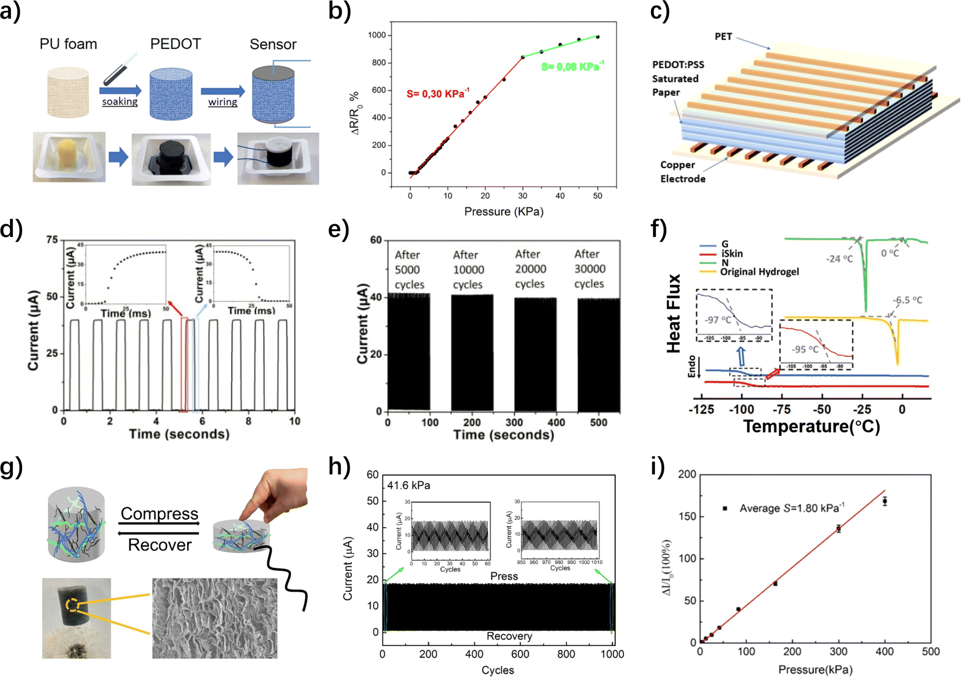
Among the surface microstructures, one of the relatively easier designs is the surface curvature and folding structure. Kim et al. introduced a serpentine pattern that allowed for diverse responses to different stresses through curvature control.153 Smaller curvature serpentine strips were found to exhibit greater strains under the same stress when compared to their larger curvature counterparts. The design with larger curvature is better for reducing induced strain and high-stress monitoring, while the design with smaller curvature provides higher sensitivity, reaching 0.41% kPa−1 within the 0% to 10% strain range, capable of detecting pressures as low as 0.87 kPa. Conversely, the design with larger curvature provided a wider linear working range, offering a sensitivity of 0.075% kPa−1 within the 0% to 30% strain range. Additionally, Xiang et al. utilized the rough structure of sandpaper, coated it with PDMS and Alk-Ti3C2, to create a flexible pressure-sensitive sensor with surface folding (Fig. 6a).154 The implementation of this structure resulted in a notable enhancement of sensitivity across the operating range of 0–800 kPa. More precisely, it exhibited a sensitivity of 95.26 kPa−1 in the low-pressure range of 0–1 kPa and a sensitivity of 543.66 kPa−1 in the high-pressure range from 200 to 600 kPa (Fig. 6b). Surface biomimetic structures and interlocking structures can also provide excellent performance to sensors.155 Although both often involve technologies like 3D printing and UV exposure, which pose challenges in terms of construction difficulties and technical complexity, their high sensitivity and reliability have still led many researchers to conduct extensive studies in this area. For instance, Chun et al. developed a flexible piezoresistive sensor with high adhesion and water-resistant stretchability by inscribing octopus-like patterns (OPs) on carbon-based conductive polymer composite (CPC) films (Fig. 6c).151 The sensor possessed good stability with an estimated sensitivity (S1) of 0.14% kPa−1 by linear fitting for vertical pressure values below 60 kPa, and an improved sensitivity (S2) of 0.01% kPa−1 when the pressure increased from 60 kPa to 1000 kPa (Fig. 6d). Particularly, flexible tactile sensors based on interlocking structures exhibit significant potential. When interlocking fibers come into contact under external force, they deform slightly starting from the tip. The greater the external force, the tighter the interlocking between the fibers, and the more pronounced the deformation becomes. These minor distortions generated by the contact between interlocking fibers can significantly enhance the sensor's sensing ability, allowing it to detect extremely low pressures. Lu et al. proposed a new method to prepare highly sensitive flexible pressure sensors using interlocking nanocone arrays (IOCA) (Fig. 6e).156 Under different pressures, the IOCA squeezed each other due to the interlocking structure, and the height and contact area between the cells changed, which in turn led to the change of IOCA conductivity and affected the pressure sensitivity. The theoretically derived results in Fig. 6f were in high agreement with the experimental results, confirming the rationality of the interlocking structure and the high sensitivity of the sensor. Pang et al. utilized interlocked nanofiber arrays coated with platinum to fabricate a sensor capable of detecting extremely low pressures, approximately around 5 Pa. It could also discern shear stress as low as 0.001 N (with a sensing limit of about 1 N), and torsional loads as small as 0.0002 N m.159 Additionally, within the strain range of 0% to 5%, it exhibited gauge factors of roughly 11.5 for pressure, 0.75 for shear, and 8.53 for torsion. This sensor proved adept at detecting minute pressures from falling droplets and repetitive jumps, enabling real-time monitoring of heartbeat pulses.
 | ||
| Fig. 6 Different structures for optimizing the sensor performance. (a) The fabrication process of PDMS films syn-thesis and flexible pressure sensors.154 Reproduced with permission. Copyright at Advanced Materials Technologies, 2021 (b) the relative current variations ΔI/I0 of the sensor under different pressures.154 Reproduced with permission. Copyright at Advanced Materials Technologies, 2021 (c) process steps and application scenarios of OP-CPC electronic placement.151 Reproduced with permission. Copyright at Advanced Functional Materials, 2018 (d) piezoresistive responses of the device subjected to static vertical pressures.151 Reproduced with permission. Copyright at Advanced Functional Materials, 2018 (e) schematic showing resistance composition and the pressure sensing mechanism of the IOCA.156 Reproduced with permission. Copyright at ACS Applied Materials & Interfaces, 2020 (f) experimental resistance changes of the IOCA fitted to theoretically calculated resistance changes.156 Reproduced with permission. Copyright at ACS Applied Materials & Interfaces, 2020 (g) schematic of opening structure: (i) opening structure. (ii) The ΔR/R versus magnetic flux density.157 Reproduced with permission. Copyright at ACS Applied Materials & Interfaces, 2018 (h) schematic of flexible piezoresistive sensor with closed-cell structures structure.158 Reproduced with permission. Copyright at Polymer Engineering & Science, 2023 (i) response and recovery times158 (1 mm s−1 compression speed and 5% strain) reproduced with permission. Copyright at Polymer Engineering & Science, 2023. | ||
In addition to their role in surface microstructures, biomimetic and interlocking structures are widely applied within the internal microstructures as well. Whether it's the spider microcrack structure and lobster exoskeleton structure, or the interlocking fiber-like magnetically induced column forest, they all significantly enhance the sensitivity of sensors.160 Among these, Zhao and colleagues drew inspiration from the geometric shape of lobster shells.93 They introduced fragmented Gr sponges and silver nanoparticles into the sensor. Under tensile strain, the stacked Gr inside the sensor underwent mutual sliding rather than direct separation, thereby maintaining the conductive pathway without breaking, which significantly enlarges the operational range of the sensor.
Likewise, researchers have concentrated on internal porous structures, which provide benefits such as reduced density and lower elastic modulus. Depending on whether their internal voids are open or closed, porous structures can be classified into open-cell and closed-cell structures. Open-cell structures often refer to internal frameworks with high porosity and strong deformability, such as sponge-like structures.161 Under external forces, the framework of an sponge-like structures.161 Under external forces, the framework of an open-cell structure begins to undergo compressive deformation, leading to the creation of new contact points between the framework elements, thereby reducing overall resistance. Additionally, the good resilience of sponge-like structures has led many researchers to explore their applications.162 For instance, sacrificial templates were used by Ding and colleagues to create porous sponge-like flexible tactile sensors capable of detecting pressure, stress, and magnetic fields (Fig. 6g).157 As the magnetic flux density changes, the GFm of the sensor changed, ranging from 0.07 to 0.14 and 0.14 to 0.25 in the ranges of 0.086 to 0.115 T and 0.115 to 0.15 T, respectively.
Closed-cell structures, on the other hand, refer to structures with discontinuous bubbles surrounded by solid pore walls, which offer better load-bearing capabilities than open-cell structures. Closed-cell structures provide the sensor with derived structural elasticity and lower elastic modulus. Furthermore, due to the increase in contact area between the microstructured film caused by bubble deformation and the electrodes under external compression, closed-cell structures can achieve ultra-high sensitivity for low-pressure detection. Pan and colleagues employed PPy to fabricate hollow sphere microstructures through a multiphase reaction, resulting in a flexible resistive tactile sensor capable of detecting pressures below 1 Pa with a response time as low as 50 ms.111 Li et al. achieved tunable piezoresistance and high resilience of the sensor by foaming to form a tunable closed-pore structure in the nanocomposite (Fig. 6h).158 Owing to the closed-pore structure and the mixed conductive nanofillers (1D MWCNTs and 2D GNPs) distributed in the cell walls, the sensor exhibited shorter response and recovery times, with fast response and recovery within 160 ms after both pressure application and pressure removal (Fig. 6i).
Compared to conventional structures, enhancements have been made in both surface and internal microstructures, enabling the optimal utilization of conductive materials' properties. These structural improvements significantly enhance the performance of flexible resistive tactile sensors, particularly in terms of response and relaxation times and resolution. The performance comparison of flexible resistive tactile sensors with varying structural designs is summarized in Table 3.
| Type | Structural designs | Response and relaxation time | Resolution | Reference |
|---|---|---|---|---|
| Surface microstructures | Curved folds structures | 10 ms, 20 ms | 1 Pa | 147 |
| Biomimetic structures | 1 ms, 0.5 ms | 0.1 Pa | 155 | |
| Interlocking structures | 48 ms, 56 ms | 0.98 Pa | 156 | |
| Cone-like structures | 24 ms, 36 ms | 16 Pa | 148 | |
| Hemispherical structures | 35 ms, 50 ms | 0.4 Pa | 149 | |
| Internal porous structures | Biomimetic structures | 20 ms, 20 ms | — | 93 |
| Interlocking structures | 20 ms, 20 ms | — | 93 | |
| Open-cell structures | 200 ms, 200 ms | 4.1 Pa | 161 | |
| Closed-cell structures | 160 ms, 160 ms | 0.8 kPa | 158 |
Common array structures frequently adopt the checkerboard pattern, comprising an x × y array of flexible resistive tactile sensor units, offering a straightforward and dependable approach to monitoring. Researchers have used this checkerboard structure to detect the magnitude and distribution of gravitational forces acting on objects. Zhao and colleagues utilized laser direct writing (LDW) technology to create a high-performing, flexible asymmetric pressure sensor from multi-walled carbon nanotubes (MWCNTs) and laser-induced graphene (LIG). This sensor boasted an integrated sensing array with multi-point recognition, enabling the detection of objects with varying loads (Fig. 7a).165 Furthermore, magneto-sensitive flexible tactile sensor units have been employed by scholars to construct checkerboard arrays that can detect non-contact gesture actions through force sensing, enhancing their applicability in human–machine interaction.160
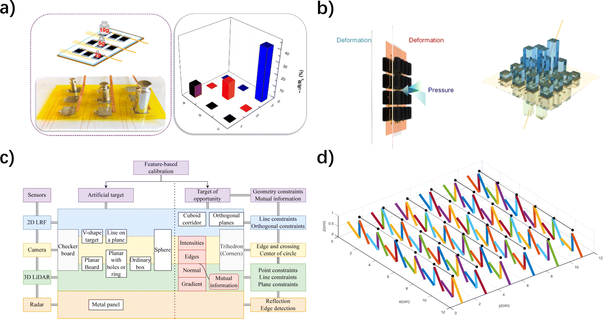 | ||
| Fig. 7 Integration, arrays, optimized calibration and decoupling algorithms are all important ways to ensure the stability of the information acquired and transmitted by the sensor. (a) Optical image and the relative resistance response mapping of pressure of a 3 × 3 pixel sensory array positioned different loads (2, 5 and 10 g).165 Reproduced with permission. Copyright at Sensors and Actuators A: Physical, 2021 (b) the schematic diagram of the fin-like double-side CNTs sponge-based sensor array subjects to external force changes and identifies force direction.166 Reproduced with permission. Copyright at ACS Applied Materials & Interfaces, 2020 (c) different feature-based calibration methods for different sensors according to their individual constraints.167 Reproduced with permission. Copyright at Information Fusion, 2023 (d) the deformation of the sensitive units when the force components are applied on it.168 Reproduced with permission. Copyright at Micromachines, 2018. | ||
Multi-unit monitoring in an array structure can also be used to perceive the magnitude and direction of three-dimensional forces. Typically, the pressures experienced by each stimulated sensor unit follow a pattern from small to large in the direction of force application, and analyzing the distinct response signals from each unit to directional forces enables data decoupling into magnitude and direction.166 However, this structure might not provide very precise resolution for the direction of three-dimensional forces (for example, the response difference between forces at 30° and 60° directions is not significant). Common solutions include increasing the number of sensor units, reducing the sensor unit's area, and optimizing the decoupling algorithm. Furthermore, Zhao and colleagues were inspired by fish fins to create a fin-like double-sided array of flexible electronic skin (Fig. 7b).166 The fin-like double-sided array structure offered more accurate perception of force direction because when one side of this electronic skin experienced external force, the other side responded synchronously.
Calibration, in the context of flexible resistive touch sensors, involves employing standard equipment to ascertain the sensor's input–output relationship. Due to variations between theoretical ideals and real-world characteristics, often stemming from the choice of flexible substrate and conductive materials, calibration experiments are essential for identifying and addressing these discrepancies. Through calibration experiments, discrepancies can be pinpointed, their origins analyzed, and the sensor's structure and model can be refined and enhanced accordingly (Fig. 7c).167 Currently, calibration methods often involve using weights for loading.
Decoupling methods entail using algorithms to correct the input–output relationship of a multivariable system, reducing or eliminating interdependencies to convert the system into multiple single-input single-output systems.169 Presently, decoupling techniques can be broadly classified into three categories: methods utilizing the least squares approach, those employing fuzzy reasoning, and those relying on neural networks.170 H However, these decoupling techniques come with their respective limitations. For instance, the least squares method is constrained by linear estimation. Fuzzy reasoning can be subjective when determining weight vectors for indicators, particularly with large sets of indicators, potentially resulting in reduced resolution. Neural networks, while proficient in swiftly finding optimal solutions thanks to their self-learning ability, can be critiqued for their lack of transparent reasoning processes and explanations, as they heavily rely on data for learning. Among these methods, neural networks excel due to their self-learning capabilities and high-speed optimization, which result in improved decoupling accuracy. For instance, Song et al. employed a backpropagation (BP) neural network to successfully decouple three-dimensional forces on a 6 × 6 flexible array touch sensor.168 The sensor consists of numerous sensitive units, each with two upper tactile sensing electrodes at its top. When force components along the X and Z directions are applied to the sensor, the tactile sensor array undergoes deformation. If the original position of one of the two upper tactile sensing electrodes is represented by a black dot, Fig. 7d illustrates a deformation state of the sensor. According to the established mathematical model of the sensor, coupled with the backpropagation (BP) neural algorithm, the force state applied to the sensor can be accurately inferred. Consequently, the decoupling accuracy of force sensing is effectively enhanced.
3.4. Optimization and applications in extreme scenarios
The lifespan of flexible resistive tactile sensors is influenced by various factors, including material durability, design quality, working environment, frequency of use, maintenance, and upkeep. Among these factors, the working environment is a crucial element that cannot be overlooked. In extreme environments, the durability and reliability of conventional flexible resistive tactile sensors significantly decrease, leading to impaired signal output. Besides mechanical extreme conditions such as excessive stretching, compression, and bending, environmental factors like cold temperatures, high heat, polluted conditions, and varying humidity also profoundly impact sensor performance. An excellent flexible resistive tactile sensor must undergo targeted optimizations to ensure proper functionality in such extreme conditions.4. Applications of flexible resistive tactile sensor
With increasing demands for intelligent machinery and complex working environments, the role of sensors in acquiring extensive information becomes crucial. Flexible resistive tactile sensors, renowned for their sensitivity, biocompatibility, and flexibility, excel in applications where rigid sensors fall short. Their robust tensile properties and ductility make them suitable for large-scale deformation data monitoring, while their precision and flexibility are advantageous in medical testing. Additionally, their stress perception and self-healing capabilities find applications in robotic contact sensing, leading to diverse and convenient applications that enhance our daily lives.4.1. Tactile perception and human–machine interaction
Human touch sensors grant us the ability to perceive numerous object characteristics, including shape, size, texture, and more, enabling sensory cognition and informed actions.176 Just as essential as they are to humans, imparting machines with tactile perception capabilities is a primary research goal to enhance their capabilities in industries like manufacturing, healthcare, and services.177With the emergence and application of various flexible resistive tactile sensors, flexible devices have replaced some rigid sensors, leading to improved safety and deformation resistance of machines.178 For instance, flexible tactile sensors simulated from human fingerprints, as developed by Wang et al., could detect various stresses such as compression, stretching, vibrations, and sliding (Fig. 8a).179 The strain signal of this sensor was highly synchronized with the applied pressure, which contributed to the real-time perception of the machine (Fig. 8b). In addition to tactile perception, flexible resistive tactile sensors hold significant potential in the field of human–machine interaction. By integrating flexible resistive tactile sensors into smart gloves or electronic skins, thanks to mature control systems, advantages in information acquisition, from gravity analysis to gesture recognition and motion monitoring, can be fully exploited. Amjadi et al., for example, utilized Ag NW-based flexible strain sensors to create a smart glove system for monitoring finger bending and various hand gestures.87 This smart glove integrated a chip combining data collection and wireless communication systems to acquire sensor data and perform corrections. Furthermore, Yuan et al.'s electronic skin model could monitor the location of applied force as well as movements of the knee joint, such as walking, jumping, and squatting.145
 | ||
| Fig. 8 The application of flexible resistive tactile pressure sensors for tactile sensing, human–computer interaction, healthcare and e-skin. (a) Schematic diagram of the preparation process of pressure-sensing.179 Reproduced with permission. Copyright at Advanced Materials Technologies, 2019 (b) output signal and pressure as the function of time.179 Reproduced with permission. Copyright at Advanced Materials Technologies, 2019 (c) the fully-integrated wearable electronic device attached to the skin and signals acquisition with a cellphone.182 Reproduced with permission. Copyright at Sensors and Actuators A: Physical, 2021 (d) resistance changes versus different pressures.182 Reproduced with permission. Copyright at Sensors and Actuators A: Physical, 2021 (e) schematics and the optical images of the E-skins attached to human fingers. Three red-colored E-skins are affixed on different fingers and detect temperature, pressure, and tension, respectively.183 Reproduced with permission. Copyright at Proceedings of the National academy of Sciences of the United States of America, 2020 (f) the real-time signals of the variation of resistance of the E-skin under different test conditions.183 Reproduced with permission. Copyright at Proceedings of the National academy of Sciences of the United States of America, 2020. | ||
4.2. Medical and health monitoring
The growing societal demands have led to an increased prevalence of cardiovascular diseases, posing health risks to a larger population. In today's fast-paced lives, individuals are placing greater emphasis on monitoring physiological parameters like heart rate, pulse, and blood pressure.180 As a result, smart wristbands for heart rate monitoring have gained popularity. Yet, traditional methods like power-hungry photoplethysmography and complex ECG signal testing fall short of meeting people's needs. Hence, researchers are turning their attention to the vibration measurement method employing flexible resistive tactile sensors.181The vibration measurement method captures the body's vibration signals caused by each heartbeat using high-precision flexible tactile sensors and obtains the heart rate through signal processing. A highly sensitive pressure sensor developed by Liu's research team could detect three peaks of the arterial pulse wave (anacrotic wave, tidal wave, and dicrotic wave).110 Furthermore, Zhang and colleagues have created a washable, breathable, and flexible tactile sensor, capable of monitoring breathing, pulse, and temperature (Fig. 8c).182 This sensor exhibited high sensitivity, registering at 19.47 kPa−1 within the small pressure range of 0–4 kPa, and 3.66 kPa−1 within the larger pressure range of 4–20 kPa (Fig. 8d). The data collected by this sensor could be conveniently and efficiently transmitted to mobile devices via Bluetooth for further analysis and ease of access.
Apart from heart rate monitoring, to address the challenge of environment perception for people with disabilities, some researchers are exploring how to directly convey collected signals to biological neural networks. Kim et al., for example, have worn their developed smart prosthetic skin capable of monitoring stress, temperature, and humidity on a prosthetic limb.153 They connected the signals collected by this electronic skin to multiple electrodes connected to the sciatic nerve of rats' hind legs, successfully transmitting the signals to the rats' central nervous system.
4.3. Electronic skins
With the rise of the field of artificial intelligence in recent years, multifunctional electronic skins for both humans and robots have become a focus of attention.184,185 E-skin, utilizing state-of-the-art flexible piezoresistive haptic sensors, closely emulates real skin in its sensing approach, providing a comprehensive record of environmental and object data through its exceptional flexibility and snug fit in contact sensing. Zhang et al. proposed an e-skin based on a composite liquid crystal hydrogel of hydroxypropyl cellulose (HPC), polyacrylamide-acrylic acid (PACA) and CNTs (Fig. 8e).183 The addition of HPC and PACA gave the e-skin the ability to feedback the stimulation site, and the addition of CNTs made the ability even more powerful, allowing it to provide quantitative feedback on environmental data such as temperature, pressure, bending angle, etc. (Fig. 8f). Zou et al. also doped conductive silver nanoparticles in a dynamic covalent thermosetting material to develop a repairable, recyclable and malleable electronic skin.186 In addition to its basic function of sensing environmental forces, the skin achieved advanced functionalities including full recyclability and multiple sensing of temperature, humidity, and flow. Likewise, Jo et al. explored multifunctional flexible resistive tactile sensors and converted them into electronic skins, granting robots human-like tactile sensing capabilities as well as functions like electromagnetic shielding and heat insulation.187Although these applications are conceptual, some companies have already introduced commercial products of flexible resistive tactile sensors in practice. Companies such as Tekscan and Pressure Profile Systems are notable examples. They specialize in providing flexible tactile sensor solutions for measuring and analyzing pressure distribution, widely applied in the fields of healthcare, industry, human–machine interaction, and research. These products, by real-time and accurate measurement of pressure distribution, have the potential to enhance medical treatments, improve industrial production efficiency, and bring more convenience and innovation to society.
5. Conclusions and perspectives
Flexible resistive tactile sensors, in recent decades, have made significant strides and hold promise for electronic skin applications. High-flexibility materials like PDMS, PET, PU, and Ecoflex as substrates, paired with excellent conductive materials such as AgNW, Gr, CNTs, PPy, MXene, and conducting polymers as sensing elements were employed, resulting in sensors with remarkable properties including stretchability, sensitivity, and linearity. Furthermore, sensors featuring microstructures, particularly those with interlocking and porous designs, exhibit heightened sensitivity, enabling the detection of subtle stresses within an extremely low range. Nonetheless, the integration of these sensors into electronic skin poses challenges, necessitating further research and development.Firstly, a balance between high flexibility and high sensitivity is still challenging to achieve in flexible resistive tactile sensors. While some sensors have achieved high sensitivity within a low operating range, extending this performance to a broader operating range remains limited. Secondly, flexible resistive tactile sensors are difficult to tailor and join, and large-area coverage remains challenging. Although the performance of individual sensing units has improved, their scalability is poor, and they cannot be easily tailored or joined to different carrier conditions. Furthermore, the fabrication process of electronic skin is complex, and the high cost of production hampers its scalability. Additionally, the large-scale array of sensing units and complex signal readout circuits with limited sampling rates can hinder real-time data acquisition. Finally, the high power consumption of electronic skin and shortcomings in intelligent algorithms are also limiting factors. High power consumption in the circuits requires significant electrical energy, and the high-density tactile image demands specialized intelligent algorithms for intelligent recognition and perception of tactile objects.
However, the existing limitations of electrically flexible resistive tactile sensors also indicate the future directions for improvement. With the rapid development of intelligent robots and the widespread adoption of mobile smart devices, flexible resistive tactile sensors are showing great potential for applications in machine tactile sensing systems and flexible human–machine interaction networks. Additionally, tactile sensing systems can enhance a robot's self-learning and adaptability, allowing them to operate not only in safe workspaces but also in extreme and harsh environments, making greater contributions in manufacturing, services, healthcare, and defence industries. Moreover, integrated electronic skin with flexible resistive tactile sensors can develop towards multi-functionality, anti-interference, self-healing, and self-cleaning, achieving a comprehensive tactile perception performance more akin to human skin. This will enable robots to better adapt to complex external environments. Electronic skin will hold substantial practical value in fields such as biomimetic robotics, healthcare, and more.
Currently, flexible resistive tactile sensors not only need to mimic the comprehensive sensing abilities of human skin, such as pressure, stress, temperature, humidity, proximity, and surface roughness, but also need to evolve towards flexibility, lightweight, transparency, integration, multi-functionality, low power consumption, self-healing, self-cleaning, and high resolution. However, due to issues such as the trade-off between high flexibility and high sensitivity, poor compatibility, limited scalability, difficulties in large-scale coverage, complex manufacturing processes, high costs, and high energy consumption, the widespread implementation of flexible resistive tactile sensors in human society remains challenging.
In the future, the most critical research for flexible resistive tactile sensors should focus on achieving a balance between high flexibility, wide operating range, high sensitivity, multi-functionality, self-healing, self-cleaning, interference resistance, and transparency. This will enable them to make significant contributions to protecting against external interferences, monitoring human health, perceiving posture and motion, assisting in mechanical operations, and sensing the external environment for the benefit of human society. In the future, electrically flexible resistive tactile sensors will continue to face new challenges, such as transparency, biocompatibility, biodegradability, neural interface control, high integration, and miniaturization. Developing high-performance, easily manufacturable, and scalable electrically flexible resistive tactile sensors for widespread integration across various domains of human society is the direction of its future development.
Conflicts of interest
The authors declare that they have no known competing financial interests or personal relationships that could have appeared to influence the work reported in this paper.Acknowledgements
This work was financially supported by the Natural Science Research Start-up Foundation of Recruiting Talents of Nanjing University of Posts and Telecommunications (Grant No. NY221111, NY221003), Natural Science Foundation of the Higher Education Institutions of Jiangsu Province (Grant No. 22KJB430038), and Natural Science Foundation of Jiangsu Province of China (BK20230359) and National Natural Science Foundation of China (62304112) awarded to Yang Li. Zhiwei Liu thanks the support from the National Natural Science Foundation Program of China (No. 52204370).Notes and references
- S. Yuan, C. Ma, E. Fetaya, T. Mueller, D. Naveh, F. Zhang and F. Xia, Science, 2023, 379, eade1220 CrossRef CAS PubMed.
- J. T. Atkinson, L. Su, X. Zhang, G. N. Bennett, J. J. Silberg and C. M. Ajo-Franklin, Nature, 2022, 611, 548–553 CrossRef CAS PubMed.
- H. C. Ates, P. Q. Nguyen, L. Gonzalez-Macia, E. Morales-Narváez, F. Güder, J. J. Collins and C. Dincer, Nat. Rev. Mater., 2022, 7, 887–907 CrossRef PubMed.
- Y. Li, B. Cui, S. Zhang, B. Li, J. Li, S. Liu and Q. Zhao, Small, 2022, 18, 2107413 CrossRef CAS PubMed.
- J.-C. Hsieh, Y. Li, H. Wang, M. Perz, Q. Tang, K. W. K. Tang, I. Pyatnitskiy, R. Reyes, H. Ding and H. Wang, J. Mater. Chem. B, 2022, 10, 7260–7280 RSC.
- X. Li, Q. Xu, Y. Tang, C. Hu, J. Niu and C. Xu, IEEE Sens. J., 2023, 23, 14773–14780 Search PubMed.
- H. Ikeuchi and H. Saito, IEEE Sens. J., 2022, 22, 10740–10750 Search PubMed.
- M. Chen, D. Cui, Z. Zhao, D. Kang, Z. Li, S. Albawardi, S. Alsageer, F. Alamri, A. Alhazmi, M. R. Amer and C. Zhou, Nano Res., 2022, 15, 5510–5516 CrossRef CAS PubMed.
- J.-C. Hsieh, H. Alawieh, Y. Li, F. Iwane, L. Zhao, R. Anderson, S. I. Abdullah, K. W. Kevin Tang, W. Wang, I. Pyatnitskiy, Y. Jia, J. d. R. Millán and H. Wang, Biosens. Bioelectron., 2022, 218, 114756 CrossRef CAS PubMed.
- S. Zhang, Y. Li, G. Tomasello, M. Anthonisen, X. Li, M. Mazzeo, A. Genco, P. Grutter and F. Cicoira, Adv. Electron. Mater., 2019, 5, 1900191 CrossRef.
- Y. Li, S. Zhang, X. Li, V. R. N. Unnava and F. Cicoira, Flexible Printed Electron., 2019, 4, 044004 CrossRef CAS.
- S. Pyo, J. Lee, K. Bae, S. Sim and J. Kim, Adv. Mater., 2021, 33, 2005902 CrossRef CAS PubMed.
- S. Sim, E. Jo, Y. Kang, E. Chung and J. Kim, Small, 2021, 17, 2105334 CrossRef CAS PubMed.
- B. Ji, Q. Zhou, M. Lei, S. Ding, Q. Song, Y. Gao, S. Li, Y. Xu, Y. Zhou and B. Zhou, Small, 2021, 17, 2103312 CrossRef CAS PubMed.
- S. Hamaguchi, T. Kawasetsu, T. Horii, H. Ishihara, R. Niiyama, K. Hosoda and M. Asada, IEEE Robot. Autom. Lett., 2020, 5, 4028–4034 Search PubMed.
- F. Qi, L. Xu, Y. He, H. Yan and H. Liu, Cryst. Res. Technol., 2023, 58, 2300119 CrossRef CAS.
- X. Qu, J. Xue, Y. Liu, W. Rao, Z. Liu and Z. Li, Nano Energy, 2022, 98, 107324 CrossRef CAS.
- G. Canavese, S. Stassi, C. Fallauto, S. Corbellini, V. Cauda, V. Camarchia, M. Pirola and C. F. Pirri, Sens. Actuators, A, 2014, 208, 1–9 CrossRef CAS.
- A. P. Gerratt, H. O. Michaud and S. P. Lacour, Adv. Funct. Mater., 2015, 25, 2287–2295 CrossRef CAS.
- Y. Gao, H. Ota, E. W. Schaler, K. Chen, A. Zhao, W. Gao, H. M. Fahad, Y. Leng, A. Zheng, F. Xiong, C. Zhang, L.-C. Tai, P. Zhao, R. S. Fearing and A. Javey, Adv. Mater., 2017, 29, 1701985 CrossRef PubMed.
- C. Wu, T. W. Kim, J. H. Park, B. Koo, S. Sung, J. Shao, C. Zhang and Z. L. Wang, ACS Nano, 2020, 14, 1390–1398 CrossRef CAS PubMed.
- J. Wang, P. Cui, J. Zhang, Y. Ge, X. Liu, N. Xuan, G. Gu, G. Cheng and Z. Du, Nano Energy, 2021, 89, 106320 CrossRef CAS.
- D. Lu, T. Liu, X. Meng, B. Luo, J. Yuan, Y. Liu, S. Zhang, C. Cai, C. Gao, J. Wang, S. Wang and S. Nie, Adv. Mater., 2023, 35, e2209117 CrossRef PubMed.
- Y. Lu, D. Kong, G. Yang, R. Wang, G. Pang, H. Luo, H. Yang and K. Xu, Adv. Sci., 2023, 2303949 CrossRef PubMed.
- L. Chen, Y. Xu, Y. Liu, J. Wang, J. Chen, X. Chang and Y. Zhu, ACS Appl. Mater. Interfaces, 2023, 15, 24923–24932 CrossRef CAS PubMed.
- H. Hu, C. Zhang, C. Pan, H. Dai, H. Sun, Y. Pan, X. Lai, C. Lyu, D. Tang, J. Fu and P. Zhao, ACS Nano, 2022, 16, 19271–19280 CrossRef CAS PubMed.
- M. Yang, Y. Cheng, Y. Yue, Y. Chen, H. Gao, L. Li, B. Cai, W. Liu, Z. Wang, H. Guo, N. Liu and Y. Gao, Adv. Sci., 2022, 9, 2200507 CrossRef CAS PubMed.
- D. Yu, Z. Zheng, J. Liu, H. Xiao, G. Huangfu and Y. Guo, Nano-Micro Lett., 2021, 13, 117 CrossRef CAS PubMed.
- R. Yu, T. Xia, B. Wu, J. Yuan, L. Ma, G. J. Cheng and F. Liu, ACS Appl. Mater. Interfaces, 2020, 12, 35291–35299 CrossRef CAS PubMed.
- W. Du, Z. Li, Y. Zhao, X. Zhang, L. Pang, W. Wang, T. Jiang, A. Yu and J. Zhai, Chem. Eng. J., 2022, 446, 137268 CrossRef CAS.
- J. Tolvanen, J. Hannu and H. Jantunen, IEEE Sens. J., 2017, 17, 4735–4746 CAS.
- Y. Zou, J. Liao, H. Ouyang, D. Jiang, C. Zhao, Z. Li, X. Qu, Z. Liu, Y. Fan, B. Shi, L. Zheng and Z. Li, Appl. Mater. Today, 2020, 20, 100699 CrossRef.
- L. Huang, S. Wang, P. Zhang, K. Zhang, Y. Li and Z. Chen, Adv. Mater. Technol., 2024, 9, 2301377 CrossRef CAS.
- M. Kong, Z. Xiang, X. Xu, S. Ma, C. Chen, X. Wang, K. Li and D. Wei, Adv. Electron. Mater., 2023, 9, 2300197 CrossRef CAS.
- D. Guo, P. Guo, Y. Yao, L. Ren, M. Jia, W. Wang, Y. Wang, Y. Zhang, A. Yu and J. Zhai, Nano Energy, 2022, 100, 107508 CrossRef CAS.
- H. R. Na, H. J. Lee, J. H. Jeon, H.-J. Kim, S.-K. Jerng, S. B. Roy, S.-H. Chun, S. Lee and Y. J. Yun, npj Flexible Electron., 2022, 6, 2 CrossRef.
- J. Cai, Y. He, Y. Zhou, H. Yu, B. Luo and M. Liu, Composites, Part A, 2022, 155, 106813 CrossRef CAS.
- R. Chen, Z. He, X. Luo, M. Wang, Z. Liu, Y. Wu, X. Dong, D. Zhang and J. Liu, ACS Appl. Polym. Mater., 2022, 5, 391–399 CrossRef.
- W. B. Han, G. J. Ko, K. G. Lee, D. Kim, J. H. Lee, S. M. Yang, D. J. Kim, J. W. Shin, T. M. Jang, S. Han, H. Zhou, H. Kang, J. H. Lim, K. Rajaram, H. Cheng, Y. D. Park, S. H. Kim and S. W. Hwang, Nat. Commun., 2023, 14, 2263 CrossRef CAS PubMed.
- L. Zhang, T. Song, L. Shi, N. Wen, Z. Wu, C. Sun, D. Jiang and Z. Guo, J. Nanostruct. Chem., 2021, 11, 323–341 CrossRef CAS PubMed.
- J. Miao and T. Fan, Carbon, 2023, 202, 495–527 CrossRef CAS.
- S. He, Y. Hong, M. Liao, Y. Li, L. Qiu and H. Peng, Aggregate, 2021, 2, e143 CrossRef CAS.
- B. C. Zhang, Y. H. Shi, J. Mao, S. Y. Huang, Z. B. Shao, C. J. Zheng, J. S. Jie and X. H. Zhang, Adv. Mater., 2021, 33, e2008171 CrossRef PubMed.
- Y. Zhang, Q. Lu, J. He, Z. Huo, R. Zhou, X. Han, M. Jia, C. Pan, Z. L. Wang and J. Zhai, Nat. Commun., 2023, 14, 1252 CrossRef CAS PubMed.
- Y. Li, J. Long, Y. Chen, Y. Huang and N. Zhao, Adv. Mater., 2022, 34, e2200517 CrossRef PubMed.
- Y. Yan, Z. Hu, Z. Yang, W. Yuan, C. Song, J. Pan and Y. Shen, Sci. Robot., 2021, 6, eabc8801 CrossRef PubMed.
- Y.-R. Ding, C.-H. Xue, Q.-Q. Fan, L.-L. Zhao, Q.-Q. Tian, X.-J. Guo, J. Zhang, S.-T. Jia and Q.-F. An, Chem. Eng. J., 2021, 404, 126489 CrossRef CAS.
- L. Wang, F. Xia, W. Xu, G. Wang, S. Hong, F. Cheng, B. Wu and N. Zheng, Adv. Funct. Mater., 2023, 33, 2215127 CrossRef CAS.
- K. Liu, C. Yang, S. Zhang, Y. Wang, R. Zou, Alamusi, Q. Deng and N. Hu, Mater. Des., 2022, 218, 110689 CrossRef CAS.
- A. Rivadeneyra, A. Marín-Sánchez, B. Wicklein, J. F. Salmerón, E. Castillo, M. Bobinger and A. Salinas-Castillo, Compos. Sci. Technol., 2021, 208, 108738 CrossRef CAS.
- M. Korrapati, S. Chauhan, Y. Tang and D. Gupta, Macromol. Mater. Eng., 2022, 307, 2200132 CrossRef CAS.
- X. Li, J. Liu, Q. Guo, X. Zhang and M. Tian, Small, 2022, 18, e2201012 CrossRef PubMed.
- K.-X. Hou, S.-P. Zhao, D.-P. Wang, P.-C. Zhao, C.-H. Li and J.-L. Zuo, Adv. Funct. Mater., 2021, 31, 2107006 CrossRef CAS.
- H. S. Cho, H. Y. Moon, H. S. Lee, Y. T. Kim and S. C. Jeoung, Bull. Korean Chem. Soc., 2021, 42, 1225–1231 CrossRef CAS.
- J. Vaicekauskaite, P. Mazurek, S. Vudayagiri and A. L. Skov, J. Mater. Chem. C, 2020, 8, 1273–1279 RSC.
- K. Anasiewicz and J. Kuczmaszewski, Materials, 2022, 15, 8060 CrossRef CAS PubMed.
- Y. Tai, T. Chen and G. Lubineau, ACS Appl. Mater. Interfaces, 2017, 9, 32184–32191 CrossRef CAS PubMed.
- S. Wang, K. Chen, M. Wang, H. Li, G. Chen, J. Liu, L. Xu, Y. Jian, C. Meng, X. Zheng, S. Liu, C. Yin, Z. Wang, P. Du, S. Qu and C. W. Leung, J. Mater. Chem. C, 2018, 6, 4737–4745 RSC.
- Z.-X. Li, X.-Y. Gao, P. Huang, Y.-Q. Li and S.-Y. Fu, J. Mater. Chem. A, 2022, 10, 9114–9120 RSC.
- Q. Lin, F. Zhang, X. Xu, H. Yang, Q. Mao, D. Xian, K. Yao and Q. Meng, Nanomaterials, 2023, 13, 2630 CrossRef CAS PubMed.
- A. Mokhtari, N. Tala-Ighil and Y. A. Masmoudi, J. Mater. Eng. Perform., 2021, 31, 2715–2722 CrossRef.
- X. Hong, L. Zou, J. Zhao, C. Li and L. Cong, IOP Conf. Ser.: Mater. Sci. Eng., 2018, 439, 042011 Search PubMed.
- Y. Lou, H. Liu and J. Zhang, Chem. Eng. J., 2020, 399, 125732 CrossRef CAS.
- T.-L. Chu, C. Ha-Minh and A. Imad, Composites, Part B, 2016, 95, 144–154 CrossRef CAS.
- F. Xu, W. Fan, Y. Zhang, Y. Gao, Z. Jia, Y. Qiu and D. Hui, Composites, Part B, 2017, 116, 398–405 CrossRef CAS.
- S. Fu, B. Yu, W. Tang, M. Fan, F. Chen and Q. Fu, Compos. Sci. Technol., 2018, 163, 141–150 CrossRef CAS.
- M. S. H. Al-Furjan, L. Shan, X. Shen, M. S. Zarei, M. H. Hajmohammad and R. Kolahchi, J. Mater. Res. Technol., 2022, 19, 2930–2959 CrossRef CAS.
- S. Wang, S. Xuan, M. Liu, L. Bai, S. Zhang, M. Sang, W. Jiang and X. Gong, Soft Matter, 2017, 13, 2483–2491 RSC.
- D. Yang, H. K. Nam, T.-S. D. Le, J. Yeo, Y. Lee, Y.-R. Kim, S.-W. Kim, H.-J. Choi, H. C. Shim, S. Ryu, S. Kwon and Y.-J. Kim, ACS Nano, 2023, 17, 18893–18904 CrossRef CAS PubMed.
- G. Tian, L. Zhan, J. Deng, H. Liu, J. Li, J. Ma, X. Jin, Q. Ke and C. Huang, Chem. Eng. J., 2021, 425, 130682 CrossRef CAS.
- K. Liu, J. Yu, Y. Li, X. Yan, D. Bai, X. Liao, Z. Zhou, Y. Gao, X. Yang and L. Li, Adv. Mater. Technol., 2019, 4, 1900475 CrossRef CAS.
- P. M. Pataniya, C. K. Sumesh, M. Tannarana, C. K. Zankat, G. K. Solanki, K. D. Patel and V. M. Pathak, Nanotechnology, 2020, 31, 435503 CrossRef CAS PubMed.
- J. Huang, Y. Cai, C. Xue, J. Ge, H. Zhao and S.-H. Yu, Nano Res., 2021, 14, 3636–3642 CrossRef CAS.
- X. Dai, P. Li, Y. Sui and C. Zhang, J. Polym. Sci., 2021, 59, 627–637 CrossRef CAS.
- A. Dallinger, K. Keller, H. Fitzek and F. Greco, ACS Appl. Mater. Interfaces, 2020, 12, 19855–19865 CrossRef CAS PubMed.
- T.-P. Huynh and H. Haick, Adv. Mater., 2016, 28, 138–143 CrossRef CAS PubMed.
- M. Zhang, X. Xia, L. Zhang, G. Zhao, C. Liu, N. Li, J. Xu, Y. Chen and X. Jian, Eur. Polym. J., 2023, 191, 112018 CrossRef CAS.
- G. Dias, M. Prado, R. Ligabue, M. Poirier, C. Le Roux, P. Micoud, F. Martin and S. Einloft, Polym. Polym. Compos., 2018, 26, 127–140 CAS.
- Y. Luo, L. Zhao, G. Luo, L. Dong, Y. Xia, M. Li, Z. Li, K. Wang, R. Maeda and Z. Jiang, Microsyst. Nanoeng., 2023, 9, 113 CrossRef CAS PubMed.
- T. Yao, S. Han, C. Men, J. Zhang, J. Luo and Y. Li, Constr. Build. Mater., 2021, 292, 123434 CrossRef CAS.
- X. Ye, Y. Li, Y. Zhang and P. Wang, J. Bionic Eng., 2022, 19, 853–876 CrossRef.
- M. Wang, Y. Yu, Y. Liang, Z. Han, C. Liu, S. Ma, Z. Lin and L. Ren, J. Bionic Eng., 2022, 19, 1439–1448 CrossRef.
- M. Paolieri, Z. Chen, F. Babu Kadumudi, M. Alehosseini, M. Zorrón, A. Dolatshahi-Pirouz and H. Maleki, ACS Appl. Nano Mater., 2023, 6, 5211–5223 CrossRef CAS.
- N. Yang, X. Yin, H. Liu, X. Yan, X. Zhou, F. Wang, X. Zhang, Y. Zhao and T. Cheng, ACS Appl. Mater. Interfaces, 2023, 15, 42992–43002 CrossRef CAS PubMed.
- Y. Xiong, Y. Zhao, Y. Tao, F. Yao, D. Li and D. Xu, Nano Lett., 2020, 20, 8576–8583 CrossRef CAS PubMed.
- L. Meng, M. Zhang, H. Deng, B. Xu, H. Wang, Y. Wang, L. Jiang and H. Liu, CCS Chem., 2021, 3, 2194–2202 CrossRef CAS.
- M. Amjadi, A. Pichitpajongkit, S. Lee, S. Ryu and I. Park, ACS Nano, 2014, 8, 5154–5163 CrossRef CAS PubMed.
- C. Peng, P. Wei, X. Li, Y. Liu, Y. Cao, H. Wang, H. Yu, F. Peng, L. Zhang, B. Zhang and K. Lv, Nano Energy, 2018, 53, 97–107 CrossRef CAS.
- B. C. Tee, C. Wang, R. Allen and Z. Bao, Nat. Nanotechnol., 2012, 7, 825–832 CrossRef CAS PubMed.
- F. Han, J. Li, S. Zhao, Y. Zhang, W. Huang, G. Zhang, R. Sun and C.-P. Wong, J. Mater. Chem. C, 2017, 5, 10167–10175 RSC.
- L. Zhao, P. Yang, S. Shi, X. Wang and S. Yu, Chem. Eng. J., 2023, 473, 145156 CrossRef CAS.
- T. Hu, S. Xuan, L. Ding and X. Gong, Sens. Actuators, B, 2020, 314, 128095 CrossRef CAS.
- S. Zhao, L. Guo, J. Li, N. Li, G. Zhang, Y. Gao, J. Li, D. Cao, W. Wang, Y. Jin, R. Sun and C.-P. Wong, Small, 2017, 13, 1700944 CrossRef PubMed.
- C. E. Owens, R. J. Headrick, S. M. Williams, A. J. Fike, M. Pasquali, G. H. McKinley and A. J. Hart, Adv. Funct. Mater., 2021, 31, 2100245 CrossRef CAS.
- X. Fu, J. Li, D. Li, L. Zhao, Z. Yuan, V. Shulga, W. Han and L. Wang, ACS Appl. Mater. Interfaces, 2022, 14, 12367–12374 CrossRef CAS PubMed.
- M. Sang, K. Kang, Y. Zhang, H. Zhang, K. Kim, M. Cho, J. Shin, J. H. Hong, T. Kim, S. K. Lee, W. H. Yeo, J. W. Lee, T. Lee, B. Xu and K. J. Yu, Adv. Mater., 2022, 34, e2105865 CrossRef PubMed.
- G.-H. Lee, H. Kim, J. Lee, J.-Y. Bae, C. Yang, H. Kim, H. Kang, S. Q. Choi, S. Park, S.-K. Kang, J. Kang, Z. Bao, J.-W. Jeong and S. Park, Mater. Today, 2023, 67, 84–94 CrossRef CAS.
- V. Rana, P. Gangwar, J. S. Meena, A. K. Ramesh, K. N. Bhat, S. Das and P. Singh, Nanotechnology, 2020, 31, 385501 CrossRef CAS PubMed.
- Y. Jung, K. Jung, B. Park, J. Choi, D. Kim, J. Park, J. Ko and H. Cho, Micro Nano Lett., 2019, 7, 20 CrossRef.
- J. Li, S. Zhao, X. Zeng, W. Huang, Z. Gong, G. Zhang, R. Sun and C.-P. Wong, ACS Appl. Mater. Interfaces, 2016, 8, 18954–18961 CrossRef CAS PubMed.
- X. Hui, S. Sharma, M. Sharifuzzaman, M. A. Zahed, Y. D. Shin, S. K. Seonu, H. S. Song and J. Y. Park, Small, 2022, 18, e2201247 CrossRef PubMed.
- S. Wang, S. Xuan, W. Jiang, W. Jiang, L. Yan, Y. Mao, M. Liu and X. Gong, J. Mater. Chem. A, 2015, 3, 19790–19799 RSC.
- W. Zhao, D. Zhang, Y. Yang, C. Du and B. Zhang, J. Mater. Chem. A, 2021, 9, 22082–22094 RSC.
- L. Ding, L. Sun, Z. Wang, D. Li, Q. Zhang, X. Liu, Y. Ren and Y. Li, Chem. Eng. J., 2023, 474, 145859 CrossRef CAS.
- Y. Wang, Y. Yue, F. Cheng, Y. Cheng, B. Ge, N. Liu and Y. Gao, ACS Nano, 2022, 16, 1734–1758 CrossRef CAS PubMed.
- Q. Li, X. Zhi, Y. Xia, S. Han, W. Guo, M. Li and X. Wang, ACS Appl. Mater. Interfaces, 2023, 15, 19435–19446 CrossRef CAS PubMed.
- M. Zhang, W. Yang, Z. Wang, H. Liu, R. Yin, C. Liu and C. Shen, Appl. Phys. Lett., 2023, 122, 043507 CrossRef CAS.
- H.-H. Tai, B.-A. Chen, Y.-H. Liu, Y.-J. Lu and J.-C. Wang, IEEE Sens. J., 2022, 22, 6418–6425 CAS.
- T. Yin, Y. Cheng, Y. Hou, L. Sun, Y. Ma, J. Su, Z. Zhang, N. Liu, L. Li and Y. Gao, Small, 2022, 18, e2204806 CrossRef PubMed.
- K. Liu, Z. Zhou, X. Yan, X. Meng, H. Tang, K. Qu, Y. Gao, Y. Li, J. Yu and L. Li, Polymers, 2019, 11, 1120 CrossRef PubMed.
- L. Pan, A. Chortos, G. Yu, Y. Wang, S. Isaacson, R. Allen, Y. Shi, R. Dauskardt and Z. Bao, Nat. Commun., 2014, 5, 3002 CrossRef PubMed.
- X. Wang, O. Yue, X. Liu, M. Hou and M. Zheng, Chem. Eng. J., 2020, 392, 123672 CrossRef CAS.
- Y. Li, X. Zhou, B. Sarkar, N. Gagnon-Lafrenais and F. Cicoira, Adv. Mater., 2022, 34, 2108932 CrossRef CAS PubMed.
- Y. Li, X. Li, S. Zhang, L. Liu, N. Hamad, S. R. Bobbara, D. Pasini and F. Cicoira, Adv. Funct. Mater., 2020, 30, 2002853 CrossRef CAS.
- Y. Li, S. Zhang, N. Hamad, K. Kim, L. Liu, M. Lerond and F. Cicoira, Macromol. Biosci., 2020, 20, 2000146 CrossRef CAS PubMed.
- V. Panwar and G. Anoop, Measurement, 2021, 168, 108406 CrossRef.
- Y.-T. Tseng, Y.-C. Lin, C.-C. Shih, H.-C. Hsieh, W.-Y. Lee, Y.-C. Chiu and W.-C. Chen, J. Mater. Chem. C, 2020, 8, 6013–6024 RSC.
- X. Zhao, W. Wang, Z. Wang, J. Wang, T. Huang, J. Dong and Q. Zhang, Chem. Eng. J., 2020, 395, 125115 CrossRef CAS.
- Y. Xia, Y. Cui, P. Huang, L. Wu, S. Du, Y. Zhu, J. Lin, X. Liu and G. Zhong, Appl. Phys. Lett., 2022, 120, 203302 CrossRef CAS.
- M. Beccatelli, M. Villani, F. Gentile, L. Bruno, D. Seletti, D. M. Nikolaidou, M. Culiolo, A. Zappettini and N. Coppedè, ACS Appl. Polym. Mater., 2021, 3, 1563–1572 CrossRef CAS.
- E. S. Muckley, J. Lynch, R. Kumar, B. Sumpter and I. N. Ivanov, Sens. Actuators, B, 2016, 236, 91–98 CrossRef CAS.
- Z. Tan, H. Li, Y. Huang, X. Gong, J. Qi, J. Li, X. Chen, D. Ji, W. Lv, L. Li and W. Hu, Composites, Part A, 2021, 143, 106299 CrossRef CAS.
- J. J. Lee, S. Gandla, B. Lim, S. Kang, S. Kim, S. Lee and S. Kim, NPG Asia Mater., 2020, 12, 65 CrossRef CAS.
- T. Yang and J. M. Mativetsky, ACS Appl. Mater. Interfaces, 2019, 11, 26339–26345 CrossRef CAS PubMed.
- M. Galliani, L. M. Ferrari, G. Bouet, D. Eglin and E. Ismailova, APL Bioeng., 2023, 7, 016101 CrossRef CAS PubMed.
- X. Meng, L. Mo, S. Han, J. Zhao, Y. Pan, F. Wang, Y. Fang and L. Li, Adv. Mater. Interfaces, 2023, 10, 2201927 CrossRef CAS.
- R. Yin, S. Yang, Q. Li, S. Zhang, H. Liu, J. Han, C. Liu and C. Shen, Sci. Bull., 2020, 65, 899–908 CrossRef CAS PubMed.
- Y. Li, P. Ding, Y. Gu, S. Qian, Y. Pang, L. Wang, J. Feng, B. Liu, Q. Wan, P. Li and Z. Liu, Nano Res., 2024, 17, 3302–3323 CrossRef CAS.
- C. Guo, A. Zhu, X. Wang, J. Dai, L. Luo, Y. Xu, B. Zeng, G. Chen and L. Dai, Sens. Actuators, B, 2022, 362, 131796 CrossRef CAS.
- M. Wu, M. Pan, C. Qiao, Y. Ma, B. Yan, W. Yang, Q. Peng, L. Han and H. Zeng, Chem. Eng. J., 2022, 450, 138212 CrossRef CAS.
- Y. Zhao, N. Yang, X. Chu, F. Sun, M. U. Ali, Y. Zhang, B. Yang, Y. Cai, M. Liu, N. Gasparini, J. Zheng, C. Zhang, C. Guo and H. Meng, Adv. Mater., 2023, 35, e2211617 CrossRef PubMed.
- B. Ying, R. Z. Chen, R. Zuo, J. Li and X. Liu, Adv. Funct. Mater., 2021, 31, 2104665 CrossRef CAS.
- Y. Yuan and N. Solin, ACS Appl. Bio Mater., 2022, 5, 3360–3370 CrossRef CAS PubMed.
- S. Dai, S. Wang, X. Dong, X. Xu, X. Cao, Y. Chen, X. Zhou, J. Ding and N. Yuan, J. Mater. Chem. C, 2019, 7, 14581–14587 RSC.
- R. Fu, L. Tu, Y. Zhou, L. Fan, F. Zhang, Z. Wang, J. Xing, D. Chen, C. Deng, G. Tan, P. Yu, L. Zhou and C. Ning, Chem. Mater., 2019, 31, 9850–9860 CrossRef CAS.
- J. Wang, X. Yue, X. Li, J. Dong, Q. Zhang and X. Zhao, ACS Appl. Polym. Mater., 2022, 4, 3205–3216 CrossRef CAS.
- L. Chen, Z. G. Yu, D. Liang, S. Li, W. C. Tan, Y.-W. Zhang and K.-W. Ang, Nano Energy, 2020, 76, 105020 CrossRef CAS.
- X. Pang, Q. Zhang, Y. Shao, M. Liu, D. Zhang and Y. Zhao, Sensors, 2021, 21, 1130 CrossRef CAS PubMed.
- Z. Kang, X. Li, X. Zhao, X. Wang, J. Shen, H. Wei and X. Zhu, Nanomaterials, 2023, 13, 4 CrossRef CAS PubMed.
- Y.-F. Fu, Y.-Q. Li, Y.-F. Liu, P. Huang, N. Hu and S.-Y. Fu, ACS Appl. Mater. Interfaces, 2018, 10, 35503–35509 CrossRef CAS PubMed.
- H. S. Jo, C.-W. Park, S. An, A. Aldalbahi, M. El-Newehy, S. S. Park, A. L. Yarin and S. S. Yoon, NPG Asia Mater., 2022, 14, 23 CrossRef CAS.
- M. Zhang, W. Yang, Z. Wang, H. Liu, R. Yin, C. Liu and C. Shen, Appl. Phys. Lett., 2023, 122, 043507 CrossRef CAS.
- Y. Yuan and N. Solin, ACS Appl. Bio Mater., 2022, 5, 3360–3370 CrossRef CAS PubMed.
- H.-H. Jang, J.-S. Park and B. Choi, Sens. Actuators, A, 2019, 286, 107–114 CrossRef CAS.
- F. Yuan, S. Wang, S. Zhang, Y. Wang, S. Xuan and X. Gong, J. Mater. Chem. C, 2019, 7, 8412–8422 RSC.
- S. R. A. Ruth, V. R. Feig, H. Tran and Z. Bao, Adv. Funct. Mater., 2020, 30, 2003491 CrossRef CAS.
- D. Du, X. Ma, W. An and S. Yu, Measurement, 2022, 201, 111645 CrossRef.
- Y. Wang, W. Zhu, Y. Deng, P. Zhu, Y. Yu, S. Hu and R. Zhang, J. Mater. Sci. Technol., 2022, 103, 1–7 CrossRef.
- S. Song, C. Zhang, W. Li, J. Wang, P. Rao, J. Wang, T. Li and Y. Zhang, Nano Energy, 2022, 100, 107513 CrossRef CAS.
- Y. Wang, S. Dai, D. Mei and J. Jin, IEEE Trans. Instrum. Meas., 2022, 71, 1–10 Search PubMed.
- S. Chun, D. W. Kim, S. Baik, H. J. Lee, J. H. Lee, S. H. Bhang and C. Pang, Adv. Funct. Mater., 2018, 28, 1805224 CrossRef.
- N. Bai, L. Wang, Y. Xue, Y. Wang, X. Hou, G. Li, Y. Zhang, M. Cai, L. Zhao, F. Guan, X. Wei and C. F. Guo, ACS Nano, 2022, 16, 4338–4347 CrossRef CAS PubMed.
- J. Kim, M. Lee, H. J. Shim, R. Ghaffari, H. R. Cho, D. Son, Y. H. Jung, M. Soh, C. Choi, S. Jung, K. Chu, D. Jeon, S.-T. Lee, J. H. Kim, S. H. Choi, T. Hyeon and D.-H. Kim, Nat. Commun., 2014, 5, 5747 CrossRef CAS PubMed.
- Y. Xiang, L. Fang, F. Wu, S. Zhang, H. Ruan, H. Luo, H. Zhang, W. Li, X. Long, B. Hu and M. Zhou, Adv. Mater. Technol., 2021, 6, 2001157 CrossRef CAS.
- S. Chun, W. Son, C. Choi, H. Min, J. Kim, H. J. Lee, D. Kim, C. Kim, J. S. Koh and C. Pang, ACS Appl. Mater. Interfaces, 2019, 11, 13608–13615 CrossRef CAS PubMed.
- Y. Lu, Y. He, J. Qiao, X. Niu, X. Li, H. Liu and L. Liu, ACS Appl. Mater. Interfaces, 2020, 12, 55169–55180 CrossRef CAS PubMed.
- L. Ding, S. Xuan, L. Pei, S. Wang, T. Hu, S. Zhang and X. Gong, ACS Appl. Mater. Interfaces, 2018, 10, 30774–30784 CrossRef CAS PubMed.
- X.-D. Li and H.-X. Huang, Polym. Eng. Sci., 2023, 63, 1678–1690 CrossRef CAS.
- C. Pang, G.-Y. Lee, T.-I. Kim, S. M. Kim, H. N. Kim, S.-H. Ahn and K.-Y. Suh, Nat. Mater., 2012, 11, 795–801 CrossRef CAS PubMed.
- L. Ding, L. Pei, S. Xuan, X. Fan, X. Cao, Y. Wang and X. Gong, Adv. Electron. Mater., 2020, 6, 1900653 CrossRef CAS.
- S. Zhao and R. Zhu, Adv. Mater. Technol., 2019, 4, 1900414 CrossRef CAS.
- F. Lin and W. Cheng, Adv. Electron. Mater., 2023, 9, 2300334 CrossRef CAS.
- X. Fu, Z. Zhuang, Y. Zhao, B. Liu, Y. Liao, Z. Yu, P. Yang and K. Liu, ACS Appl. Mater. Interfaces, 2022, 14, 44792–44798 CrossRef CAS PubMed.
- E. Karner-Petritz, A. Petritz, T. Uemura, N. Namba, T. Araki, T. Sekitani and B. Stadlober, Adv. Electron. Mater., 2023, 9, 2201333 CrossRef CAS.
- J. Zhao, J. Luo, Z. Zhou, C. Zheng, J. Gui, J. Gao and R. Xu, Sens. Actuators, A, 2021, 323, 112658 CrossRef CAS.
- X.-F. Zhao, C.-Z. Hang, X.-H. Wen, M.-Y. Liu, H. Zhang, F. Yang, R.-G. Ma, J.-C. Wang, D. W. Zhang and H.-L. Lu, ACS Appl. Mater. Interfaces, 2020, 12, 14136–14144 CrossRef CAS PubMed.
- Z. Qiu, J. Martínez-Sánchez, P. Arias-Sánchez and R. Rashdi, Inf. Fusion, 2023, 97, 101806 CrossRef.
- Y. Song, F. Wang and Z. Zhang, Micromachines, 2018, 9, 236 CrossRef PubMed.
- T. Yuan, R. Yin, C. Li, C. Wang, Z. Fan and L. Pan, Chem. Eng. J., 2023, 473, 145475 CrossRef CAS.
- W. Zhang, H. Ma and S. X. Yang, Expert Syst. Appl., 2015, 42, 1039–1049 CrossRef.
- Z. Wang, Z. Qin, B. Zhao, H. Zhu and K. Pan, J. Mater. Chem. C, 2023, 11, 14641–14651 RSC.
- C. Huang, Y. Wang, Y. Cheng, Z. Qi, A. Liu, Q. Deng and N. Hu, Compos. Sci. Technol., 2022, 226, 109549 CrossRef CAS.
- A. Kaidarova, N. Alsharif, B. N. M. Oliveira, M. Marengo, N. R. Geraldi, C. M. Duarte and J. Kosel, Glob. Chall., 2020, 4, 2000001 CrossRef PubMed.
- G. Tian, K. Xu, Y. Huang, X. You, W. Yu, H. Liu, J. Li, J. Liu, X. Jin, H. Li, Q. Ke and C. Huang, J. Mater. Chem. A, 2023, 11, 21333–21344 RSC.
- C. Zhao, F. M. Li, X. Y. Pan, S. Li, Y. F. Zhai, S. Fahad, H. Y. Yu and M. Wang, IEEE Sens. J., 2023, 23, 12607–12617 CAS.
- Y. Li, M. Zhao, Y. Yan, L. He, Y. Wang, Z. Xiong, S. Wang, Y. Bai, F. Sun, Q. Lu, Y. Wang, T. Li and T. Zhang, npj Flexible Electron., 2022, 6, 46 CrossRef.
- P. Xiao, W. Zhou, Y. Liang, S.-W. Kuo, Q. Yang and T. Chen, Adv. Funct. Mater., 2022, 32, 2201812 CrossRef CAS.
- J. Qu, B. Mao, Z. Li, Y. Xu, K. Zhou, X. Cao, Q. Fan, M. Xu, B. Liang, H. Liu, X. Wang and X. Wang, Adv. Funct. Mater., 2023, 33, 2306249 CrossRef CAS.
- H. Wang, H. Yang, S. Zhang, L. Zhang, J. Li and X. Zeng, Adv. Mater. Technol., 2019, 4, 1900147 CrossRef CAS.
- M. Amit, L. Chukoskie, A. J. Skalsky, H. Garudadri and T. N. Ng, Adv. Funct. Mater., 2020, 30, 1905241 CrossRef CAS.
- Y. Park, K. Kwon, S. S. Kwak, D. S. Yang, J. W. Kwak, H. Luan, T. S. Chung, K. S. Chun, J. U. Kim, H. Jang, H. Ryu, H. Jeong, S. M. Won, Y. J. Kang, M. Zhang, D. Pontes, B. R. Kampmeier, S. H. Seo, J. Zhao, I. Jung, Y. Huang, S. Xu and J. A. Rogers, Sci. Adv., 2020, 6, eabe1655 CrossRef CAS PubMed.
- H. Zhang, R. He, H. Liu, Y. Niu, Z. Li, F. Han, J. Li, X. Zhang and F. Xu, Sens. Actuators, A, 2021, 322, 112611 CrossRef CAS.
- Z. Zhang, Z. Chen, Y. Wang and Y. Zhao, Proc. Natl. Acad. Sci. U.S.A., 2020, 117, 18310–18316 CrossRef CAS PubMed.
- C. Zhang, S. Liu, X. Huang, W. Guo, Y. Li and H. Wu, Nano Energy, 2019, 62, 164–170 CrossRef CAS.
- C. Zhong, S. Zhao, Y. Liu, Z. Li, Z. Kan and Y. Feng, Robotica, 2022, 41, 1025–1038 CrossRef.
- Z. Zou, C. Zhu, Y. Li, X. Lei, W. Zhang and J. Xiao, Sci. Adv., 2018, 4, eaaq0508 CrossRef PubMed.
- H. S. Jo, S. An, C.-W. Park, D.-Y. Woo, A. L. Yarin and S. S. Yoon, ACS Appl. Mater. Interfaces, 2019, 11, 40232–40242 CrossRef CAS PubMed.
| This journal is © The Royal Society of Chemistry 2024 |