
 Open Access Article
 This Open Access Article is licensed under a Creative Commons Attribution-Non Commercial 3.0 Unported Licence
Paper-based sensing of pancreatic-cancer biomarker α-chymotrypsin through turn-on lanthanide-luminescence†
Ananya
Biswas
and
Uday
Maitra
 *
*
Department of Organic Chemistry, Indian Institute of Science, Bangalore, Karnataka 560012, India. E-mail: maitra@iisc.ac.in
First published on 18th July 2024
Abstract
We report the facile detection of a pancreatic cancer biomarker α-chymotrypsin (Chy) by turn-on, time-gated lanthanide luminescence for the first time. To the best of our knowledge, the non-peptide probe we designed is the simplest one currently available. The probe undergoes Chy-induced release of the sensitizing antenna (2,3-dihydroxynaphthalene), leading to enhanced lanthanide luminescence. The detection protocol was further modified to develop a paper-based sensor and was used to detect Chy in commercial tablets, and to rapidly screen Chy-inhibitors.
α-Chymotrypsin (Chy) is a well-known peptidase associated with protein digestion, cell proliferation, gene expression, cystic fibrosis, necrosis, inflammatory arthritis, and apoptosis of digestive proteins.1,2 It preferentially cleaves peptide amide bonds or small ester substrates where the N-terminus to the scissile amide/ester bond is a large hydrophobic amino acid (Tyr, Trp, and Phe).3 The enzyme has been used to treat rhinitis, pharyngitis, otorhinolaryngologic diseases, lung abscesses, tissue repair, sinusitis, redness, and for reduction of swelling in various situations like infection and surgery.4 An abnormal expression of Chy, on the other hand, could lead to pancreatic fibrosis, diabetes mellitus,5 hyperinsulinemia,6 Crohn's Disease,7 maldigestions, and pancreatic cancer.4,8 It has been recognized as a potential biomarker for pancreatic function studies.4,8,9 Therefore, developing a simple analytical protocol for detecting and quantifying chymotrypsin activity is important in clinical diagnostics, drug discovery, and therapy of associated disorders.10
Among the previously reported analytical protocols,6,11,12 enzyme-linked immunosorbent assay (ELISA) and Western blot methods are less attractive because of their higher cost and time-consuming protocols. Peptide substrates13,14 used in colorimetric and fluorimetric assays require special storage to prevent rapid degradation. Non-peptide probe-based fluorescence-sensing of Chy has recently received much attention due to the greater intrinsic sensitivity of fluorescence technique and simple synthetic procedure of non-peptide molecules (Table 1).15–25


However, interference (autofluorescence) from endogenous components and background scattering in biological samples may obscure the output signal.
In this context, lanthanide luminescence-based sensors with long radiative lifetimes and large Stokes shift are more advantageous since they allow time-gated luminescence measurement which eliminates short-lived fluorescence from biological systems.26
Our group has developed a non-covalent lanthanide sensitization approach without tedious synthesis.27–32 An appropriate antenna (2,3-dihydroxynaphthalene for Tb3+) doped in lanthanide cholate hydrogels enhanced the corresponding lanthanide luminescence in the gel matrix. When covalently modified with enzyme-cleavable groups, the resulting masked-sensitizer (termed as ‘pro-sensitizer’) did not sensitize the lanthanide.33,34 Only in the presence of the appropriate enzyme, the pro-sensitizer was cleaved, with the released sensitizer enhancing lanthanide emission.
In the present work, we have combined the advantages of non-peptide probes and lanthanide-based pro-sensitizer strategies to develop a sensor for α-chymotrypsin. We reasoned that a bis-phenylalanine ester derivative of 2,3-dihydroxy naphthalene (1) could be the simplest non-peptide substrate that would not sensitize Tb3+. Furthermore, if α-chymotrypsin cleaves the substrate completely, the liberated 2,3-dihydroxynaphthalene will trigger Tb(III) sensitization (Scheme 1). This was indeed observed in practice. A paper-based protocol was subsequently explored to develop a low-cost, paper-based sensor. To the best of our knowledge, this work represents the first example of a time-gated detection of Chy using lanthanide luminescence and one of the rare examples of its paper-based sensing. To date, there is only one report of a paper-based flow sensor for detecting chymotrypsin and its inhibitors which utilized the viscosity change of gelatin.35
 | ||
| Scheme 1 Non-peptide substrate 1 for Chy. | ||
Pro-sensitizer 1 was easily synthesized (Scheme S1†) by esterification of the two hydroxyl groups of 2,3-DHN with N-Ac-L-Phe. Tb-cholate gels were prepared by simply mixing equal volumes of Tb acetate (10 mM) and Na cholate (30 mM) solutions and sonicating the mixture for 3–5 s. When 2,3-DHN (37.5 μM) was doped in the gel, it showed bright green emission under UV lamp (365 nm). On the other hand, Tb-cholate gel doped with probe 1 (37.5 μM) was non-luminescent under the UV lamp. A time-dependent luminescence profile showed that the luminescence intensity of Tb(III) in the presence of pro-sensitizer 1 remained constant even after incubation at 25 °C for 60 min. The same gel, in the presence of 2.5 μg mL−1 of α-chymotrypsin,36 developed a bright green emission, with the intensity increasing with time (Fig. S4†). Control experiments performed with denatured α-chymotrypsin (90 °C/20 min) showed no luminescence enhancement, indicating that luminescence enhancement was indeed due to α-chymotrypsin action (Fig. S7†). Time-delayed excitation spectra showed a new band with λmax at 335 nm (Fig. S3a†), like the absorption band of DHN (Fig. S1†), only in the presence of Chy, providing further evidence that the enzyme action released free DHN. Simulated binding modes of probe 1 in the active site of α-chymotrypsin were analysed and the mode with the most interactions is shown in Fig. 1. The ester carbonyl of 1 interacts with Ser-195 (which is expected to facilitate the nucleophilic attack on the substrate) in the active site. The affinity calculated from the docking experiment was found to be −11.1 kcal mol−1.
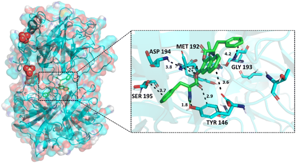 | ||
| Fig. 1 Simulated binding model of probe 1 in the active site of Chy. | ||
AFM images of 1-doped TbCh gel with or without enzyme incubation (30 min) indicated that the gel fibres were intact even upon enzyme action on the substrate. Clearly, Chy did not affect the gel morphology (Fig. 2) suggesting the gel's robustness and stability (Fig. 3).
 | ||
| Fig. 2 AFM images of (a) 1 doped TbCh gel (b) (1 + enzyme incubated at 25 °C for 30 min) doped TbCh gel. | ||
 | ||
| Fig. 3 Lineweaver–Burk plot for α-chymotrypsin assay. | ||
To optimize the assay, two methods were tested. In method 1, the enzyme assay was carried out in the gel medium wherein both the stock solutions of Chy and substrate 1 were prepared in 30 mM sodium cholate solutions, and simple mixing of the two solutions with 10 mM aqueous Tb(OAc)3 formed a gel that was incubated at 25 °C for 20 min. The gel turned luminescent with time, indicating the release of DHN upon the enzyme action. In method 2, both the stock solutions of Chy and 1 were prepared in 30 mM sodium cholate solution. This mixture was incubated at 25 °C, then mixed with 10 mM Tb(OAc)3 to form the gel, which was luminescent indicating the formation of DHN.
Both methods involved incubation at 25 °C. Method 1 being more straightforward (enzyme incubation and gel processing in a single step) was followed for enzyme activity measurements. The KM value was found to be 85 μM, comparable with previous reports (Table S1†). The lower value of KM denotes significant affinity of α-chymotrypsin towards the synthetic non-peptide substrate.
To check the selectivity of the assay, compound 1 was incubated with α-chymotrypsin and several other enzymes such as lipase, urease, β-glucuronidase, trypsin, and alkaline phosphatase that could co-exist with α-chymotrypsin in biological samples. Tb(III)-luminescence enhancement was observed only with α-chymotrypsin, and no luminescence changes could be detected with other enzymes even when the interfering enzyme concentration was 25-fold higher (Fig. 4).
 | ||
| Fig. 4 Extent of emission enhancement at 545 nm (λex 335 nm) after incubating 1 with Chy (0.4 U mL−1) and other enzymes (10 U mL−1) for 30 min at 25 °C. Final incubation concentration of 1 was 37.5 μM. | ||
The limit of detection (LOD) for Chy calculated using the formula LOD = 3σ/b (σ = standard deviation of the signal obtained from the blank and b = slope of the titration plot) were found to be 0.014 U mL−1 and 0.037 U mL−1, for method 1 and method 2, respectively (Fig. 5). The response was generated within 15 min, indicating the assay's sensitivity, selectivity, rapidness, and practicality.
 | ||
| Fig. 5 Emission at 545 nm (λex 335 nm) for α-chymotrypsin assay a) method 1 and b) method 2. | ||
A rapid enzyme assay should be ideally suited for rapid screening of inhibitors, and we have explored this possibility too. 1-Naphthol, a known inhibitor37 of α-chymotrypsin is associated with reduced testosterone levels in adult men.35 It is a metabolite of carbaryl and naphthalene that is an intermediate in the metabolism of xenobiotics by cytochrome P450. The IC50 value for 1-napthol calculated using our methodology was 0.58 mM (Fig. S12†). Further refinement of our gel-based assay using 1 can lead to rapid screening of α-chymotrypsin inhibitors and may greatly help drug discovery processes and detailed enzyme activities in real systems in the presence of potentially interfering metabolites.
To further improve the protocol, we have designed paper-based sensors.38–44 TbCh hydrogel (5/15 mM) doped with the pro-sensitizer 1 (37.5 μM) was stabilized at RT for 10 min, sonicated again (5–6 s) to reduce its viscosity. A 20 μL aliquot of this weak gel was drop casted on 3.5 mm diameter discs cut from Whatman 3 paper using a standard one-hole punch. The paper absorbed the gel in 30 min and was transferred to a 96-well plate. α-Chymotrypsin solution (10 μL, prepared in NaCh) was added on each disc, and emission measurements were recorded using a plate reader after 15 min of air drying. The LOD using this paper-based strategy was found to be 330 ng mL−1 or 0.012 U mL−1 (Fig. 6a). This value is comparable to previous literature-reported values (Table S2†). The presence of Chy in a commercial tablet was readily detected using the paper-based method (Fig. 6b). One essential criterion for a sensor to smoothly reach end-users is its low cost which generally reflects its practicability in the real diagnostic field. In resource-limited areas, such a protocol can be very useful where fully equipped lab facilities are unavailable. Material cost for our developed paper-based sensor was calculated and was estimated to be less than GBP 0.01 for a single paper disc.
 | ||
| Fig. 6 (a) LOD measurement for paper-based α-chymotrypsin assay, (b) detection of Chy in a commercial tablet. | ||
In conclusion, we developed an innovative luminogenic supramolecular framework for highly selective and sensitive sensing of α-chymotrypsin. A non-peptide-based small-molecule probe was designed, which released the sensitizer molecule upon the action of Chy. As the sensing strategy involved time-gated luminescence of sensitized Tb(III), it has a competitive advantage over the prevalent methodologies that commonly employ fluorogenic probes. The technique was subsequently utilized in the testing an inhibitor of α-chymotrypsin. Furthermore, the gel's soft, solid-like characteristics facilitated enhanced immobilization on a passive substrate like paper, thus leading to the development of a simple, inexpensive paper-based sensor. This marks the first demonstration of merging the benefits of both paper-based sensing technologies and a delayed luminescence output for α-chymotrypsin assay. Considering these attributes, this probe framework emerges as an appealing prospect for forthcoming analytical applications in drug exploration and the clinical diagnosis of ailments related to the pancreas in pharmaceutical and pathological investigations.45
Data availability
The data supporting this article have been included as part of the ESI.†Conflicts of interest
There are no conflicts to declare.Acknowledgements
Science and Engineering Research Board, India, supported this work through grant no. CRG/2020/001140. UM also thanks SERB for the award of the J. C. Bose fellowship (SR/S2/JCB-68/2007). Mr. Arnab Ghosh (MBU, IISc) is acknowledged for help with the docking studies. AB thanks the Council of Scientific and Industrial Research, New Delhi, for a research fellowship.Notes and references
- H. Tsukada and D. M. Blow, J. Mol. Biol., 1985, 184, 703–711 CrossRef CAS PubMed.
- A. Kumar and P. Venkatesu, Chem. Rev., 2012, 112, 4283–4307 CrossRef CAS PubMed.
- H. U. Bergmeyer, Methods of enzymatic analysis, Academic Press, London, 1974 Search PubMed.
- S. Yamashita, M. Sakabe, T. Ishizawa, K. Hasegawa, Y. Urano and N. Kokudo, Br. J. Surg., 2013, 100, 1220–1228 CrossRef CAS PubMed.
- G. Montalto, A. Carroccio, G. Marino, M. Soresi, C. Di Marco and A. Notarbartolo, Acta Diabetol. Lat., 1990, 27, 157–163 CrossRef CAS PubMed.
- S. H. Bharmal, S. A. Pendharkar, R. G. Singh, M. O. Goodarzi, S. J. Pandol and M. S. Petrov, Pancreatology, 2017, 17, 876–883 CrossRef CAS PubMed.
- J. P. Van De Merwe and G. J. J. Mol, Digestion, 1982, 24, 1–4 CAS.
- J. Zhou and M. Sahin-Tóth, J. Gastroenterol. Hepatol., 2011, 26, 1238–1246 CrossRef CAS PubMed.
- Y. Chen, J. Cao, X. Jiang, Z. Pan and N. Fu, Sens. Actuators, B, 2018, 273, 204–210 CrossRef CAS.
- W. Appel, Clin. Biochem., 1986, 19, 317–322 CrossRef CAS PubMed.
- G. R. Schonbaum, B. Zerner and M. L. Bender, J. Biol. Chem., 1961, 236, 2930–2935 CrossRef CAS PubMed.
- K. Xu, F. Liu, J. Ma and B. Tang, Analyst, 2011, 136, 1199–1203 RSC.
- L. Liu, C. Liu and L. Gao, Biosensors, 2023, 13(2), 263–272 CrossRef CAS PubMed.
- D. Milićević and J. Hlavác, ACS Omega, 2024, 9, 17481–17490 Search PubMed.
- P. J. Brynes, P. Bevilacqua and A. Green, Anal. Biochem., 1981, 116, 408–413 CrossRef CAS PubMed.
- L. Wu, S. H. Yang, H. Xiong, J. Q. Yang, J. Guo, W. C. Yang and G. F. Yang, Anal. Chem., 2017, 89, 3687–3693 CrossRef CAS PubMed.
- H. Xiong, R. R. Li, S. Y. Liu, F. S. Wu, W. C. Yang and G. F. Yang, ACS Appl. Bio Mater., 2018, 1, 310–317 CrossRef CAS PubMed.
- S. Mu, Y. Xu, Y. Zhang, X. Guo, J. Li, Y. Wang, X. Liu and H. Zhang, J. Mater. Chem. B, 2019, 7, 2974–2980 RSC.
- Y. Qu, Z. Xu, J. Wang, W. Liu, A. Iqbal, K. Iqbal, Y. Su, Y. Cao, J. Yang, W. Qin and Y. Liu, Sens. Actuators, B, 2023, 382, 133552 CrossRef CAS.
- X. Zou, Y. Zhao, C. Lai, Y. Liang and W. Lin, J. Mater. Chem. B, 2021, 9, 8417–8423 RSC.
- C. Fan, J. Gao, Y. Gao, X. Yang, G. Li, X. Wang, F. Li, J. Zhou, H. Yu, Y. Huang, J. Chen, Y. Shan, L. Chen, C. Fan, J. Gao, Y. Gao, X. Yang and G. Li, Chin. Chem. Lett., 2024, 109838 CrossRef CAS.
- Y. Chen, J. Cao, X. Jiang, Z. Pan and N. Fu, Sens. Actuators, B, 2018, 273, 204–210 CrossRef CAS.
- Y. Zhao, X. Zou, X. Liang, L. Huang and W. Lin, Sens. Actuators, B, 2023, 382, 133553 CrossRef CAS.
- H. Shi, C. Liu, J. Cui, J. Cheng, Y. Lin, L. Gao and R. Luo, New J. Chem., 2020, 44, 20921–20929 RSC.
- K. Kuromizu, Y. Shimokawa, O. Abe and N. Izumiya, Anal. Biochem., 1985, 151, 534–539 CrossRef CAS PubMed.
- M. C. Heffern, L. M. Matosziuk and T. J. Meade, Chem. Rev., 2014, 114, 4496–4539 CrossRef CAS PubMed.
- S. Bhowmik and U. Maitra, Chem. Commun., 2012, 48, 4624–4626 RSC.
- A. Biswas and U. Maitra, RSC Adv., 2022, 12, 26106–26110 RSC.
- S. Bhowmik, S. Banerjee and U. Maitra, Chem. Commun., 2010, 46, 8642–8644 RSC.
- A. Biswas and U. Maitra, Chem. Commun., 2024, 60, 6765–6768 RSC.
- R. Laishram, S. Bhowmik and U. Maitra, J. Mater. Chem. C, 2015, 3, 5885–5889 RSC.
- T. Gorai, S. Sakthivel and U. Maitra, Chem. – Asian J., 2020, 15, 4023–4027 CrossRef CAS PubMed.
- T. Gorai and U. Maitra, J. Mater. Chem. B, 2018, 6, 2143–2150 RSC.
- T. Gorai and U. Maitra, ACS Sens., 2016, 1, 934–940 CrossRef CAS.
- S. Liu, X. Wang, Q. Hu, Y. Geng and H. Dong, J. Food Compos. Anal., 2023, 116, 105064–105070 CrossRef CAS.
- Chy did not act on the bis-D-phenylalanine ester derivative of DHN.
- R. A. Wallace, A. N. Kurtz and C. Niemann, Biochemistry, 1963, 2, 824–836 CrossRef CAS PubMed.
- Q. Ju, M. O. Noor and U. J. Krull, Analyst, 2016, 141, 2838–2860 RSC.
- K. Tenda, B. van Gerven, R. Arts, Y. Hiruta, M. Merkx and D. Citterio, Angew. Chem., Int. Ed., 2018, 57, 15369–15373 CrossRef CAS PubMed.
- S. K. Mahadeva, K. Walus and B. Stoeber, ACS Appl. Mater. Interfaces, 2015, 7, 8345–8362 CrossRef CAS PubMed.
- F. Güder, A. Ainla, J. Redston, B. Mosadegh, A. Glavan, T. J. Martin and G. M. Whitesides, Angew. Chem., Int. Ed., 2016, 55, 5727–5732 CrossRef PubMed.
- B. Liu, D. Du, X. Hua, X. Y. Yu and Y. Lin, Electroanalysis, 2014, 26, 1214–1223 CrossRef CAS.
- C. Parolo and A. Merkoçi, Chem. Soc. Rev., 2013, 42, 450–457 RSC.
- A. M. López-Marzo and A. Merkoçi, Lab Chip, 2016, 16, 3150–3176 RSC.
- A detailed validation with clinical samples will be published after completing the assay-development of other relevant enzymes for whole pancreatic function studies.
Footnote |
| † Electronic supplementary information (ESI) available. See DOI: https://doi.org/10.1039/d4sd00124a |
| This journal is © The Royal Society of Chemistry 2024 |